APPLIED AND ENVIRONMENTAL MICROBIOLOGY, Oct. 2003, p. 5907–5913 0099-2240/03/$08.00⫹0 DOI: 10.1128/AEM.69.10.5907–5913.2003 Copyright © 2003, American Society for Microbiology. All Rights Reserved.
Vol. 69, No. 10
Effects of Iron Limitation on Adherence and Cell Surface Carbohydrates of Corynebacterium diphtheriae Strains Lílian de Oliveira Moreira,1,2 Arnaldo Feitosa Braga Andrade,1 Ma´rcio Damasceno Vale,1 So ˆnia Maria Silva Souza,1,3 Raphael Hirata, Jr.,1 Lídia Maria Oliveira Buarque Asad,3 Nasser Ribeiro Asad,3 Luiz Henrique Monteiro-Leal,3 Jose´ Osvaldo Previato,2 and Ana Luiza Mattos-Guaraldi1* Faculdade de Cieˆncias Me´dicas1 and Instituto de Biologia Prof. Roberto Alca ˆntara Gomes,3 Universidade do Estado do Rio de Janeiro, and Instituto de Biofísica Carlos Chagas Filho, Universidade Federal do Rio de Janeiro,2 Rio de Janeiro, Brazil Received 2 December 2002/Accepted 18 July 2003
Iron limitation may cause bacterial pathogens to grow more slowly; however, it may also stimulate these microorganisms to produce greater tissue damage, given that many virulence factors are controlled by the iron supply in the environment. The present study investigated the influence of low iron availability on the expression of proteins and surface sugar residues of two toxigenic strains of Corynebacterium diphtheriae subsp. mitis and evaluated their adherence to human group B erythrocytes and HEp-2 cells. A comparison was made between bacteria grown in (i) Trypticase soy broth (TSB), (ii) TSB treated with dipyridyl to deplete free iron, and (iii) TSB enriched with FeCl3. The effects of iron concentration on adhesive properties were different for strains 241 and CDC-E8392, of the sucrose-fermenting and non-sucrose-fermenting biotypes, respectively. Iron-limited conditions enhanced interaction of strain 241 with erythrocytes and HEp-2 cells. Inhibition assays suggested the involvement of nonfimbrial protein combination 67-72p on hemagglutination of diphtheria bacilli grown under iron-limited conditions. Conversely, iron limitation inhibited adherence to glass and expression of electron-dense material on the bacterial surface. Lectin binding assays demonstrated a reduction in the number of sialic acid residues and an increase in D-mannose and D-galactose residues on the surfaces of both strains. Thus, iron exerts a regulatory role on adhesive properties of diphtheria bacilli, and low iron availability modulates the expression of C. diphtheriae surface carbohydrate moieties. The significant changes in the degree of lectin binding specific for D-mannose, D-galactose and sialic acid residues may have an effect on binding of host cells. The expression of dissimilar microbial virulence determinants may be coordinately controlled by common regulatory systems. For C. diphtheriae, the present results imply regulation of adherence and slime production as part of a global response to iron-limited environmental conditions that includes derepression of genes for the synthesis of cytotoxin and siderophores and for transport of the Fe(III)siderophore complexes. hanced ability to colonize and spread (14). Attachment of bacteria is a critical step in the pathogenesis of many infections, particularly in cases where the pathogen is confined to mucosal surfaces. Investigations carried out with Brazilian diphtheria bacillus isolates demonstrated that non-sucrose-fermenting strains preferentially colonize skin lesions and show higher cell surface hydrophobicity than throat-colonizing sucrose-fermenting strains (12, 13). C. diphtheriae strains adhere to human erythrocytes and solid surfaces at various intensities. The hemagglutinating activity of nonfimbrial adhesion protein combination 67-72p is influenced by the concentrations of sugar residues expressed on bacterial surface (5). Sialic acid terminal moieties are expressed mainly on the surface of the nonhemagglutinating and highly glass-adherent sucrose-fermenting strain 241 (11). C. diphtheriae, which is generally considered to be an extracellular colonizer, also exhibits the ability to survive within cultured epithelial cells (9). The molecular differences observed in bacterial adherence to host cells might correlate with maintenance and dissemination of specific C. diphtheriae clones. The expression of adherence factors by several species is influenced by the iron supply in the environment. Iron restriction in the growth medium promotes slime production by
The examination of environmental signals controlling virulence is an essential step in comprehension of the underlying strategies that microbes have adopted to become successful pathogenic organisms (15). Iron is essential for bacterial growth, but its availability in the human body is very limited because it is almost entirely complexed with metalloproteins or glycoproteins (3, 24). During infection, the restriction of iron is stronger because the host response to the invading bacteria includes hypoferremia (4). Corynebacterium diphtheriae is able to overcome host conditions, in part by producing siderophores or other iron uptake mechanisms that allow it to express virulence factors such as toxins and enzymes. Cytotoxins, siderophores, heme oxygenase (HmuO), and cell surface lipoproteins are regulated by both iron and DtxR protein (18, 20–22). Epidemic or invasive clones, as well as the atypical sucrosefermenting biotype, of diphtheria bacilli seem to possess some selective advantage, such as increased virulence or an en-
* Corresponding author. Mailing address: Disciplina de Microbiologia e Imunologia, Faculdade de Cieˆncias Me´dicas, Universidade do Estado do Rio de Janeiro, Av. 28 de Setembro, 87 Fundos, no. 3 andar, Vila Isabel, RJ CEP 20 551-030, Brazil. Phone: 55 (21) 2587-6380. Fax: 55 (21) 2587-6476. E-mail:
[email protected]. 5907
5908
MOREIRA ET AL.
APPL. ENVIRON. MICROBIOL.
TABLE 1. Number of viable bacterial cells in supernatant and HEp-2 cell monolayer lysates and adherence index for C. diphtheriae strains grown under standard and iron-limited conditions No. of viable bacterial cells (106)a Strain and incubations time (min)
Adherence index (%)b
TSB ⫺ Fe
TSB
TSB
TSB ⫺ Fe
P valuec
1.23 ⫾ 0.24 0.51 ⫾ 0.09
61.86 32.98
91.75 57.20
0.001 0.001
0.03 ⫾ 0.01 0.03 ⫾ 0.01
18.29 59.11
15.21 58.02
0.779 0.443
Total
Cell lysate
Total
Cell lysate
Sucrose-fermenting 241 30 120
0.84 ⫾ 0.12 1.06 ⫾ 0.28
0.50 ⫾ 0.16 0.36 ⫾ 0.13
1.33 ⫾ 0.32 0.85 ⫾ 0.11
Non-sucrose-fermenting CDC-E8392 30 120
0.28 ⫾ 0.08 0.17 ⫾ 0.11
0.05 ⫾ 0.02 0.12 ⫾ 0.11
0.18 ⫾ 0.03 0.07 ⫾ 0.01
a TSB, bacterial growth in standard TSB; TSB ⫺ Fe, TSB medium with 0.5 mM dypiridyl; total, number of viable bacterial cells in supernatant plus number of viable bacterial cells obtained from lysate of infected HEp-2 monolayers during specific period of interaction; cell lysate, number of viable bacteria obtained from lysate of infected HEp-2 monolayers during specific period of interaction. Values are medians ⫾ standard deviations from three experiments, with differences lower than 10%. b Number of viable bacterial cells in lysate of infected monolayers during specific period of interaction ⫻ (number of viable bacterial cells in supernatant plus number of viable bacterial cells in lysate of monolayers during specific period of interaction)⫺1 ⫻ 100. c Statistically significant at a P value of ⬍0.01.
Staphylococcus aureus (1, 17) and Staphylococcus epidermidis (7), production of mucin-binding adhesins by Pseudomonas aeruginosa (19), and hydrophobicity of and adherence to HEp-2 cells by Vibrio parahaemolyticus (6). Although studies have reported regulatory roles of iron in the pathogenesis of C. diphtheriae infection, little is known about the actual connection between iron availability and the adhesive properties of diphtheria bacilli. The present study investigated the influence of low iron availability on the expression of proteins and surface sugar residues of two toxigenic strains of C. diphtheriae and evaluated their adherence to human erythrocytes and HEp-2 cells. MATERIALS AND METHODS Bacterial strains and culture conditions. The toxigenic, sucrose-fermenting C. diphtheriae subsp. mitis strain 241 (from Diphtheria Laboratory of Faculdade de Cieˆncias Me´dicas, Universidade do Estado do Rio de Janeiro, Rio de Janeiro, Brazil) and the non-sucrose-fermenting strain CDC-E8392 (from the Centers for Disease Control and Prevention, Atlanta, Ga.) were used in this study (14). Microorganisms were stored in GC medium base (Difco Laboratories, Detroit, Mich.) with 20% glycerol at ⫺20°C. Microorganisms were grown in three different media at 37°C for 24 h without shaking: Trypticase soy broth (TSB) (Difco) (iron content [measured by atomic absorption spectrophotometry] of 0.74 g ml⫺1), TSB deprived of iron by addition of the chelating agent 2,2⬘-dipyridyl (Sigma Chemical Co., St. Louis, Mo.) at a final concentration of 0.5 mM (TSB ⫺ Fe) (19) and TSB enriched with 4.0 mM to 1.0 M FeCl3 (TSB ⫹ FeCl3) (21). Experiments were also done with microorganisms initially cultured in iron-limited TSB and subcultured in TSB [(TSB ⫺ Fe)TSB]. Bacterial cells were harvested by centrifugation at 10,000 ⫻ g, washed with 0.01 M Na–phosphate-buffered saline (PBS) (pH 7.2) to deplete the chelated-iron residues, and subjected to further studies. Bacterial growth was monitored by counting CFU on solid media (6). Determination of adherence to HEp-2 cells. Bacterial adherence to a cell line derived from a human epidermoid larynx carcinoma (ATCC CCL23 HEp-2; American Type Culture Collection, Manassas, Va.) was assayed as previously described (9). Briefly, HEp-2 cells were grown in Dulbecco’s modified Eagle medium (Sigma) supplemented with 5% fetal calf serum (Gibco BRL, Grand Island, N.Y.), 50 g of gentamicin ml⫺1, 2.5 g of amphotericin B ml⫺1, and 0.5% L-glutamine at 37°C in 5% CO2 atmosphere. Microorganisms were washed twice with Dulbecco’s mineral salt solution (PBS-D; Sigma) and resuspended in Dulbecco’s modified Eagle medium without antibiotics to a final concentration of 1.0 ⫻ 106 bacteria ml⫺1. Monolayers grown to about 95% confluence in 24-well tissue culture plates were infected with 250 l of bacterial suspension per well. Infected monolayers were washed six times and lysed with PBS-D plus 0.1% Triton X-100 after incubation in 5% CO2 at 37°C for periods of 30 and 120 min. Bacterial viable counts (CFU) of supernatant and HEp-2 monolayer lysates for
each incubation period were determined and expressed as the mean ⫾ standard deviation from three independent experiments performed in triplicate. The adherence index for each incubation period represented the percentage of adherence, calculated as lysate CFU ⫻ (lysate CFU ⫹ supernatant CFU)⫺1 ⫻ 100. Hemagglutination and hemagglutination inhibition assays. Hemagglutination and hemagglutination inhibition assays were performed with a 0.5% suspension of human group B erythrocytes as previously described (5, 12). The nonfimbrial adhesion protein combination 67-72p were obtained from C. diphtheriae strain CDC-E8392 grown in TSB by mechanical blending and precipitation by ammonium sulfate (25 and 45% saturation) (5). Hemagglutination inhibition assays were performed in the presence of dilutions of a solution of these protein adhesins (300 g/ml) at 37°C for 1 h. Bacterial autoaggregation assays. Briefly, microorganisms that remained clumped in TSB medium were considered to be spontaneously autoaggregating bacteria. Aliquots (50 l) of bacterial suspensions prepared in PBS (pH 6.8) were dropped on a glass slide and observed for aggregation. Nonaggregating bacteria produced turbid suspensions. Spontaneously autoaggregating strains were considered to be highly hydrophobic, and aggregating strains were considered to be moderately hydrophobic (13). Assays for adherence to glass. Briefly, microorganisms were inoculated in glass tubes (13 by 100 mm) containing 4 ml of TSB and incubated for 48 h at 37°C without shaking. The tubes were gently shaken for 5 s, and the supernatants containing bacterial cells that were nonadherent to the surfaces of the glass tubes were discarded. TSB (4 ml) was then added, and the tubes were reincubated for 48 h. This procedure was repeated twice. The glass-adherent bacteria created a confluent coat of cells on sides of the tube (12). SDS-PAGE analysis of crude cells lysates. Bacterial cells were lysed by treatment with 5 mg of lysozyme (grade III; Sigma) ml⫺1 in PBS for 2 h at 37°C (16). Protein samples were solubilized in cracking buffer (0.5 mM Tris-HCl [pH 6.8], 4% sodium dodecyl sulfate [SDS], 20% glycerol, 10% -mercaptoethanol, and 0.001% bromophenol blue) by heating at 100°C for 15 min. After cell debris and unbroken cells were cleared by centrifugation at 14,000 ⫻ g for 5 min, supernatants were collected and used as crude cell lysates. The protein profiles were observed by SDS–10% polyacrylamide gel electrophoresis (SDS–10% PAGE) (Bio-Rad, Richmond, Calif.) with the Laemmli buffer system (10), and the gels were stained with Coomassie brilliant blue R-250 (Bio-Rad). Western blot analysis of bacterial surface proteins. Bacterial cell surface proteins were labeled with biotin (Sigma) at 37°C for 1 h by previously described methods (8) and washed with PBS (pH 9.0) before preparation of crude cell lysates as described above. Proteins bands obtained in SDS-PAGE were transferred to a nitrocellulose membrane at 100 V and 400 mA for 90 min with a Mini Trans-Blot cell (Bio-Rad). Protein blots were blocked in PBS containing 0.5% Tween 20 with 5% skim milk for 2 h at room temperature, washed three times with PBS-D (pH 7.3), and incubated with streptavidin-peroxidase conjugate (Sigma) diluted 1:5,000 in PBS–0.5% Tween 20 for 30 min at room temperature. The nitrocellulose membrane was washed three times and then reacted with 0.3% hydrogen peroxide, 1 mg of 3,3⬘-diaminobenzidine ml⫺1, and 1 mg of imidazole ml⫺1 for color development (25).
IRON LIMITATION AND ADHERENCE OF C. DIPHTHERIAE
VOL. 69, 2003
5909
TABLE 2. Adherence to glass and autoaggregating activity of C. diphtheriae strains grown under different iron conditionsa Strain
Sucrose-fermenting 241 Non-sucrose-fermenting CDC-E8392
Glass adherence
Autoaggregation
TSB
TSB ⫺ Fe
(TSB ⫺ Fe)TSB
TSB ⫹ FeCl3
TSB
TSB ⫺ Fe
(TSB ⫺ Fe)TSB
TSB ⫹ FeCl3
⫹ ⫺
⫺ ⫺
⫹ ⫺
⫹ ⫺
⫺ ⫹
⫹⫹ ⫹⫹
⫺ ⫹
⫺ ⫹
a TSB, bacterial growth in standard TSB medium; TSB ⫺ Fe, TSB medium with 0.5 mM dypiridyl; (TSB ⫺ Fe)TSB, initial incubation in TSB with 0.5 mM dypiridyl and reincubation in standard TSB; TSB ⫹ FeCl3, TSB with 4.0 mM to 1 M FeCl3; ⫹, positive result; ⫹⫹, strong positive result; ⫺, negative result.
Lectin binding studies. Briefly, a bacterial suspension (15 l) containing 3 ⫻ 107 bacteria ml⫺1 was placed on glass slides, air dried, and fixed in methanol at 22°C for 10 min. The slides were washed in PBS–5% bovine serum albumin (BSA) for 5 min and then incubated with 15 l of increasing dilutions of fluorescein isothiocyanate-labeled lectins (FITC-lectin) (Sigma) in PBS–5% BSA at 22°C for 30 min. The slides were then washed three times in PBS–5% BSA for 5 min each time, mounted in PBS-glycerol (50%), and observed under a fluorescence microscope (Universal Photomicroscope; Zeiss, Oberkochen, Germany). FITC-lectin binding to bacterial cells was inhibited by preincubation with the specific sugar hapten (0.1 M) (11). Lectin radioiodination and binding studies. The lectins from Sambucus nigra (SNA) and Canavalia ensiformis (ConA) were conjugated with 125I-labeled acylating agent in the presence of 0.1 M specific inhibitory sugars (2). Specific activities ranged from 1 ⫻ 104 to 3 ⫻ 104 cpm of lectin g⫺1. For the lectin binding studies, 106 bacterial cells were incubated with a range of concentrations of iodinated lectin in 150 l of PBS–0.5% BSA. The amount of iodinated lectin bound to the cells was determined with a gamma counter (Beckman Instruments, Inc., Palo Alto, Calif.). The specificity of binding was ascertained by performing parallel binding determinations in the presence of a 0.1 M concentration of the specific sugar inhibitor for each concentration of lectin used (11).
Transmission electron microscopy (TEM). Bacterial cells (strain 241) were fixed with 0.2% glutaraldehyde at 4°C for 15 min, placed on Formvar-coated grids (200 mesh; Sigma), negatively stained with 1% potassium phosphotungstate, and viewed in an EM 906 Zeiss transmission electron microscope (5). Statistical analysis. Results were statistically analyzed with Student’s t test.
RESULTS HEp-2 cell adherence. Higher levels of adherence to HEp-2 cells by strains 241 (62%) and CDC-E8392 (59%) cultured in standard medium (TSB) were observed within 30 and 120 min of incubation, respectively (Table 1). Iron-limited (TSB ⫺ Fe) cultures of strain 241 showed significantly higher levels of adherence to HEp-2 cells than standard (TSB) cultures within 30 and 120 min of incubation (P ⫽ 0.001). The CDC-E8392 strain showed similar adherence levels when cultured under standard and iron-limited conditions (P ⬎ 0.01). The total number of
FIG. 1. Protein profiles of strains CDC-E8392 (A) and 241 (B) of C. diphtheriae grown under standard and iron-limited conditions. Total protein was analyzed by SDS–10% PAGE. Lane 1, molecular mass markers; lanes 2 and 4, strains CDC-E8392 and 241, respectively, from standard cultures; lanes 3 and 5, strains CDC-E8392 and 241, respectively, from iron-limited cultures. Arrows indicate proteins bands expressed at high levels, and dots indicate proteins bands that were absent or expressed at low levels in iron-limited cultures.
5910
MOREIRA ET AL.
APPL. ENVIRON. MICROBIOL.
TABLE 3. Activities of lectins of various specificities for C. diphtheriae strains grown under standard and iron-limited conditionsa Minimum lectin conc (g ml⫺1) for detectable fluorescence of strain: Lectin specificity
CDC-E8392 (non-sucrose fermenting) TSB
D-GlcNac-binding
lectins Triticum vulgaris (wheat germ agglutinin) Bandeiraea simplicifolia (BS-II) Solanum tuberosum (STA)
D-GalNAc-binding
lectins Glycine max (SBA) Wisteria floribunda (WFH) Artocarpus integrifolia (JACA)
D-Gal-binding
lectins Euonymus europaeus (EEL) Arachis hypogaea (PNA) Ricinus communis I (RCA-I)
D-Man-binding
lectins Canavalia ensiformis (ConA) Lens culinaris (LCL) Pisum sativum (PSA)
Sialic acid-binding lectin, Sambucus nigra (SNA) a b
TSB ⫺ Fe
241 (sucrose fermenting) TSB
Effect of iron depletion on lectin binding
TSB ⫺ Fe
62.5 31.2 250.0
250.0 31.2 250.0
15.6 3.9 125.0
250.0 3.9 125.0
Decrease No effect No effect
31.2 250.0 31.2
31.2 62.5 31.2
125.0 250.0 125.0
125.0 31.5 125.0
No effect Increase No effect
⬎500b ⬎500 62.5
125.0 62.5 15.6
⬎500 ⬎500 250.0
125.0 15.6 31.2
Increase Increase Increase
62.5 125.0 125.0
7.8 31.2 31.2
125.0 250.0 250.0
3.9 15.6 15.6
Increase Increase Increase
62.5
250.0
15.6
250.0
Decrease
Results are average from three experiments. TSB, bacterial growth in standard TSB medium; TSB ⫺ Fe, TSB medium with 0.5 mM dypiridyl. No fluorescence was seen at the maximum concentration used.
cells of strain 241 remained essentially unchanged during 120 min of incubation with HEp-2 cells, while the number of CDCE8392 cells dropped sharply to between 7 and 17% of the inoculums. This seemed to be partially due to medium sensitivity of strain CDC-E8392. However, a differential killing effect by the host cells is also a possibility, which remains under investigation. Hemagglutination. The hemagglutinating activity (titer of 32) of strain CDC-E8392 was independent of iron availability. For strain 241, binding to erythrocytes (titer of 4) occurred only when the bacteria were grown in iron-limited medium (TSB ⫺ Fe) (data not shown). The hemagglutinating activity of strain 241 grown under iron-limited conditions was completely inhibited by subsequent growth of the strain in standard medium [(TSB ⫺ Fe)TSB]. For strain CDC-E8392, there were no differences between the hemagglutinating titers of microorganisms grown in TSB ⫺ Fe and TSB. The addition of 4.0 to 1 M FeCl3 to TSB did not result in any significant changes in the hemagglutinating properties of strains 241 (negative) and CDC-E8392 (titer of 32) compared to those seen with TSB. The results of inhibition assays showed that the heterologous 67-72p, obtained from strain CDC-E8392 grown in TSB, inhibited hemagglutination of strain 241 at a minimum concentration of 0.5 g ml⫺1. 67-72p also inhibited hemagglutination of strain CDC-E8392 grown in both TSB and TSB ⫺ Fe. The data suggest involvement of 67-72p in the hemagglutinating activity of diphtheria bacilli grown under iron-limited conditions. Adherence to glass and hydrophobic properties. The results in Table 2 demonstrate that strain 241 grown in iron-limited medium (TSB ⫺ Fe) did not exhibit binding to glass surfaces. Strain 241 cultured in TSB ⫺ Fe became adherent to glass
after its subsequent growth in standard TSB. Conversely, iron limitation led to spontaneous autoaggregating of both strains 241 and CDC-E8392. Expression of iron-related peptides. Figure 1 shows the SDS-PAGE whole-cell protein profiles of strains CDC-E8392 and 241 grown under standard (lanes 2 and 4) and iron-limited (lanes 3 and 5) conditions. The expression of crude proteins with unknown functions was enhanced in both strains CDCE8392 (54- and 69 kDa peptides) and 241 (54-kDa peptide) cultured in iron-limited medium. Western blotting analysis of nitrocellulose sheets containing biotinylated bacterial surface proteins of strain 241 revealed six to eight distinct biotinlabeled surface proteins. The number and relative labeling levels were the same in cells of strain 241 grown under standard and iron-limited culture conditions, identifying minor differences in cell wall protein patterns due to iron. The presence of biotin-labeled surface proteins in the 67- to 72-kDa range suggests expression of 67-72p adhesins by strain 241 grown under both standard and iron-limited culture conditions (data not shown). Fluorescent lectin binding assay analysis. The data present in Table 3 demonstrate that (i) the cells bound representatives of all of the classes of lectins tested; (ii) all of the D-galactose (D-Gal), D-mannose (D-Man), and N-acetylneuraminic (sialic) acid lectins that were bound reacted differentially to bacterial cells depending on the iron conditions of growth; (iii) these differential reactions (when present) were similar in both strains; (iv) N-acetylglucosamine (D-GlcNAc) and N-acetylgalactosamine (D-GalNAc) showed iron-sensitive reactivity only in some cases; and (v) while the D-Gal, D-Man, and DGalNAc lectins showed an increase, the sialic acid and DGlcNAc lectins showed a decrease in binding at low iron con-
VOL. 69, 2003
IRON LIMITATION AND ADHERENCE OF C. DIPHTHERIAE
5911
cose-binding lectins did not react with the C. diphtheriae strains tested (data not shown). 125 I-lectin binding analysis. As shown in Fig. 2A, increasing concentrations of 125I-lectin resulted in binding of the labeled lectin in a saturable fashion. The bacterial growth in ironlimited medium resulted in a marked reduction in 125I-SNA binding, confirming that these bacteria were poorly sialylated. However, a significant increase was observed in the 125I-ConA binding assay. The binding was specific, because it could be reversed with a 0.1 M concentration of the corresponding specific saccharide (data not shown). The binding data in Fig. 2A demonstrate that the differential interaction of the C. diphtheriae sucrose-fermenting strain with the SNA and ConA lectins (Table 3) correlated with the number of exposed 125I-lectin receptor sites on them. As shown in Fig. 2B, the average number of lectin-binding sites (n) and the lectin association constant (Ko) were determined. For binding of SNA to bacterial cells grown under standard iron conditions, n was approximately 3.0 ⫻ 105/cell and Ko was approximately 3.9 ⫻ 106 M⫺1; for binding of ConA to bacterial cells grown under ironlimited conditions, n was approximately 5.5 ⫻ 105/cell and Ko was approximately 1.8 ⫻ 106 M⫺1. TEM analysis. Figure 3 demonstrates the structural differences in strain 241 cultured in TSB and TSB ⫺ Fe, respectively. The experimental conditions used did not allow obser-
FIG. 2. (A) Binding of iodinated lectins SNA (F, E) and ConA (Œ, ‚) to C. diphtheriae strain 241 grown under standard (F, ‚) and iron-limited (E, Œ) conditions. (B) For each lectin, the binding data for high-affinity receptors (filled symbols from panel A have been plotted by the method of Steck and Wallach) (23) according to the equation C/[lectin] bound ⫽ 1/Kn ⫻ 1/[lectin] free ⫹ 1/n, where C is the concentration of bacteria, n is the number of lectin molecules bound per cell, K is the lectin association constant, and [lectin] is the concentration of lectin in molar. All points represent means from triplicate experiments; the standard deviations is less than 10%.
centrations. The most significant feature of the D-Man-binding lectins is the striking of ConA for C. diphtheriae strains grown under iron-limited conditions, in that this lectin interacted strongly with sucrose-fermenting and non-sucrose-fermenting strains at minimum concentrations of 3.9 and 7.8 g ml⫺1, respectively. The sialic acid-binding lectin from S. nigra (SNA), as with wheat germ agglutinin lectin, reacted specifically with sucrose-fermenting and non-sucrose-fermenting strains grown in standard TSB at minimum concentrations of 15.6 and 62.5 g ml⫺1, respectively. The FITC-SNA binding to bacterial cells was inhibited by N-acetylneuraminic acid (5 mg ml⫺1). L-Fu-
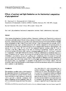
FIG. 3. Electron micrographs showing structural differences on the surface of the sucrose-fermenting C. diphtheriae strain 241 grown under standard (A) and iron-limited (B) conditions. Magnifications, ⫻40,000 and ⫻70,500, respectively.
5912
MOREIRA ET AL.
vation of fimbrial structures. There was no electron-dense material on the bacterial surface of strain 241 grown in ironlimited medium (Fig. 3B). DISCUSSION Iron has a regulatory role in the adhesion of diphtheria bacilli to cells of the human respiratory tract (HEp-2 cells) and blood (erythrocytes). As demonstrated in this study, iron restriction in the growth medium enhanced the adhesive properties of the sucrose-fermenting strain 241. This phenomenon was not observed with the non-sucrose-fermenting strain CDCE8392, suggesting that some, but not all, toxigenic diphtheria bacilli may exhibit an increased ability to colonize epithelial surfaces and spread within the low-iron environment of the host. Analysis of whole-cell protein profiles showed that iron limitation is able to both inhibit and stimulate protein expression, as previously observed with S. epidermidis (26) and S. aureus (17, 24), respectively. The roles of most iron-regulated proteins, other than toxin, in the pathogenicity of C. diphtheriae remain unknown. Previous transmission electron microscopy studies with immunolabeled colloidal gold-protein A revealed a diffuse distribution of 67-72p on the surfaces of both the hemagglutinating CDC-E8392 and nonhemagglutinating 241 C. diphtheriae strains grown in iron-containing medium (5). Here, the results of inhibition assays suggest that the 67-72p protein combination also act as hemagglutinins of C. diphtheriae, including strain 241. Western blot analysis demonstrated the binding of 67-72p to membranes of HEp-2 cells, as previously observed with erythrocytes by immunoblotting (5). Prior investigations demonstrated that differences in degrees of hemagglutination and adherence to glass were related to differences in the expression of surface carbohydrates of C. diphtheriae. For strains 241 and CDC-E8392, lectin receptors containing terminal D-GlcNAc, D-GalNAc, D-Gal, D-Man, and sialic acid were identified on surfaces of cells grown in ironcontaining medium. Sialic acid residues were expressed mainly on the surface of the nonhemagglutinating and highly glassadherent strain 241. It is known that sugar residues, particularly sialic acid residues, contribute to the expression of hydrophilic characteristics and adherence to glass by C. diphtheriae (11). Here we have demonstrated that iron has a regulatory role in the expression of surface carbohydrate moieties of C. diphtheriae strains. We have observed an absolute reduction in the sialic acid residues and a significant increase in the amounts of D-GlcNAc, D-GalNAc, D-Gal, and D-Man residues in both strains. Sialic acid terminal constituents of cell moieties seemed to raise difficulties in C. diphtheriae adherence to HEp2 cells, as previously observed with erythrocytes (11). Hydrophobic interaction is expected to provide the driving force for host-parasite interaction through the displacement of water and formation of adhesive bonds (6). In the present study, for strain CDC-E8392, iron limitation enhanced bacterial hydrophobicity (autoaggregation) but did not influence the adhesion to human cells. For strain 241, low-iron conditions enhanced bacterial autoaggregation and adherence to erythrocytes and HEp-2 cells but inhibited adherence to glass surfaces. In the same way, previous studies demonstrated that the hydrophobicity and HEp-2 cell adherence of the gram-negative
APPL. ENVIRON. MICROBIOL.
species V. parahaemolyticus in iron-limited culture were significantly increased. Those authors suggested that the enhancement of cell adherence of V. parahaemolyticus was probably due to the formation of lateral flagella, a cytotoxic factor, or other, unknown factors (6). However, for C. diphtheriae, iron limitation intensified autoaggregation and adherence to erythrocytes and HEp-2 cells, possibly by reduction of electrostatic repulsion and/or by increased exposure of 67-72p due to the removal of the sialic acid. Similar observations during bacterial treatment with neuraminidase were previously made (5, 11, 13). Iron limitation inhibited slime production by strain 241, as suggested by TEM and corroborated by glass and lectin binding assays. Among the structures involved in virulence, bacteria have developed the production of slime, a higher-molecularmass polymer of carbohydrate that encourages biofilm formation. In the gram-positive species S. epidermidis, iron limitation promotes slime production (7). S. aureus slime-producing strains preferentially accumulate on surfaces and are responsible for chronic colonization, whereas non-slime-producing strains are responsible for acute infection (24). C. diphtheriae slime-producing strains preferentially accumulate on glass surfaces and are responsible for acute respiratory infection, whereas non-slime-producing strains show higher cell surface hydrophobicity and are responsible for colonization of skin lesions (12, 13). Similar to the case for other human pathogens, under ironrestrictive circumstances C. diphtheriae develops alternative metabolic strategies to overcome the environmental conditions. Low iron availability modulates the adhesive properties and expression of surface carbohydrate moieties of strains and consequently may influence the course of C. diphtheriae infection. ACKNOWLEDGMENTS This work was supported by CNPq, CAPES, FAPERJ, SR-2/UERJ, and Programa de Nu ´cleo de Exceleˆncia (PRONEX) of the Brazilian Ministry of Science and Technology. We are grateful to Gabriel Oliver from LAQAM, Instituto de Química, Universidade do Estado do Rio de Janeiro, for technical assistance with iron measurement by atomic absorption spectrophotometry. REFERENCES 1. Baldassari, L., L. Bertuccini, C. R. Ammendolia, C. R. Ariciola, and L. Montanaro. 2001. Effect of iron limitation on slime production by Staphylococcus aureus. Eur. J. Clin. Microbiol. Infect. Dis. 20:343–345. 2. Bolton, A. E., and W. M. Hunter. 1973. The labelling of proteins to high specific radioactivities by conjugation to a 125I-containing acylating agent. Biochem. J. 133:529–540. 3. Bullen, J. J. 1985. Iron and infection. Eur. J. Clin. Microbiol. 4:537–539. 4. Carniel, E. 2001. The Yersinia high-pathogenicity island: an iron-uptake island. Microbes Infect. 3:561–569. 5. Colombo, A. V., R. Hirata, Jr., C. M. R. Souza, L. H. Monteiro-Leal, J. O. Previato, L. C. D. Formiga, A. F. B. Andrade, and A. L. Mattos-Guaraldi. 2001. Corynebacterium diphtheriae surface protein as adhesin to human erythrocytes. FEMS Microbiol. Lett. 197:235–239. 6. Dai, J. A., Y. Lee, and H. Wong. 1992. Effects of iron limitation on production of a siderophore, outer membrane proteins, and hemolysin and on hydrophobicity, cell adherence, and lethality for mice of Vibrio parahaemolyticus. Infect. Immun. 60:2952–2956. 7. Deighton, M., and R. Bordland. 1993. Regulation of slime production in Staphylococcus epidermidis by iron limitation. Infect. Immun. 61:4473–4479. 8. Harlow, E., and D. Lane. 1988. Antibodies: a laboratory manual, p. 353–355. Cold Spring Harbor Laboratory Press, Cold Spring Harbor, N.Y. 9. Hirata, R., Jr., F. Napolea ˜o, L. H. Monteiro-Leal, A. F. B. Andrade, P. E. Nagao, L. C. D. Formiga, L. S. Fonseca, and A. L. Mattos-Guaraldi. 2002. Intracellular viability of toxigenic Corynebacterium diphtheriae strains in HEp-2 cells. FEMS Microbiol. Lett. 215:115–119.
VOL. 69, 2003
IRON LIMITATION AND ADHERENCE OF C. DIPHTHERIAE
10. Laemmli, U. K. 1970. Cleavage of structural proteins during the assembly of the head of bacteriophage T4. Nature 227:680–685. 11. Mattos-Guaraldi, A. L., E. A. Cappelli, J. O. Previato, L. C. D. Formiga, and A. F. B. Andrade. 1999. Characterization of surface saccharides in two Corynebacterium diphtheriae strains. FEMS Microbiol. Lett. 170:159–166. 12. Mattos-Guaraldi, A. L., and L. C. D. Formiga. 1991. Relationship of biotype and source to the hemagglutination and adhesive properties of Corynebacterium diphtheriae. Brazilian J. Med. Biol. Res. 24:399–406. 13. Mattos-Guaraldi, A. L., L. C. D. Formiga, and A. F. B. Andrade. 1999. Cell surface hydrophobicity of sucrose fermenting and nonfermenting Corynebacterium diphtheriae strains by different methods. Curr. Microbiol. 38:37–42. 14. Mattos-Guaraldi, A. L., L. C. D. Formiga, and G. A. Pereira. 2000. Cell surface components and adhesion in Corynebacterium diphtheriae. Microbes Infect. 2:1507–1512. 15. Mekalanos, J. J. 1992. Environmental signals controlling expression of virulence determinants in bacteria. J. Bacteriol. 174:1–7. 16. Merquior, V. L. C., J. M. Peralta, R. R. Facklam, and L. M. Teixeira. 1994. Analysis of electrophoretic whole cell protein profiles as a tool for characterization of Enterococcus sp. Curr. Microbiol. 28:149–153. 17. Morrissey, J. A., A. Cockayne, J. Hammacott, K. Bishop, A. Denman-Johnson, P. J. Hill, and P. Williams. 2002. Conservation, surface exposure, and in vivo expression of the Frp family of iron-regulated cell wall proteins in Staphylococcus aureus. Infect. Immun. 70:2399–2407. 18. Russel, L. M., S. J. Cryz, and R. K. Holmes. 1984. Genetic and biochemical evidence for a siderophore-dependent iron transport system in Corynebacterium diphtheriae. Infect. Immun. 45:143–149. 19. Scharfman, A., H. Kroczynski, C. Carnoy, E. V. Brussel, G. Lamblin, R.
20.
21.
22. 23. 24. 25.
26.
5913
Ramphal, and P. Roussel. 1996. Adhesion of Pseudomonas aeruginosa to respiratory mucins and expression of mucin-binding proteins are increased by limiting iron during growth. Infect. Immun. 64:5417–5420. Schmitt, M. P. 1997. Utilization of host iron source by Corynebacterium diphtheriae: identification of gene whose product is homologous to eukaryotic heme oxygenases and is required for acquisition of iron from heme and hemoglobin. J. Bacteriol. 179:838–845. Schmitt, M. P., and K. K. Holmes. 1991. Characterization of a defective diphtheria toxin repressor (dtxR) allele and analysis of dtxR transcription in wild-type and mutant strains of Corynebacterium diphtheriae. Infect. Immun. 59:3903–3908. Schmitt, M. P., B. G. Talley, and K. K. Holmes. 1997. Characterization of lipoprotein IRP1 from Corynebacterium diphtheriae, which is regulated by diphtheria toxin repressor (DtxR) and iron. Infect. Immun. 65:5364–5367. Steck, T. L., and D. F. H. Wallach. 1965. The binding of kidney-bean phytohaemagglutinin by Ehrlich acites carcinoma. Biochim. Biophys. Acta 97: 510–522. Trivier, D., and R. J. Courcol. 1996. Iron depletion and virulence in Staphylococcus aureus. FEMS Microbiol. Lett. 141:117–127. Tsang, V. C. W., J. M. Peralta, and A. R. Simmons. 1983. Enzyme-linked immunoelectrotransfer blot techniques (EITB) for study of the specificities of antigens and antibodies separated by electrophoresis. Methods Enzymol. 92:377–391. William, P., S. P. Denyer, and R. G. Finch. 1988. Protein antigens of Staphylococcus epidermidis grown under iron-restricted conditions in human peritoneal dialysate. FEMS Microbiol. Lett. 50:29–33.