Ornithine decarboxylase activity was assessed in serum-deprived quiescent NIH-3T3 murine fibroblasts after exposure to a variety of growth-promoting fac- tors.
T H E JOURNAL OF BIOLOGICAL CHEMISTRY C 1986 by The American Society of Biological Chemists, Inc.
Vol. 261, No. 22, Issue of August 5, pp. 10380-10386, 1986 Printed in U.S.A.
Effects of Mitogens on Ornithine Decarboxylase Activity and Messenger RNA Levels in Normal and Protein Kinase C-deficient NIH-3T3 Fibroblasts* (Received for publication, November 21, 1985)
John G. Hovis, Deborah J. StumpoS, DavidL. Halseys, and Perry J. Blackshear7 From the Howard Hughes Medical Institute Laboratories, Durham,North Carolina 27710 and the Section of Diabetes and Metabolism, Division of Endocrinolmv. Metabolism. and Genetics, Departmentof Medicine, Duke University Medical Center, Durham, North Carolina 27710 -”I
Ornithinedecarboxylaseactivitywasassessedin activating protein kinase C in quiescent murine fibroblasts. serum-deprived quiescent NIH-3T3 murine fibroblasts For example, PDGF can promote phosphorylation of an acidic after exposure to a variety of growth-promoting fac- multicomponent proteinof M, 80,000 with identical phosphotors. Ornithine decarboxylase activity increased after amino acid specificity and peptide maps to those obtained treatment with phorbol 12-myristate 13-acetate after exposure of the cells to active phorbol esters such as (PMA), fetal calf serum, bovine pituitary fibroblast phorbol 12-myristate 13-acetate (PMA) or synthetic diacylgrowth factor (FGF), platelet-derived growth factor glycerols (1-3). Phosphorylation of this protein in response (PDGF), and the synthetic diacyglycerol sn-1,2-dioc- to PDGF or FGF is abolished by pretreatment of the cells tanoylglycerol but not after treatment with epidermal with high concentrations of PMA, which makes the cells growth factor, insulin, 4a-phorbol 12,13-didecanoate, transiently protein kinasedeficient (3-5). The presumedsigsn- 1,2-dibutyrylglycerol,orthe calciumionophore nal for PDGF-mediated activation of protein kinase C is the A23187. Activity peaked at 3-4 h and returned to of diacylglycerols from endogenous membrane inositol release basal levels after 8 h. To determine the importance of protein kinaseC in this increase,cells were pretreated phospholipids in response to hormone-receptor interactions with PMA for 16 h to make the cells effectively defi- (6) and possibly growth factor-mediated increases in cytosolic cient in protein kinase C; this deficiency was docu- Ca2+ aswell (7, 8). In general, growth-promoting agentswhich activate protein mented by direct measurement of enzyme activity and immunoreactivity.Theornithinedecarboxylase re- kinase C in these cells, such as PMA, PDGF, and FGF, also promote rapid increases in the transcription rate of certain sponse to each mitogen was then compared in cells pretreated withPMA or control conditions. PMA pre- genes, whereas EGF is muchless potent and insulin is apparently ineffective at activating protein kinase C and the rapid treatment abolished the increase in ornithine decarboxylase activity due to additional PMA and decreased transcriptional response (3, 9-15). However, it seems clear but did not eliminate the ability of serum, FGF, and that not allof the biological effects of PDGF, FGF, and serum PDGF to cause increases in ornithine decarboxylase are mediated through protein kinase C. For example, PDGF activity. Similarly, pretreatment withPMA abolished has been shown to stimulate DNA synthesis in quiescent the ability of additional PMA to increase ornithine fibroblastsafter phorbol ester-induced down-regulation of decarboxylase mRNA levels but did not prevent the protein kinaseC (16). Both PDGF and FGF, well as as insulin, increasesinthese mRNA levelscaused by FGF or can promote normal phosphorylation of proteins of M , 22,000 serum. These data suggest that the increases in orni- and 31,000 (the ribosomal protein S6) and activate an apparthinedecarboxylaseactivity and mRNA levels that ently specific ribosomal S6 protein kinase in 3T3-Ll fibrooccur in quiescent fibroblasts in response to serum, blastsand adipocytesmade proteinkinase Cdeficient by FGF, or PDGF are due to activation of at least two phorbol ester-induced down-regulation of protein kinaseC (3, separate pathways, one involving protein kinase C and 17). the other independentof protein kinase C. One of the most striking responses to growth-promoting agents in quiescent fibroblasts is a rapid increase in ornithine decarboxylase activity (EC4.1.1.17), the rate-limitingenzyme in the polyamine biosynthetic pathway (for review see Refs. Several lines of evidence suggest that certain polypeptide 18-22). The rapidity and large magnitude of this response growth factors, such as platelet-derived growth factor make ita particularly sensitive andreadily quantifiable index (PDGF’) and fibroblast growth factor (FGF), act in part by of growth factor action. In the present study, we evaluated the importance of protein kinase C in the response of orni* The costs of publication of this article were defrayed in part by thine decarboxylase to various growth-promoting agents in the payment of page charges. This article must therefore be hereby marked “advertisement” in accordance with 18 U.S.C. Section 1734 quiescent NIH-3T3 murine fibroblasts by studying this response in normal cells and thosewhich had been made protein solely to indicate this fact. $ Associate of the Howard Hughes Medical Institute. kinase C deficient by prior exposure to high concentrations § Supported by National Institutes of Health Grant AM 07012. of PMA.
11 Investigator of the Howard Hughes Medical Institute. The abbreviations used are: PDGF, platelet-derived growth factor; EXPERIMENTAL PROCEDURES PMA, phorbol 12-myristate 13-acetate; Me2S0, dimethyl sulfoxide; Materiak-~-[l-’*C]Ornithine (52.6 Ci/mol), [y-32P]adenosine5’FGF, fibroblast growth factor from bovine pituitary; EGF, epidermal growth factor; EGTA, [ethylenebis(oxyethylenenitrilo)]tetraacetic triphosphate, [a-32P]deoxycytidine5”triphosphate (-3000 ci/mmol) and Liquifluor 2,5-diphenyloxazole-1,4-bis[2-(5-phenyloxazolyl)]benacid; SDS, sodium dodecyl sulfate.
10380
Ornithine Decarboxylase Growthand zene toluene concentratewere from New England Nuclear. Dulbecco's modified Eagle's medium, glutamine, ~nicillin/streptomycinsolution, calf serum, fetal calf serum, and trypsin with EDTA were from GIBCO, Grand Island, NY. Epidermal growth factor (EGF) andFGF (basic form from bovine pituitary, estimated purity, 10-20%) were from Collaborative Research Inc., Waltham, MA. PDGF was a generous gift from Dr. L. T. Williams, Department of Cardiology, University of California at San Francisco, San Francisco, CA. Synthetic diacylglycerols were generous gifts from Drs. R. M. Bell and B. R. Ganong, Department of Biochemistry, Duke University Medical Center, Durham, NC. PMA, 4a-phorbol 12,13-didecanoate and acridine orange were from Sigma. Dimethyl sulfoxide (Me'SO) and formaldehyde (37%) were from Fisher. Actrapid pork insulin was obtained from Novo Industries, Bagsvaerd, Denmark. Cesium chloride (optical grade), formamide, and nick translation kits were purchased from Bethesda Research Laboratories. Dextran sulfate was from Pharmacia. Guanidinium thiocyanate (purumgrade) was obtained from Fluka Chemical Corp., Hauppauge, NY. Nitrocellulose (BA85,0.45-pm pore diameter) was from Schleicher and Schuell. All other chemicals were from standard suppliers. CeUs-NIH-3T3 cells (CRL 1658) were obtained from the American Type Culture Collection, Rockville, MD. Cells weregrown to confluence in Dulbecco's modified Eagle's medium supplemented with 2 mM glutamine, 100 units/ml penicillin, 100 pg/ml streptomycin, and 10% (v/v) calf serum. Cells were maintained in 75-cm2flasks and split 1:lO-15 into 100-mm diameter plates every week using 0.25% trypsin plus EDTA. Feedings were performed 3 times/week. Cells were incubated in humidified 5% C o t atmosphere at 37 "C. Approximately 18h prior to use in an experiment, each plate of confluent cells was washed three times with 4 ml of serum-free Dulbecco's modified Eagle's medium and left overnight in 4 mlof serum-free Dulbecco's modified Eagle's medium containing 1%(w/v) bovine serum albumin (crystallized and lyophilized, Sigma). Hormone Additions-Hormones were added directly to the plates without change of medium. Final concentrations were as follows: PMA (1.6 p~ in 0.01% (v/v) MezSO for induction of ornithine decarboxylase, 16 p~ (in 0.1% Me2SO)for down-regulation of protein kinase C), MezSO (0.01% (v/v) as control for the induction studies, 0.1% as control inthe down-regulation experiments), fetalcalf serum (10% (vlv)), FGF (125 ng/ml), EGF (1 pg/ml), insulin (1 milliunit/ ml), PDGF (approximately 10" M). Other concentrations and exposure times were as listed. PMA (16 mM) and Me's0 were diluted 1:lOO in Dulbecco's modified Eagle's medium containing 1%bovine serum albumin i m m ~ i a t e l ybefore use in the short-term experiments. Measurement of Ornithine Decarboxylase Activity-Cells wereharvested and homogenized following a procedure similar to that of Erwin et al. (23). Each plate was rapidly washed three times with 4 ml of ice-cold phosphate~bufferedsaline. The cells were then scraped into 1 mi of PBS with a rubber policeman, centrifuged a t 2000 X g for 1 min at 4"C, and the cell pellet was resuspended in 300 pl of an ice-cold hypotonic homogenization buffer consisting of 25 mM TrisHCL, pH 7.5, 5 mM dithiothreitol, 0.1 mM EDTA, and 0.1 mM pyridoxal 5"phosphate. Homogenization was achieved by 2 rapid freeze-thaw cycles using liquid N,. The homogenate was centrifuged at 7000 X g for 10 min a t 4 "C, and the resulting supernatant was used immediately for ornithine decarboxylase assay and protein determination by the method of Bradford (24). Ornithine decarboxylase activity was measured essentially as previously described (23). Briefly, enzyme assays were carried out in a total volume of 250 pl, of which 100 p1 was cell extract; duplicate assays were performed for each plate of cells. The assay mixture contained (final concentration) 50 mM Tris-HC1, pH 7.5, 2.5 mM dithiothreitol, 0.04 mM pyridoxal 5'-phosphate, 0.1 mM EDTA, and 0.38 mM [I4C]ornithine (4.72 mCi/mmol). Assays were performed in Falcon 2018 polypropylene tubesfitted with special stopper and center well assemblies (Kontes Co., Vineland, NJ) containing 0.2 ml of hyamine hydroxide for trapping released "COZ. The reaction was begun by adding the [I4C]ornithine,and incubation was carried out for 2 h in a 37 "C shaker bath. To stop the reaction, 0.5 ml of 2 M citrate was injected through the stopper, and CO, was collected during an additional 3-4 h at 37 "C. The wells were removed and placed in liquid scintillation vials containing 10 ml of 2,5-diphenyloxazole-1,4bis[2-(5-phenyloxazolyl)]benzenetoluene and 2 ml of 95% ethanol and counted on a Beckman LS3801 liquid scintillationcounter. Results were expressed as nmol of CO, released per 30-min assay time per mg of protein. In preliminary studies the assay was shown to be linear with time for at least 2 h and protein concentrations well above those used in these experiments.
Factors
10381
Measurement of Protein Kinase C Activity in Cell Extracts-In previous studies, we determined that protein kinase C activity could be measured conveniently in column eluates of 3T3-Ll fibroblast supernatants, eluted from a DE52 column with a step of0-0.1 M NaCl (3). Briefly, 25 plates (IO-cm diameter) of confluent NIH-3T3 cells were exposed to either 0.1% Me&O or 16 p M PMA in serumfree DMEM for 16 h; the latter treatment has been shown to result ina virtually complete absence of protein kinase C activity and immunoreactivity in 3T3-Ll fibroblasts (3). Each set of plates was washed three times, and thecells were pelleted in ice-cold PBS and homogenized in 15 ml of ice-cold buffer (20 mM Tris-HC1 (pH 7.5 at 4 "C), 2 mM EDTA, 10 mM EGTA, 0.25 M sucrose) using three 20-9 bursts with a Polytronhomogenizer a t setting 5 with cooling between each burst. The crude homogenates were centrifuged at 105,000 X g for 75 min at 4 "C, and 10 ml of the resulting supernatants (0.62 and 0.70 mgof protein/ml for cells treated with MelSO and PMA, respectively) were applied to columns (4 mi) of DE52 cellulose previously equilibrated with buffer 1 (20 mM Tris-HC1 (pH 7.5), 2 mM EDTA, 5 mM EGTA, 50 mM 2-mercaptoethanol). The columns were washed with 3-4 column volumes of buffer 1 and then washed with 3-4 column volumes of buffer 2 (20 mM Tris-HC1 (pH 7.5), 1 mM EDTA, 1 mM EGTA, 50 mM 2-mercaptoethanol). Protein kinase C activity was then eluted with 8 ml of buffer 2 containing 0.1 M NaC1; it was assayed as described (25), except that the final phosphatidylserine concentration was 100 pg/ml. In a second study, protein kinase C activity was measured directly in supernatantand extractedparticulate fractions, using the N bromosuccinimide cleavage fragment of histone 111-S as a substrate for the kinase, as described by Glynn et al. (26). We have recently characterized this assay in detail in the related 3T3-Ll fibroblast line'; under the conditions employed in the current study, the assay was linear with respect to time andto the protein concentration of the enzyme source? As described above, confluent cells in 100-mm diameter plates were preincubated with either 0.1% Me's0 or 16 p~ PMA in 0.1% Me's0 for 16 h; they were then washed three times with ice-cold phosphate-buffered saline and scraped into 0.8 ml of a homogenization buffer slightly modified from that described in Ref. 26 containing 100 mM @-glycerophosphate(pH 7.4), 2 mM EDTA, 2 mM EGTA, 2 mM dithiothreitol, and 0.5 M sucrose. They were homogenized with 20 strokes of a Dounce homogenizer and then centrifuged a t 200,000 X g for 60 min. The supernatantfraction was used directly for the assay of protein kinase C (see below), and the particulate fraction was resuspended in 0.8 ml of homogenization buffer containing 0.3% (v/v) Triton X-100 by passing the material 10 times through a 25-gauge needle and incubated for 30 min on ice with frequent mixing, followed by centrifugation at 12,000 X g for 15 min at 4 "C. Experiments in 3T3-L1 murine fibroblasts had shown that this concentration of this detergent resulted in maximum extraction of membrane-associated active protein kinase C.2 Kinase reactions of both fractions were carried out essentially as described (261, using a final histone fragment concentration of 0.5 mg/ml in the presence or absence of 600 PM phosphatidyiserine, 80 p~ diolein, 1.5 mM CaC1,; all reactions contained final concentrations of 0.5 mM EDTA and 0.5 mM EGTA. In a final study, whole cell homogenates from cells exposed to Me,SO (0.1%) or PMA (16 p~ in 0.1% Me,SO) for 16 h in serumfree DMEM were analyzed for immunoreactive protein kinase C exactly as described (3); this analysis was kindly performed by Drs. Peggy R. Girard and J. F. Kuo, Department of Pharmacology, Emory University School of Medicine, Atlanta, GA. Measurement of Ornithine Decarboxylase mRNA Leuek-Total cellular RNA was isolated from control and PMA-pretreated confluent cells by extraction with guanidinium thiocyanate and purified by ultracentrifugation through a cesium chloride cushion as described (27) with the following changes: the guanidinium thiocyanate stock solution did not contain antifoamA, and it was added directly to the plates of cells, which had been placed on ice. After cell lysis, the extract was scraped into a centrifuge tube, Cesium chloride was added to the extract at 1 g/2.5 ml; then the mixture was centrifuged in a Beckman SW 41 rotor for 20 h at 32,000 rpm at 20 "C. RNA pellets were dissolved in a buffer containing 10 mM Tris-HC1 (pH 7.4,0.35 M NaCl, 10 mM EDTA, 0.01% SDS and stored a t -70 "C. Prior to use, the appropriate amount of RNA was precipitated overnight with 0.1 volume of 3 M sodium acetate (pH 5.2) and 2.5 volumes of 95% ethanol, and then redissoived in water. RNA (15 pg) was fractionated on a1.2% formaldehyde-agarose gel "_
D. L. Halsey and P. J. Blackshear, manuscript in preparation.
Ornithine Decarboxylase and Factors Growth
10382
as described (28). Following electrophoresis the gel was stained for 10 min in 10 mM Na2HP04 (pH 6.8) containing 33 pg/ml acridine orange andthen destained with three 20-min washes in 10 mM Na2HP04 (pH6.8). The gel was photographed under ultraviolet light to assess the integrity of the RNA, assure that the amounts of RNA in each lane were identical, and to determine the position of 28 and 18 S rRNA. The gel was then soaked for 30 min in 50 mM NaOH, 10 mM NaCl, neutralized with two 20-min washes of 10 X SSC, and transferred to nitrocellulose in 20 X SSC for 20 h. After transfer, the RNA blot was rinsed in 2 X SSC, airdried, and baked for 2 hat 80 "C in uacuo. The blot was prehybridized for 2 days at 42"C in 50% formamide, 5 X SSC, 50 mM Nap04 (pH6.5), 1X Denhardt's solution, 0.5% SDS, and 150 pg/ml heat-denatured salmon sperm DNA. Hybridization was carried out for 30 h a t 42 "C in a medium containing 4 parts prehybridization solution, 1 part 50% dextran sulfate, and 5 X lo6cpm of heat-denatured nick-translated ornithinedecarboxylase cDNA (pODC 54, containing sequences complementary to mouse ornithine decarboxylase mRNA, was generously provided by Dr. 0. A. Janne, The Population Council Center for Biomedical Research, New York, NY 10021). The RNA blot was washed at room temperature with four 5-min changes of 2 X SSC, 0.1% SDS, and then a t 50 "C with two 15-min changes of0.1 X SSC, 0.1% SDS. The blot was exposed to Kodak X-Omat AR film at -70 "C using an intensifying screen (DuPont Cronex Lightning Plus). RESULTS
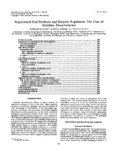
Time Course of Ornithine Decarboxylase Induction by Various Agents in NZH-3T3 Cells-Addition of PMA, FGF, or serum to serum-deprived NIH-3T3 cells resulted in marked increases in ornithinedecarboxylase activity, withpeak activities occurringa t 3-4 h (Fig. 1).Similar results were obtained in preliminary studies with PDGF (not shown). Ornithine decarboxylase activity then decreased rapidly in all cases to reach near base-line values by 8 h. Under similar conditions, the inactive phorbol analogue 4a-phorbol 12,13-didecanoate (1.6 p ~ had ) no effect on ornithine decarboxylase activity when assayed 3 h after addition (not shown). The synthetic when added at hourly diacylglycerol sn-1,2-dioctanoylglycerol,
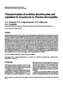
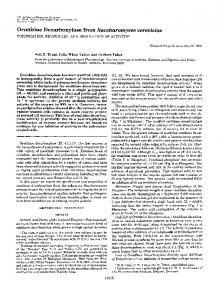
intervals (at 100 pM each time) for 2h, caused a%fold increase in ornithinedecarboxylase activity; no change was seen after identical addition of the inactive analogue sn-1,2-dibutyrylglycerol (not shown). The addition of the calcium ionophore A23187 for3hcaused no detectable changes in ornithine decarboxylase activity a t final concentrations of 0.15, 0.38, 0.75, 1.5, and 6 p ~ it ;has been shown recently that concentrations of 0.5 and 1 p M A23187 cause marked increases incmyc mRNA levels in the relatedSwiss mouse 3T3 cells (29). Effects of 16 H of Exposure to PMA or MezSO on Protein Kinase CActiuity-We assessed the effect of 16 h of exposure to 16PM PMA on protein kinase C in threeways: by fractionating cell extracts on DE52 cellulose and measuring kinase activity, as described (3); by measuring the activity directly in cell fractions, using a histone fragment as a specific substrate for the kinase (26); and by measuring immunoreactive kinase in total cellular homogenates (3). Using the column fractionationmethod, high speed supernatants from NIH3T3 cells preincubatedwith 0.1% MezSOin serum-free DMEM contained readily measurable protein kinaseC activity (Fig.2a). However, afterpreincubation with PMA,no detectable protein kinase C activity was present in extracts fractionated in parallel with control extracts (Fig. 2b). Furthermore, no increase in phospholipid/Ca2+-independentkinase activity was noted in extracts from the PMA-pretreated cells. C activity directly, withWe next measured protein kinase outcolumnfractionation,bothin high speed supernatant fractions and in particulate fractions extracted with 0.3% Triton X-100, which we have found to maximally extract the membrane-associated kinase in active form.' Using this sensitive assay, we were able to detectresidual protein kinase C activity in both the supernatant and particulate fractions of PMA-pretreated cells (Fig. 3). However, in the supernatant fraction,thisrepresented only about 1.6% of theactivity found in the control cells pretreated with MezSO; about 7% of control activity was detected in the extractsof the particulate fraction from PMA-pretreated cells (Fig. 3). Again, no change inphospholipid/Ca2+-independentkinase activitywas noted in either the supernatant or particulate fractions from cells pretreated with PMA(Fig. 3). Finally, as we have shown previously in 3T3-Ll fibroblasts b. P M A
o.CONTROL
0
O
O
1
2
3 4 TIME ( h )
5
6
7
8
FIG. 1. Time course of ornithine decarboxylase ( O D 0 induction by various agents in NIH-3T3 cells. Confluent NIH3T3 cells were incubated in serum-free medium for 16 h prior to the addition of growth factors, which were added at time zero. Cells were harvested at the times indicated, with each point representing the average of duplicate ornithine decarboxylase determinations from 1.6) p~ , PMA two plates. Agents added were 0.01% Me2S0 (U in 0.01% Me2S0 ( O " X ) ) , 125 ng/ml FGF ( I C - . ) , 10% (v/v) fetal See the text for further details. calf serum (M).
6
12
18
24 0 6 18 TIME (min)
12
24
FIG. 2. Effect of preincubation with PMA on protein kinase C activity in column-fractionated cell supernatants. Twentyfive plates (100-mm diameter) of confluent NIH-3T3 cells were exposed to 0.1% Me2S0 in serum-free medium (4.Control) or 16 p~ PMA in 0.1% Me2S0 (b. P M A ) for 16 h. The cells were then homogenized, the homogenate centrifuged, thesupernatant chromatographed, and the column eluates assayed for protein kinase activity or) absence (M of) phosphatidylserine in the presence (M and Ca2+as described in the text. Each point represents the mean of duplicate determinations; results are expressed as pmol of 32Ptransferred to histone 111-S (Sigma) per pg of protein in the column eluate as a function of time. See the text for further details.
Factors Growth Decarboxylase Ornithine and 0.
Soluble Fraction
b. Porticulote Fraction 600-
500400-
'I
300200 -
10383 cells (Fig. 5). In the PMA-pretreated cells, there was an initial inhibitory effect of PMA at 1h followed by a rapid rise in ornithine decarboxylase activity to relatively high levels. This activity peaked at approximately 6 h and then decreased to control levels by 16 h. It is important to note that ornithine decarboxylase activity was negligible in both the PMA- and MezSO-pretreated cells at 16 h because subsequent experiments on down-regulated cells were performed 16 h after PMA addition.
Effects of Mitogens on OrnithineDecarboxylase Activity in Protein Kinase C-deficientCells-After 16 h of pretreatment with PMA or MezSO, mitogens were added directly to the 100medium without change, i.e. the PMA-pretreated cells were 0still exposed to 16 PM PMA. Cells exposed to 0.01% MezSO + - c + - c + - c + - c " " for a further 2 h showed low ornithine decarboxylase activity Control PMA Control PMA afterboth MezSO andPMApretreatment (Fig. 6). Cells FIG.3. Effect of PMA pretreatment on protein kinase C pretreated with PMA did not respond to further PMA addiactivity in supernatant and particulate fractions. Confluent tion, but cells preincubated with MezSO responded to PMA serum-deprived NIH-3T3 cells were exposed to 0.1% Me2S0 (control) or 16 PM PMA in 0.1% Me2S0 for 16 h; the cells were then washed, in a normal manner. Cells preincubated with PMA also did homogenized, centrifuged, and the supernatant (a. Soluble Fraction) not respond to sn-1,2-dioctanoylglycerol(not shown). The and Triton X-100-extracted particulate fraction ( b ) were assayed for response to serum, PDGF, and FGF was decreased but not protein kinase C as described in the text. Each bar represents the eliminated by PMA pretreatment (Fig. 6). Specifically, the mean & the S.D. of duplicate determinations from four plates of cells, response to serum was decreased by 66%, that to PDGF by each harvested and assayed individually. The assays were conducted by 76%. Cells exposed to EGF showed in the presence (+) or absence (-) of calcium, phosphatidylserine, 84%, and that to FGF no response after either pretreatment while cells exposed to and diolein; C refers to thedifference between the two conditions, i.e. insulin showed a slight response only in the PMA-pretreated protein kinase C activity. See the text for further details. cells (Fig. 6). zd Several experiments were alsoperformed to assess the possible effects of changes in intracellularcalcium concentrations on ornithinedecarboxylase activity. In one experiment, cells were preincubated with either PMA (16 PM) or control conditions for 16 h; to half the plates was then added EGTA (2.72 mM)at twice the concentrationof Ca2+in the incubation medium (1.36 mM). After 2 hof exposure to EGTAor control conditions, cells were then stimulated with FGF (125 ng/ml) M, 80,000 without change of medium, and ornithine decarboxylase activity was assessed 3 h later. As noted above, preincubation with PMA in medium containing Ca2+ caused a significant reduction in the FGF-inducedincrease in ornithinedecarboxylase activity (Fig. 7). However, in cells deprived of Ca2+ for 2 h before hormone addition, during which intracellular as well as extracellular Ca2+ could be expected to be significantly depleted, no effect of FGF on ornithinedecarboxylase activity was noted in either the controlcells or those pretreatedwith PMA (Fig. 7). Similarly, pretreatment with EGTA in the FIG.4. Effect of PMA pretreatment on protein kinase C same way also abolished the stimulatory effect of PMA (1.6 immunoreactivity. Confluent serum-deprived NIH-3T3 cells were exposed to either 0.1% (v/v) Me2S0 (C) or 16 PM PMA in 0.1% I.~Mfor 3 h) on ornithine decarboxylase activity (data not Me2S0 (PMA) for 16 h as described in the text. At that time, total shown). cellular homogenates were prepared, and these were subjected to As noted above, addition of the calcium ionophore at a wide electrophoresis, transfer, andimmunoblotting for protein kinase C as range of concentrations had noeffect on ornithine decarboxdescribed (3). Immunoreactive materialcontained in a rat brain ylase activity assessed 3 h later. In another experiment, adhomogenate (Rat Brain) is also shown for comparison. The arrow points to the M,80,000 band which represents immunoreactive pro- dition of1.5 PM A23187 for 3 h did not affect ornithine decarboxylase activity either in normal,serum-deprived cells, tein kinase C. See the text for further details. or in cells preincubated with PMA (notshown). Effects of Mitogens on Ornithine Decarboxylase m R N A Lev(3), 16h of PMA pretreatment removed detectable immunoreactive protein kinaseC fromthe whole cellular homogenate, els in Protein Kinase C-deficientCells-Under similar conditions, cells pretreated with either MezSO or PMA for 16 h as assessed by the Western blotting technique(Fig. 4). were exposed to mitogens for a further 3 h without medium Time Course of Ornithine Decarboxylase Activity during Down-regulation of Protein KinaseC-To determine whether change, i.e. the PMA-treatedcells were still exposed to 16I.~M PMA during the addition of the other mitogens. Evaluation ornithine decarboxylase activity would return to baseline of mitogens during "down-regulation'' of protein kinase C activity, itwas of mRNA levels 3 hafter the subsequent addition cells, PMA, FGF, and, measured after exposure of the cells to 16PM PMA in serum- showed that, in the MezSO-pretreated to alesser extent, fetal calf serum allincreased ornithine free medium. decarboxylase mRNA concentrations (Fig. 8). In the PMAOrnithine decarboxylase activity was moderately elevated immediately after serum deprivation butdecreased gradually pretreated cells, however, additional PMA had no effect on to a barely detectable level by 16 h in the MezSO-pretreated ornithine decarboxylase mRNA levels (Fig. 8);under thesame
f
L
10384
FIG. 5. Time course of ornithine decarboxylase (OBCj activity during preincubation with PMA. Con-
fluent NIH-3T3 cells were changed to serum-free medium at minus 2 h, then exposed to 0.1% MepSO (control; U) or 16 p M PMA in 0.1% Me2S0 (Cr”0) beginning at time zero. The cells were then harvested a t various times, and ornithine decarboxylase activity was measured. Each point represents the average of duplicate determinations from two plates of cells.
0 TIME AFTER ADDITION (h)
Fro. 6. Effect of protein kinase C depletion on agonist-induced increases in ornithine decarboxylase (000 activity in NIH-3T3 cells. Confluent cells were exposed to serum-free medium for 2 h followed by either 0.1% M e 8 0 or 16 p~ PMA
in 0.1% MezSO in serum-free medium for 16 h. The following agents were then added 0.01% MezSO (Control), 1.6 p t ; ~PMA in 0.01% MezSO, 10% fetal calf serum (Serum), approximately M PDGF, 125 ng/ml FGF, 1 pg/ml EGF, and 1 milliunit/ml insulin. Each agent was added to 8 plates, 4 pretreated with PMA and 4 pretreated with Me2S0.All cells were harvested after 2 h, and ornithine decarboxylase activity was measured. Open rectangles represent the mean rt S.D. of ornithine decarboxylase activity of duplicate measurementsfrom four plates of cells after the control preincubation,while crosshatched rectangles represent ornithine decarboxylase activity after the PMA preincubation. See the text
for further details.
c7
CONTROL PREINCUBATION K~NCUBATION
NTROL
residual protein kinase C activity remaining in the PMApretreated cells was still responsive to hormones; however, under these conditions, neither additional PMA nor the active synthetic diacylglycerol s~-1,Z-dioctanoylglycerolwas effecISC CUSS ION tive a t increasing or~ithine ~ecarboxy~ase activity. Thus, it appears that at least a portion of the increase in ornithine The resultspresentedheredemonstratethatcertain decarboxylase activity resulting from exposure of the cells t o growth-promoting substances such as serum, PDGF, and FGF can still cause increases in ornithine decarboxylase activity serum, PDGF, or FGF was due to activation of a protein in NIH-3T3 fibroblasts made effectively protein kinase C kinase C-independent pathway. One possible explanation for these results would be that deficient by preincubation with high concentrations of PMA. The growth factor exposures were performed under conditions increases in cytosolic calcium concentrations resulting from in which little protein kinaseC activity and no protein kinasegrowth factor-stimulatedformation of inositol phosphates C immunoreactivity could be demonstrated and in the pres- could lead to increases in ornithinedecarboxylase activity in ence of high concentrations of PMA (16 @ M ) , which are the protein kinase C-deficient cells. This possibility is supcells preincubatedin calciumsupramaximal for activating protein kinase C in the related ported by thefactthatin was abolished in both 3T3-Ll cell type (3). We cannotexclude other possible effects depleted medium, the response to FGF of the PMA preincubation on cellular processes nor can we normal and PMA-pretrea~d cells. However, incubation in entirely exclude the possibility that the smallamount of calcium-depleted medium also abolished the normal effect of conditions, FGF and, ato lesser extent, fetalcalf serum, both caused marked increases in ornithine decarboxylase mRNA levels (Fig. 8).
Ornithine Decarboxylase and Growth Factors
5
b. PMA
o CONTROL PRETREATMENT PRETREATMENT
ill"-
-
C FGF
C FGF
Ca+?free
Cn++-hee
FIG. 7. Effect of PMA pretreatment on FGF-induced increases in ornithine decarboxylase (000activity in cells incubated in normal or calcium-free medium. Serum-deprived confluent cells were pretreated with either 0.1% Me2S0 ( a ) or 16 p M PMA in 0.1% Me2S0 ( b ) for 16 h, a t which point FGF (125 ng/ml) or water (C) was added. Two h before the addition of FGF, either water or 2.72 mM EGTA (Ca*+free) was added to half the plates. The cells were harvested 3 h after the addition of FGF or control conditions, and samples were prepared for measurement of ornithine decarboxylase activity in the usual way. Each bar represents the mean f S.D. of duplicate determinations performed on four plates of cells. See the text for further details. Control PMA Pretreatment Pretreatment
'
-
ODC mRNA -18s
FIG. 8. Effect of PMA pretreatment onmitogen-induced increases in ornithine decarboxylase (ODC) mRNA levels. Confluent serum-deprived NIH-3T3 fibroblasts (10 plates of cells for each condition) were pretreated with Me2S0 or PMA for 16 h as described in the text. Total RNA was then prepared, fractionated, transferred to nitrocellulose, and allowed to hybridize to radiolabeled ornithine decarboxylase cDNA. An autoradiograph of this blot is shown here. The arrow points to the major hybridizing species of mouse ornithine decarboxylase mRNA of approximate size 2.15 kilobases. The markers 28 S and 18 S refer to thepositions of the major species of ribosomal RNA on the stained gel. See the text for further details. FCS, fetal calf serum; C, -.
PMAonornithine decarboxylase activity,arguing for an effect of this perturbation on other steps of the pathway. In addition, several previous studies have shown that depletion of extracellular calcium prevented the normal increases in ornithine decarboxylase activity seen after exposure of cells or mouse skin explants to serum (30, 31) or PMA (32). In addition, thecalcium ionophore A23187, over a wide range of concentrations, had no effect on ornithine decarboxylase activity, as has been reported previously in murine epidermal
10385
cells (33). This is in contrast to studiesof other responses in Swiss 3T3 cells, in which similar concentrations of the ionophore promoted increases in 2-deoxy-~-glucose uptake (34) and c-myc mRNA levels (29). Therefore, it seems unlikely that increased cytosolic calcium concentrations alone could be responsible for the observed increases in ornithine decarboxylase activity following growth factor stimulation of the PMA-pretreated cells. Another growth factor response which is maintained in the protein kinase C-deficient cells is activation of a ribosomal protein S6 protein kinase, which can then lead to phosphorylation of the ribosomal protein S6 in 3T3-Llfibroblasts (3, 17). Activation of this kinase might then lead to increased ornithine decarboxylase activity in the PMA-pretreated cells. However, this kinase was activated by both insulin and EGF in both normal and protein kinaseC-deficient 3T3-Ll fibroblasts (17), whereas these agonists had minimal effects on ornithine decarboxylase activity in the present experiments. It seems unlikely, therefore, that activationof this kinase by growth factors in the PMA-pretreated cells was solely responsible for the observed effects on ornithine decarboxylase activity. Our results indicate thatmitogens such as PMA and FGF caused increases in ornithine decarboxylase activity, at least in part, by increasing concentrations of ornithine decarboxylase mRNA in the normal cells; similar resultswere observed in response to FGF, but not to PMA, in the protein kinase Cdeficient cells. In previous work, Kahana and Nathans (35) and Liu et al. (36) demonstrated that serum caused rapid increases in ornithine decarboxylase mRNA levels in quiescent BALB/c 3T3 cells and Swiss 3T3 cells, respectively. Although we have not documented changes in transcription rates directly under these circumstances, others have noted that serumcaused increases in the rateof transcription of the ornithine decarboxylase gene in 3T3 cells, measured by the nuclear run-off t e ~ h n i q u e .Whatever ~ the mechanism(s), it appears that thegrowth factors can increase ornithine decarboxylase activity and mRNAlevels through a pathway which does not involve proteinkinase C, althoughactivation of protein kinaseC in the normal cells has the same effect. Thus, the interesting situation exists which in a single growth factor such as FGF can induce increases in the levels of the same mRNA species by two apparently separate pathways. Several recentstudies havedocumentedresponses to growth factors which occur in cells made effectively protein kinase C deficient by preincubation with phorbol esters. For example,Collins and Rozengurt(16) showed thatPDGF stimulated DNA synthesis in Swiss mouse 3T3 cells after preincubation with phorbol esters. In addition, intwo papers on similar cell types published after the initialsubmission of this paper, both Coughlin et al. (37) and Kaibuchi et al. (29) noted increases in c-myc mRNA levels in response to growth factors after preincubationof the cells with phorbol esters. In the former paper (37), PMA pretreatment of human fibroblasts inhibited PDGF-stimulated increases in c-myc mRNA levels by an average of 60% but did not significantly inhibit the mitogenic effect of PDGF. Similarly, Kaibuchi et al. (29) noted that PMA pretreatment of Swiss mouse 3T3 fibroblasts inhibitedPDGF-andFGF-stimulated increases in c-myc mRNA levels by 65 and 60%, respectively. Finally, we have noted that c-fos, c-myc, and @-actin mRNA levels are also increased in response to FGF or PDGF and serum in PMApretreated 3T3 cells.4 Taken together, these studies indicate that several mRNA species whose levels increase in response M. E. Greenberg and E. B. Ziff, personal communication. D. J. Stumpo and P. J. Blackshear, unpublished data.
10386
Ornithine ~ e c u r ~ o xwe y land ~ Growth Factors
to activation of protein kinase C also increase in response to growth factors in protein kinase C-deficient cells, although the response is inhibited by 60430% in most cases. It is tempting to ascribe that proportion of the growth factor-stimulated increase in ornithine decarboxylase activity which is eliminated by PMA pretreatment to the activation of protein kinase C under normal circumstances, i.e. activation of protein kinase C could lead to 60-80% of the total growth factor-mediated increase in enzyme activity. Given other possible effects of the PMA preincubation on the cells, this is only a very crude estimate. The future development of protein kinase C-deficient mutant cell lines will be necessary to address this question in more q u a n t i ~ t i v eterms. =~c~no~Zedgments-We thank Drs. R. M. Bell and B. R. Ganong for the synthetic diacylglycerolsused in thisstudy, Dr. L. T, Williams for PDGF, and Dr. 0. A. Janne for the ornithine decarboxylase cDNA probe. We are especially grateful to Drs. Peggy R. Girard and J. F. Kuo for performing the Western blot analysis of protein kinase C. We also thank Drs. M. E. Greenberg and E. B. Ziff for permission to cite their unpublished work, Dr. L. A. Witters for advice concerning his method of determining protein kinase C activity prior to its publication, and Dr. R. E. Kaufman for helpful advice and discussion. Finally, we are also grateful to Jane Tuttle and Janis Hoffman for expert technical assistance, and to Lessie Detwiler for typing the manuscript. REFERENCES 1. Rozengurt, E., Rodriguez-Pena, M., and Smith,K. A. (1983) Proc. Natl. Acad. Sci. U. S. A. 80,7244-7248 2. Rozengurt, E., Rodriguez-Pena, A., Coombs, M. D., and SinnettSmith, J . (1984) Proc. Natl. Acad. Sci. U. S. A. 8 1 , 5748-5752 3. Blackshear, P. J., Witters, L. A,, Girard, P. R., Kuo, J. F., and Quamo, S. N. (1985) J. Biol. Chem. 2 6 0 , 13304-13315 4. Collins, M. K. L., and Rozengurt, E. (1982) J. Cell. Physiol. 1 1 2 , 42-50 5. Rodriguez-Pena, A., and Rozengurt, E. (1984) Biochem. Bwphys. Res. Commun. 120,1053-1059 6. Habenicht, A. J. R., Glomset, J. A., King, W. C., Nist, C., Mitchell, C. D., and Ross, R. (1981) J. Biol. Chem. 256, 12329-12335 7. Moolenaar, W. H., Tertoolen, L. G. J., and deLaat, S. W. (1984) J. Biol. Chem. 259,8066-8069 8. Hesketh, T. R., Moore, J. P., Morris, J. D. H., Taylor, M.V., Rogers, J., Smith, G. A., and Metcalfe, J. C. (1985) Nature 313,481-484 9. Kelly, K., Cochran, B. H., Stiles, C. D., and Leder, P. (1983) Cell 35,603-610 10. Greenberg, M. E,, andZiff, E. B. (1984) Nature 3 1 1 , 433-438 11. Kruijer, W., Cooper, J. A., Hunter, T.,.and Verma, I. M. (1984) Nature 3 1 2 , 711-716
12. Muller, R., Bravo, R., Burckhardt,J., and Curran, T. (198-4) Nature 312,716-720 13. Berridge, M. J., Heslop, J. P., Imine, R. F., and Brown, K.D. Trans. 13,67-71 (1985) Biochem. SOC. 14. Tsuda, T., Kaibuchi, K., Kawahara, Y., Fukuzaki, H., and Takai, Y. (1985) FEBS Lett. 191, 205-210 15. Besterman, J. M., Watson, S. P., and Cuatrecasas, P. (1986) J. Bwl. Chem. 261,723-727 16. Collins, M. K. L., and Rozengurt, E. (1978) J. Cell. Physiol. 1 1 8 , 133-142 17. Blackshear, P. J., Wen, L., Nemenoff, R. A., Gunsalus, J. R., and Witters, L. A. (1986) in Progress in Brain Research (Gispen, G. H., and Routtenberg, A. H., eds) Elsevier Scientific Publishing Co., Amsterdam, in press 18. Bachrach, U. (1978) in P o l ~ a r n iin~ ~BiomedicalResearch (Campbell, R. A,, Morris, D. R., Bartos, D., Daves, G. D., and Bartos, F., eds) Vol. 1, pp. 81-107, Raven Press, New York 19. McCann, P. P. (1978) in P ~ l y a ~ ini ~Biomedica~ s Research, (Campbell, R. A., Morris, D. R., Bartos, D., Daves, G. D., and Bartos, F.,eds) Vol. 1, pp. 109-123, Raven Press, New York 20. Janne, J., POSO, H., and Raina, A. (1978) Biochim. Biophys. Acta 473,241-293 21. Tabor, C. W., and Tabor, H. (1984) Annu. Rev.Biochem. 53, 749-790 22. Cannelakis, E. S., Viceps-Madore, D., Kyriakidis, D.A., and Heller, J. S. (1979) Curr. Top. Cell. Regul. 1 5 , 155-202 23. Erwin, B. G., Seely, J. E., and Pegg, A. E. (1983) Biochemistry 22,3027-3032 24. Bradford, M. M. (1976) Anal. Biochem. 72,248-254 25. Parker, P. J., Stable, S., and Waterfield, M. D. (1984) EMBO J. 3,953-959 26. Glynn, B., Colliton, J., McDermott, J., and Witters, L. A. (1985) Bwchem. J. 231,489-492 27. Chirgwin, J. M., Przybyla, A. E., MacDonald, R. J., and Rutter, W. J. (1979) Biochemistry 18,5294-5299 28. Maniatis, T., Fritsch, E. F., and Sambrook, J. (1982) in Molecular Cloning, A Laboratory Manual, p. 202, Cold SpringHarbor Laboratory, Cold Spring Harbor, NY 29. Kaibuchi, K., Tsuda, T., Kikuchi, A., Tamimoto, T., Yamaskita, T., and Takai, Y. (1986) J. Bwl. Chem. 261,1187-1192 30. Gibbs, J. B., Hsu, C-Y., Terasaki, W. L., and Brooker, G. (1980) Proc. Natl. Acad. Sci. U. S. A. 7 7 , 995-999 31. Gibbs, J. B., and Brooker, G. (1984) Biochim. Biophys. Acta801, 87-98 32. Verma, A. K., and Boutwell, R. K. (1981) Biochem. Biophys. Res. Commun. 101,375-383 33. Sasakawa, N., Ishii, K., Yamamoto, S., and Kato, R. (1985) ~wchem.Biophys. Res. Commun. 128,913-920 34. Yamanishi, K., Nishino, H., and Iwashima, A. (1983) Bwchem. Bwp~ys.Res. Commun. 117,637-642 35. Kahana, C., and Nathans, D. (1984) Proc. Natl. Acad. Sci. U. S. A. 81,3645-3649 36. Liu, H. T.,Baserga, R., and Mercer, W. E. (1985) Mol. Cell. Biol. 5,2936-2942 37. Coughlin, S. R., Lee, W. M.F., Williams, P. W., Giels, G. M., and Williams, L. T. (1985) Cell 4 3 , 243-251