BIOLOGY OF REPRODUCTION 69, 1053–1059 (2003) Published online before print 28 May 2003. DOI 10.1095/biolreprod.102.013474
Effects of Pulsatile Shear Stress on Nitric Oxide Production and Endothelial Cell Nitric Oxide Synthase Expression by Ovine Fetoplacental Artery Endothelial Cells1 Yun Li,3 Jing Zheng,3 Ian M. Bird,3,4 and Ronald R. Magness2,3,4,5 Perinatal Research Laboratories, Departments of Obstetrics and Gynecology,3 Pediatrics,4 and Animal Sciences University of Wisconsin-Madison, Madison, Wisconsin 53715 ABSTRACT Placental blood flow, endothelial nitric oxide (NO) production, and endothelial cell nitric oxide synthase (eNOS) expression increase during pregnancy. Shear stress, the frictional force exerted on endothelial cells by blood flow, stimulates vessel dilation, endothelial NO production, and eNOS expression. In order to study the effects of pulsatile flow/shear stress, we adapted Cellco CELLMAX artificial capillary modules to study ovine fetoplacental artery endothelial (OFPAE) cells for NO production and eNOS expression. OFPAE cells were grown in the artificial capillary modules at 3 dynes/cm2. Confluent cells were then exposed to 10, 15, or 25 dynes/cm2 for up to 24 h. NO production by OFPAE cells exposed to pulsatile shear stress was inhibited to nondetectable levels by the NOS inhibitor L-NMMA and reversed by excess NOS substrate L-arginine. NO production and expression of eNOS mRNA and protein by OFPAE cells were elevated by shear stress in a graded fashion (P , 0.05). The rise in NO production with 25 dynes/cm2 shear stress (8-fold) was greater (P , 0.05) than that observed for eNOS protein (3.6fold) or eNOS mRNA (1.5-fold). The acute shear stress-induced rise in NO production by OFPAE cells was via eNOS activation, whereas the prolonged NO rise occurred by elevations in both eNOS expression and enzyme activation. Thus, elevations of placental blood flow and physiologic shear stress may be partly responsible for the increases in placental arterial endothelial eNOS expression and NO production during pregnancy.
female reproductive tract, nitric oxide, placenta, pregnancy, uterus
INTRODUCTION
In order to meet the increasing nutrient and metabolic needs of the growing fetus, dramatic decreases in placental and uterine vascular resistance with their associated increases in blood flow occurs during gestation. This response is one of the most striking maternal cardiovascular adaptations observed during normal pregnancy [1]. One potent vasodilator, nitric oxide (NO) plays an important physiologic role in pregnancy adaptation [2]. Because there is no innervation of fetal placental vessels, vascular relaxation Supported, in part, by National Institutes of Health Grants HL49210, HL57653, HD33255, HL64703, and HD38843. This study is in partial fulfillment of the Ph.D. degree in the Endocrinology-Reproductive Physiology Training Program (www.erp.wisc.edu). 2 Correspondence: Ronald R. Magness, Department of Obstetrics and Gynecology, University of Wisconsin, Perinatal Research Laboratories, 7E Meriter Hospital, 202 S. Park St., Madison, WI 53715. FAX: 608 257 1304; e-mail:
[email protected] 1
Received: 18 November 2002. First decision: 13 December 2002. Accepted: 8 May 2003. Q 2003 by the Society for the Study of Reproduction, Inc. ISSN: 0006-3363. http://www.biolreprod.org
5
occurs by the influence of circulating and/or locally produced vasodilators such as NO [3, 4]. NO is produced in greater amounts during gestation by both the fetoplacental and uteroplacental endothelium [2]. The level of NO and its second messenger cGMP are also elevated during pregnancy [5, 6], especially in the uteroplacental and fetoplacental units [7–9]. It has been reported that infusion of nitric oxide synthase (NOS) inhibitors decreased ovine fetoplacental blood flow [10], demonstrating a direct causeand-effect relationship between NO production and placental perfusion. Shear stress is the frictional tangential force imposed on the vessel wall when blood flows through a vessel [11]. Shear stress has a wide-range of effects on the expression of a variety of genes in endothelial cells. The proteins encoded by these genes in turn play important roles in regulating endothelial function, mediating many physiological and pathological processes, such as endothelial cell proliferation, vasodilation, vasoconstriction, and inflammatory responses. It has long been reported that, when blood flow is increased, the blood vessel dilates [12]. Removal of the endothelium, or pretreatment with L-NAME, a NOS inhibitor, greatly reduced or abolished the flow-induced dilation of isolated vessels. Moreover, NO inhibition of flow-mediated vasodilation was reversed by the NOS substrate, Larginine [13, 14]. In vivo studies using a model of chronic high blood flow or creating fistulas to elevate arterial flow strongly suggest that prolonged increases in shear stress will stimulate endothelial expression of endothelial cell nitric oxide synthase (eNOS) at both the mRNA and protein levels [15, 16]. This physiologic response was also demonstrated in vitro, showing that steady shear stress upregulated eNOS mRNA level in bovine aortic endothelial cells (BAEC) and human umbilical vein endothelial cells (HUVEC) [17, 18]. Among the various flow types being studied in vitro, steady laminar flow has been the most investigated. Steady laminar shear stress applied to cells grown in static culture conditions induces a biphasic production of NO in cultured HUVEC [19, 20]. Although pulsatile laminar flow also was found to stimulate endothelial eNOS activity [21, 22], both eNOS mRNA and protein expression have only been shown to be increased in an endothelial cell and smooth muscle cell cocultured pulsatile system [22]. However, in endothelial cells adapted to flow conditions, the dynamic time course of NO production related to the time-dependent changes in eNOS expression during various levels of pulsatile shear stress has never been elucidated. To investigate the effects of pulsatile shear stress on endothelial function, we grew the ovine fetoplacental artery endothelial (OFPAE) cells [23] in Cellco CELLMAX (Spectrum Laboratories, Rancho Dominguez, CA) artificial capillary modules and adapted them to an environment of
1053
1054
LI ET AL.
pulsatile shear stress. We hypothesized that further graded elevations in shear stress would increase NO production and eNOS expression by these cells in a time-dependent manner. We observed that pulsatile shear stress induced both acute and prolonged NO production by OFPAE cells, which was shear-stress-magnitude-dependent, and was inhibited by the NOS inhibitor L-NMMA. We also reported for the first time that the acute rises in NO production were due to elevations in shear stress that occur by increasing eNOS activity, whereas the more prolonged NO changes were related to both eNOS activation and regulation of the eNOS enzyme. MATERIALS AND METHODS Cell culture. OFPAE cells were cultured in Dulbecco modified Eagle medium (DMEM; Life Technologies, Gaithersburg, MD), supplemented with 10% calf serum, 10% fetal bovine serum, and 1% penicillin/streptomycin in T75 flasks [23]. When reaching confluence, the cells were passaged at 1:4. Cells of passage 13 were used in the experiments. Pulsatile flow system. OFPAE cells (5 3 106) were inoculated into Cellco CELLMAX artificial capillary modules (Spectrum Laboratories), which were kept in a 378C, 5% CO2 incubator. Every module contains 50 pronectin-coated permeable polyethylene capillaries, each with an internal diameter of 0.33 mm, a length of 13.0 cm, a pore size of 0.5 mm. The module and a reservoir (50 ml) were connected by silicone tubing, which functions as an O2/CO2 exchanger. The culture media (DMEM contained 400 mM L-arginine and was supplemented with 10% calf serum, 10% fetal bovine serum, and 1% penicillin/streptomycin) was circulated using a pump between the artificial capillary module and the reservoir at a desired flow rate. Shear stress (dynes/cm2) was calculated as 4Qh/pr3, in which Q is flow rate within each capillary, h is viscosity of culture media (0.7 3 1022 poise), and r is the fixed radius of the artificial capillary. The OFPAE cells were grown at a flow rate that provided a pulsatile (60 pulses/ min, 8.1 mm Hg) shear stress averaging 3 dynes/cm2 in each capillary in order to adapt the cells to pulsatile flow conditioning and to provide a continuous supply of CO2/O2. Daily lactate levels in culture media were measured spectrophotometrically using lactate kits (Sigma Chemical Co., St. Louis, MO), and the production rate of lactate was calculated to monitor the growth of OFPAE cells cultured in the modules. After 7 days, the production rate of lactate was stabilized, indicating that the cells were viable when they reached confluence. Further shear stress exposure was carried out at Days 11–13 after inoculation. In order to fully exclude the effects of serum on gene activation, the endothelial cells went through 24 h of serum starvation before shear stress was further elevated. Serum-free DMEM was changed into each reservoir bottle, and the entire flow path was also flushed gently with serum-free media. To examine the morphology of OFPAE cells after 12 days of culture inside the system, the capillaries within one module were fixed with 1.25% glutaraldehyde over night at 48C. Then a randomly selected capillary was longitudinally sliced open, stained with hematoxylin, and viewed under light microscopy. Shear stress experiments and media sample collection. The OFPAE cells were then exposed to higher pulsatile shear stresses, averaging 10 dynes/cm2 (140 pulses/min, 26.4 mm Hg), 15 dynes/cm2 (250 pulses/min, 52.5 mm Hg), or 25 dynes/cm2 (390 pulses/min, 71.7 mm Hg) for 24 h (n 5 3–6 for each level of shear stress). Culture media were obtained at 0 (3 dynes/cm2 before elevating pulsatile shear stress), 5, 10, 20, 30, 45, 60, 90 min, and 2, 3, 4, 6, 9 and 24 h of flow stimulation to evaluate both the effluent NOx and affluent NOx of each cartridge. For the eNOS inhibition experiment, 50 mM NG-monomethyl-L-arginine (L-NMMA) was added into the reservoir bottle containing either 5 mM or 400 mM Larginine in DMEM, 30 min before elevating shear stress (n 5 4 for each group). The culture media were obtained 20 min after treatment with either 15 dynes/cm2 or 25 dynes/cm2. NOX measurement. NOX measurement was conducted as described previously [24]. Effluent and affluent culture media (100 mL) at each time point under different shear stress were injected into the NO Analyzer (NOA 280, Siever Incorporation, Boulder, CO). A sodium nitrate standard curve (100 nM–100 mM) was created each day the samples were measured, and all readings were within the range of standard curve. NOX production rate was then calculated as ([NOX]effluent 2 [NOX]affluent) 3 Q; Q here is the flow rate that gives each specific shear stress. Cell recovery. In order to study the time course of eNOS protein expression, OFPAE cells were eluted from cartridges after a 7-min exposure to a trypsin (0.05%)/EDTA (0.53 mM) treatment, prior to (0 h) and after
various times (2, 6, 12, and 24 h) of shear stress (3, 10, and 25 dynes/ cm2) stimulation. To test eNOS mRNA levels, the endothelial cells were exposed to either 3 (n 5 4) or 25 (n 5 4) dynes/cm2 for 12 h and eluted off the cartridges by trypsin/EDTA. Each cell pellet was split into two parts, one was lysed for protein analysis and the other was subjected to total RNA extraction by RNAzol B. Western analysis for eNOS protein. The cell pellets were lysed by sonication in lysis buffer (50 mM Tris, 0.15 M NaCl, 10 mM EDTA [pH 7.4], 0.1% Tween-20, 0.1% b-mercaptoethanol, 5 mg/ml leupeptin, 5 mg/ ml aprotinin, and 0.1 mM phenylmethylsulfonylfluoride). Proteins (5 mg/ lane) were separated on 7.5% SDS-PAGE gels, electroblotted onto the Immobilon-P membrane (Millipore, Bedford, MA), immunoblotted with mouse monoclonal eNOS antibody (1:750, Transduction Laboratories, San Diego, CA), and visualized by the ECL system as described previously [6, 9, 23]. eNOS protein levels were quantified by scanning densitometry. As a loading control, the same blot was also probed for glyceraldehyde phosphate dehydrogenase (GAPDH) [25]. Total cellular RNA extraction. The pellets of OFPAE cells from each experiment were solubilized in 1 ml RNAzol B (Cinna Biotech, Houston, TX). After addition of 150 ml chloroform and phase separation by centrifugation (12 000 3 g, 20 min) the upper aqueous phase was removed, extracted twice with phenol/chloroform/isoamyl alcohol using heavy-grade phase lock gel (5-Prime, 3-Prime, Boulder CO) and finally mixed with 110% by volume of isopropanol. RNA was then precipitated by standing at 2208C for 1 h before recovery by centrifugation (12,000 x g, 30 min), and washing of the pellet in 75% ethanol. RNA was finally solubilized in molecular biology-grade water (5-Prime, 3-Prime) and quantified by spectrophotometry [25–27]. eNOS RT-PCR. The eNOS mRNA levels were quantified by coupled reverse transcription/polymerase chain reaction (RT/PCR) amplification in single-tube assays using AMV reverse transcriptase and Taq Polymerase as described previously [25, 26]. The forward and reverse primers designed according to the ovine eNOS protein coding region, were 59TGTGGCTGTCTGCATGG-39 and 59-TGGCTGGTAGCGGAAGG-39 [27]. Total cellular RNA of 0.1 mg was examined, data were calculated as copy number of eNOS mRNA per microgram total cellular RNA from the standard curve, which was generated by 104 to 1010 copies of eNOS cDNA plasmid run in each assay. All data were normalized to GAPDH content of each sample, determined by the same RT/PCR procedure using 0.1 mg total cellular RNA [25, 27]. Statistical analysis. Data were analyzed by Student t-test or one-way ANOVA. Data are presented as means 6 standard error of the means (SEM).
RESULTS
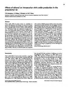
We were able to grow and adapt OFPAE cells successfully in the CELLMAX artificial capillaries under low shear stress of 3 dynes/cm2. Based on lactate production rate (data not shown), light microscopy (Fig. 1) as well as total protein and RNA harvested from the modules, the cells were viable and reached confluence by the time of the experiments. NOx production [(effluent 2 affluent) 3 flow rate] by OFPAE cells was increased rapidly when the shear stress was elevated from 3 to 15 or 25 dynes/cm2 (Fig. 2). Shear stress of 10 dynes/cm2 had no significant effect on stimulating NOx production throughout the experiment. The NOx response appeared to consist of an acute (0–30 min) and a prolonged (30 min224 h) phase. At 25 dynes/cm2, NOx production increased rapidly from 10 min, reached the first peak at 20 min, which was followed by the second peak at 2 h and then plateaued at a lower level up to 24 h. Although shear stress of 15 dynes/cm2 seemed to cause a similar trend of NOx production, the values in the acute phase (0–30 min) did not reach significance. However, with 15 dynes/cm2 the 1-, 2-, and 3-h samples showed elevations in NOx production (P , 0.05). It is noteworthy that both the initial burst and the plateaued NOx synthesis induced by the increase in pulsatile flow were shear stress level dependent. At 15 and 25 dynes/cm2, the NOS inhibitor L-NMMA completely inhibited the acute phase (20 min) of NOx production (Fig. 3). In the presence of low concentrations of
PULSATILE SHEAR STRESS AND NITRIC OXIDE PRODUCTION
1055
FIG. 1. OFPAE cells grown in CELLMAX artificial capillary modules at a shear stress of 3 dynes/cm2. The OFPAE cells were cultured in the artificial capillaries for 12 days and the capillaries were fixed by 1.25% glutaraldehyde overnight at 48C, stained with hematoxylin, and photographed at two different magnifications (340, left; 3200, right). The light micrographs indicate that the OFPAE cells attached to the lumen of the capillaries successfully and formed a confluent monolayer by the time the shear stress studies commenced.
the eNOS substrate L-arginine (5 mM, based on Km of the enzyme), L-NMMA decreased acute NOx production to nondetectable levels (ND). In contrast, this inhibition by LNMMA was fully reversed by the excess substrate (400 mM L-arginine), which is normally contained in the DMEM culture media. Because there was no further rise in NOx production in the DMEM containing 400 mM vs. 5 mM Larginine, these data suggest that substrate availability is not rate limiting for OFPAE cell NO production under shear stress. It is possible that both increased expression in eNOS as well as its activity would contribute to the observed prolonged NO production. This supposition was tested by per-
FIG. 2. Effects of elevating shear stress on NOx production by OFPAE cells in CELLMAX artificial capillary modules. OFPAE cells were inoculated into the Cellco CELLMAX cassette modules and grown at 3 dynes/ cm2 to reach confluence. After overnight serum starvation, the cells were exposed to 10, 15, or 25 dynes/cm2 for up to 24 h. Both media samples from affluent and effluent flow of the modules were collected for determination of the NOx concentration. All time-zero values are at 3 dynes/ cm2 prior to elevating shear stress. Data for all time points were compared with the 0-min value obtained from the same shear stress level. Data are means 6 standard error of the mean (SEM), n 5 3–6/time point. * P , 0.05; #P , 0.1.
forming time-course studies on protein recovered from separate cartridges with 3, 10, and 25 dynes/cm2 (Fig. 4). We noted that basal shear stress (3 dynes/cm2) throughout the experimental period of 24 h did not alter eNOS protein levels. In contrast, by increasing shear stress from 3 to 10 or 3 to 25 dynes/cm2, there was a tendency for elevations of eNOS protein levels as early as 2 h at 25 dynes/cm2 and 6 h at 10 dynes/cm2. However, increases (P , 0.05) in eNOS levels were observed after the shear force was elevated for 12 h and maintained up to 24 h. Furthermore, the effect of 25 dynes/cm2 to elevate eNOS protein expression were more prominent (P , 0.05) than 10 dynes/cm2, with the differences being observed after 12- and 24-h treatments. In contrast, the expression of the housekeeping gene GAPDH, used as a loading control, was not changed at all the time points and levels of shear forces examined (data not shown). The second more prolonged phase of NOx production described above (Fig. 2) appeared to also be related to this elevation in eNOS expression. In agreement with the
FIG. 3. Inhibition of acute NOx response of OFPAE cells to shear stress by L-NMMA. OFPAE cells were grown and reached confluence at 3 dynes/cm2. Before shear stresses were elevated to either 15 or 25 dynes/ cm2, the cells were pretreated with 50 mM L-NMMA for 30 min in the presence of either 5 mM or 400 mM L-arginine. Acute NOx production (at 20 min) was measured. ND, Nondetectable levels. Data are means 6 standard error of the mean (SEM), n 5 4/group.
1056
LI ET AL.
FIG. 4. Effects of elevating shear stress on eNOS protein expression in OFPAE cells grown in CELLMAX artificial capillary modules. After OFPAE cells reached confluence, they were exposed to 3, 10, or 25 dynes/cm2 for 24 h, and the cells were recovered from individual cartridges after 0, 2, 6, 12, and 24 h of shear stress treatment. Cell lysates were subjected to Western analysis for eNOS. Data are means 6 standard error of the mean (SEM), n 5 4/ time point. *P , 0.05 compared with 3 dynes/cm2; #P , 0.05 compared with 10 dynes/cm2.
increase in eNOS protein, eNOS mRNA levels also tended to be elevated (P , 0.1) after the cells were exposed to 25 dynes/cm2 for 12 h compared with 3 dynes/cm2 (Fig. 5). Moreover, the induction of NOx synthesis increased nearly 8-fold, while eNOS protein expression increased 3.6-fold, and eNOS mRNA was only 1.5-fold greater upon elevating shear stress. DISCUSSION
It is noteworthy that the rise in blood flow to the placenta and uterus in pregnancy is 20- to 50-fold. These elevations of placental and uterine blood flow are related directly to normal fetal growth and ultimately neonatal survivability [1]. At the same time that placental and uterine blood flows are elevated, there are also dramatic elevations in both the expression of eNOS and endothelial-derived NO production [2, 6–9, 28, 29], which contribute to the reduction of resistance of the fetoplacental and uterine circulations [1, 10, 29–31]. Flow-induced vasodilatation in uterine arteries from normal pregnant women is completely inhibited by the NOS inhibitor L-NAME [32], and flow/shear stress exposure of endothelial cells stimulates NO release from hu-
FIG. 5. Expression of eNOS mRNA (left), eNOS Protein (middle), and NOx production (right) in OFPAE cells. The OFPAE cells were exposed to shear stresses of either 3 (n 5 4) or 25 (n 5 4) dynes/cm2 for 12 h. The culture media from affluent and effluent flow of the modules were collected for NOx measurement, after which the cells were recovered to determine eNOS mRNA and protein levels. The rises in NOx production (8-fold) and eNOS protein (3.6-fold) were substantial (*P , 0.05) compared with the modest trend for an elevation of eNOS mRNA (1.5-fold; #P , 0.1). Data are means 6 standard error of the mean (SEM).
man fetal placental vasculature in vitro [33]. In addition, fluid shear stress plays a very active role in vascular adaptation to chronic physiological changes of blood flow in the cardiovascular system [34]. Therefore, because of the dramatic rises in blood flow [1, 2], it is quite plausible that shear stress is also a key element in potentiating and sustaining these dramatic changes during pregnancy. In the current studies, we developed an in vitro model to further understand the role of pulsatile shear stress in gestational adaptation utilizing our previously characterized OFPAE cell line [23]. We report the first detailed time-course studies relating the dynamic effects of graded increases in pulsatile shear stress on de novo NO production with alterations in eNOS expression. We determined that there were at least two phases of elevated OFPAE cell NO production with acute and prolonged exposure to shear stress. The acute effects of shear stress on NO production are regulated solely by eNOS activation, whereas the prolonged phase of NO production occurs via both eNOS activation and rises in eNOS expression. Various devices have been developed to study hemodynamic forces (shear stress, circumferential stress, and compressive stress) individually and combined. We are particularly interested in frictional wall shear stress. Although well-defined steady flow has been most investigated [15, 20, 21], we chose to study pulsatile flow because it is more physiologically comparable with the hemodynamic environment in vivo [1]. We adapted the commercially available Cellco CELLMAX artificial capillary module system, developed by the Ballermann group [35–37]. This system provides various irreversible pulsatile shear stresses by establishing graded settings for frequency and amplitude of flow. It has been shown that BAEC grown inside polypropylene hollow fibers perfused with venous or arterial shear stress formed an adherent, confluent monolayer, and aligned themselves in the direction of flow assessed by scanning electron microscopy [37]. This is in total agreement with endothelial morphology addressed by other investigators using a different perfusion system [38]. In the current study, we observed that the OFPAE cells cultured at 3 dynes/cm2 formed a confluent monolayer inside artificial capillaries by the time experimental treatments commenced. Because the reported in vivo systemic physiological arterial shear stress is around 15 dynes/cm2 [39–41], we elected to elevate the flow-adapted OFPAE cells from 3 dynes/cm2 to shear forces of 10, 15, or 25 dynes/cm2 for investigating NOx pro-
PULSATILE SHEAR STRESS AND NITRIC OXIDE PRODUCTION
duction and/or eNOS expression. Unfortunately, the in vivo shear stress level in placental vasculature is not yet known. In the present study, a strength of the current model is that we were able to collect media samples for NOx concentrations from both effluent and affluent flow of the same module at multiple time points after the OFPAE cells were exposed to the various levels of shear stress to calculate the rate of NOx production. We observed that, when shear stress was elevated from 3 to 15, or 3 to 25 dynes/cm2, the NOx production increased significantly and the production rate could be divided into multiple phases. There was an acute induction of NOx production within 30 min with the initial increase in shear stress, followed by a second peak at 2 h and a prolonged phase of NOx production for up to 24 h. Because we calculated NOx production rate by obtaining the difference in NOx concentrations from effluent and affluent flow of the capillary module, it required a sensitive measurement for NOx. Due to the limited sensitivity of the NO analyzer, a severe limitation of this in vitro model lies in the undetectable changes in NOx concentration at any time point with lower shear stresses because we would report this as no significant NOx production. Regardless, our data also demonstrated that the magnitude for both the acute and sustained NOx production phases were dependent on the graded level of shear stress. Responses for NOx production by HUVEC were reported in previous studies, in which endothelial cells were subjected to steady but not pulsatile laminar shear stress, and cumulative NOx levels over time were reported [19]. However, in contrast with our study, although NOx production rates followed a biphasic pattern, their acute rapid burst of NOX within 30 min was completely independent of the level of shear stress. The reason for this discrepancy is likely to be that pulsatile, not steady shear stress, was tested in our experiment. Impulse flow was able to induce NOx at a fairly high production rate even within 1 min [20]. Furthermore, in vivo studies demonstrated that pulsatile flow is more effective in lowering peripheral vascular resistance than nonpulsatile flow and that the difference is related to regulation of endothelium-derived NO [42, 43]. Because both the frequency and the amplitude of pulsatile flow in the systemic circulation are considered as potential stimuli for NO-mediated vasodilation [43], the pulsatility increase in our model, when we elevated shear stress, could be another factor responsible for the observed graded changes in NOx production. In this regard, we must point out that a limitation of the way the current pump system is connected to the cartridges does not allow for differentiation of the NO/eNOS response due to rises in shear rate with elevations in pulse number. Therefore, we cannot dissect the effects of amplitude vs. frequency of pulsatile flow on NO production and eNOS expression. It is noteworthy that the elevations in pulsatility from 60 to 140 pulse/min and 60 to 250 pulse/min (3 to 10 and 15 dynes/cm2, respectively) spans the in vivo range of ovine fetal heart rate (;180–240 pulse/min). However, the most robust rise in NOx production was observed with 25 dynes/cm2 when pulse rate was elevated from 60 to 390 pulses/min. Modifications of these experimental conditions (pulse vs. shear) will need to be made to investigate this issue in future studies. It is thought that the acute burst of NOx production relates to an increase in eNOS activity, probably via rises in intracellular Ca21 concentration and/or phosphorylation of eNOS, and that the subsequent NOx production is related to elevations in eNOS activity and expression. We observed that, by increasing shear stress from 3 to 10 or 3 to 25
1057
dynes/cm2, eNOS protein levels began to rise as early as 2–6 h and continued to increase during the 24-h treatment. Compared with 10 dynes/cm2, 25 dynes/cm2 increased eNOS levels at an earlier time point and to a higher level. Moreover, eNOS mRNA tended to be elevated at 12 h under 25 dynes/cm2 compared with 3 dynes/cm2. Ranjan et al. [18] reported that steady shear stress increased eNOS expression at the level of mRNA and protein in HUVEC and BAEC, which are endothelial cells of fetal and adult origin, respectively. Moreover, eNOS mRNA appeared to be increased by unidirectional shear stress due to increased transcription, although it is not clear whether mRNA stability was changed under the same conditions [15, 44]. We observed that elevations in eNOS mRNA level was less than that of protein, suggesting that this could be due to its gene translation efficiency as previously suggested by Xiao et al. [8]. We also observed that shear stress-induced NOx production was elevated acutely before eNOS protein level was increased and that the fold increase in NOx synthesis at 12 h of exposure to 25 dynes/cm2 was substantially more than that of eNOS protein expression. It is unlikely that the greater relative rise in NOx than eNOS expression relates to the activation of additional NOS isoforms (and/or iNOS) because we have previously shown that eNOS is the main if not sole NOS isoform in ovine placental artery endothelium [24]. These data do, however, suggest that eNOS activity of these cells is closely regulated by pulsatile shear stress both during the acute, but also prolonged exposure, to elevated shear stress. Our observations further demonstrated that, in OFPAE cells, pulsatile shear stress-induced eNOS expression was shear-level dependent and the plateaued phase of NOx production described above can also be related to an increased availability in eNOS protein for activation. Previously, our laboratory has shown that placental artery endothelial eNOS protein levels are increased markedly in vivo during the third trimester [9, 24], when fetal growth and fetoplacental blood flow are elevated [1]. In this study, we demonstrated that one of the major mechanisms for increased eNOS expression in endothelial cells is elevated shear stress or blood flow. In addition, shear stressmediated vasodilation is augmented during pregnancy [45, 46]. Therefore, these data suggest that endothelium-derived NO leads to vasodilation and placental blood flow increases, which in turn further stimulate eNOS activity and eNOS expression during gestation. The exact mechanisms by which shear stress induces endothelial NO production through elevations in both eNOS activity and protein expression need further investigation. Because the shear stress response element is present on the 59 upstream promoter region of eNOS, it is believed that this confers direct transcriptional regulation of this gene [47]. Several other endothelial-derived vasoactive factors, which are altered in concert with eNOS expression/ NO production during elevations in shear stress (e.g., adrenomedullin and prostacyclin) may interact to further augment the shear stress-induced vasodilation [48, 49]. Moreover, pregnancy is a complicated period of time when not only NO but steroid hormones (estrogen and progesterone) and numerous growth factors such as basic fibroblast growth factor, vascular endothelial growth factor, and epidermal growth factor are all elevated [1, 2, 26]. Therefore, it is very likely that some or all of these factors may synergize with shear stress to modulate endothelial function in pregnancy. Using this novel shear stress system brings us closer to the physiologic environment than conventional
1058
LI ET AL.
static culture to provide us an opportunity to study the interactions of hemodynamic force and other biologically active factors during normal pregnancy adaptation. ACKNOWLEDGMENTS
23.
The authors wish to thank Terrance M. Phernetton and Gladys Lopez for technical assistance and Cindy Goss for help in preparing this manuscript for submission.
24.
REFERENCES
25.
1. Magness RR. Maternal cardiovascular and other physiologic responses to the endocrinology of pregnancy. In: Bazer FW (ed.), The Endocrinology of Pregnancy. Totowa, NJ: Humana Press; 1998; 18:507– 539. 2. Sladek SM, Magness RR, Conrad KP. Nitric oxide and pregnancy. Am J Physiol 1997; 272:R441–R463. 3. Fox SB, Khong TY. Lack of innervation of human umbilical cord: an immunohistological study. Placenta 1990; 11:59–62. 4. Myatt L. Control of vascular resistance in the human placenta. Placenta 1992; 13:329–341. 5. Rosenfeld CR, Cox BE, Roy T, Magness RR. Nitric oxide contributes to estrogen-induced vasodilation of the ovine uterine circulation. J Clin Invest 1996; 98:2158–2166. 6. Magness RR, Shaw CE, Phernetton TM, Zheng J, Bird IM. Endothelial vasodilator production by uterine and systemic arteries. II. Pregnancy effects on NO synthase expression. Am J Physiol 1997; 272: H1730–H1740. 7. Yang D, Lang U, Greenberg SC, Myatt L, Clark KE. Elevation of nitrate levels in pregnant ewes and their fetuses. Am J Obstet Gynecol 1996; 174:573–577. 8. Xiao D, Bird IM, Magness RR, Longo LD, Zhang L. Upregulation of eNOS in pregnant ovine uterine arteries by chronic hypoxia. Am J Physiol 2001; 280:H812–H820. 9. Sheppard C, Shaw CE, Li Y, Bird IM, Magness RR. Endotheliumderived nitric oxide synthase protein expression in ovine placental arteries. Biol Reprod 2001; 64:1494–1499. 10. Chang JK, Roman C, Heymann MA. Effect of endothelium-derived relaxing factor inhibition on the umbilical-placental circulation in fetal lambs in utero. Am J Obstet Gynecol 1992; 166:727–734. 11. Randall D. Circulation of the Blood. In: Eckert R (ed.). Animal physiology, 3rd ed. New York: W.H. Freeman and Co.; 1988:443–471. 12. Lie M, Sejersted OM, Kiil F. Local regulation of vascular cross section during changes in femoral arterial blood flow in dogs. Circ Res 1970; 27:727–737. 13. Vequaud P, Freslon JL. Components of flow-induced dilation in rat perfused coronary artery. Cell Biol Toxicol 1996; 12(4–6):227–232. 14. Fukaya Y, Ohhashi T. Acetylcholine- and flow-induced production and release of nitric oxide in arterial and venous endothelial cells. Am J Physiol 1996; 270:H99–H106. 15. Harrison DG, Sayegh H, Ohara Y, Inoue N, Venema RC. Regulation of expression of the endothelial cell nitric oxide synthase. Clin Exp Pharmacol Physiol 1996; 23:251–255. 16. Nadaud S, Philippe M, Arnal JF, Michel JB, Soubrier F. Sustained increase in aortic endothelial nitric oxide synthase expression in vivo in a model of chronic high blood flow. Circ Res 1996; 79:857–863. 17. Uematsu M, Ohara Y, Navas JP, Nishida K, Murphy TJ, Alexander RW, Nerem RM, Harrison DG. Regulation of endothelial cell nitric oxide synthase mRNA expression by shear stress. Am J Physiol 1995; 269:C1371–C1378. 18. Ranjan V, Xiao Z, Diamond SL. Constitutive NOS expression in cultured endothelial cells is elevated by fluid shear stress. Am J Physiol 1995; 269:H550–H555. 19. Kuchan MJ, Frangos JA. Role of calcium and calmodulin in flowinduced nitric oxide production in endothelial cells. Am J Physiol 1995; 266:C628–C636. 20. Frangos JA, Huang TY, Clark CB. Steady shear and step changes in shear stimulate endothelium via independent mechanisms---superposition of transient and sustained nitric oxide production. Biochem Biophys Res Commun 1996; 224:660–665. 21. Noris M, Morigi M, Donadelli R, Aiello S, Foppolo M, Todeschini M, Orisio S, Remuzzi G, Remuzzi A. Nitric oxide synthesis by cultured endothelial cells is modulated by flow conditions. Circ Res 1995; 76:536–543. 22. Hendreckson RJ, Cappadona C, Yankah EN, Sitzmann JV, Cahill PA,
26.
27.
28.
29.
30. 31. 32.
33. 34. 35. 36. 37. 38. 39. 40.
41.
42.
43.
Redmond EM. Sustained pulsatile flow regulates endothelial nitric oxide synthase and cyclooxygenase expression in co-cultured vascular endothelial and smooth muscle cells. J Mol Cell Cardiol 1999; 31: 619–629. Zheng J, Magness RR, Bird IM. A cell model for studying expression of feto-placental artery endothelial cell angiotensin II type-1 receptors and nitric oxide synthase. Med Biochem 1998; 1:57–64. Zheng J, Li Y, Weiss AR, Bird IM, Magness RR. Expression of endothelial and inducible nitric oxide synthases and nitric oxide production in ovine placental and uterine tissues during late pregnancy. Placenta 2000; 21:516–524. Cale JM, Millican DS, Itoh H, Magness RR, Bird IM. Pregnancy induces an increase in the expression of glyceraldehyde-3-phosphate dehydrogenase in uterine artery endothelial cells. J Soc Gynecol Invest 1997; 4:284–292. Zheng J, Bird IM, Melsaether AN, Magness RR. Activation of the mitogen-activated protein kinase cascade is necessary but not sufficient for basic fibroblast growth factor- and epidermal growth factorstimulated expression of endothelial nitric oxide synthase in ovine fetoplacental artery endothelial cells. Endocrinology 1999; 140:1399– 1407. Bird IM, Sullivan JA, Di T, Cale JM, Zhang L, Zheng J, Magness RR. Pregnancy-dependent changes in cell signaling underlie changes in differential control of vasodilator production in uterine artery endothelial cells. Endocrinology 2000; 141:1107–1117. Magness RR, Rosenfeld CR, Hassan A, Shaul PW. Endothelial vasodilator production by uterine and systemic arteries: I. Effects of ANG II on PGI2 and NO in pregnancy. Am J Physiol 1996; 270: H1914–H1923. Magness RR, Sullivan JA, Li Y, Phernetton TM, Bird IM. Endothelial vasodilator production by uterine and systemic arteries: VI. Ovarian and pregnancy effects on eNOS and NOx. Am J Physiol Heart Circ Physiol 2001; 280:H1692–H1698. Miller SL, Jenkin G, Walker DW. Effect of nitric oxide synthase inhibition on the uterine vasculature of the late-pregnant ewe. Am J Obstet Gynecol 1999; 180:1138–1145. Chlorakos A, Langille BL, Adamson SL. Cardiovascular responses attenuate with repeated NO synthesis inhibition in conscious fetal sheep. Am J Physiol 1998; 274:H1472–H1480. Kublickiene KR, Cockell AP, Niesell H, Poston L. Role of nitric oxide in the regulation of vascular tone in pressurized and perfused resistance myometrial arteries from term pregnant women. Am J Obstet Gynecol 1997; 177:1263–1269. Wieczorek KM, Brewer AS, Myatt L. Shear stress may stimulate release and action of nitric oxide in the human fetal-placental vasculature. Am J Obstet Gynecol 1995; 173:708–713. Langille BL. Remodeling of developing and mature arteries: endothelium, smooth muscle, and matrix. J Cardiovasc Pharmacol 1993; 21: S11–S17. Ballermann BJ, Ott MJ. Adhesion and differentiation of endothelial cells by exposure to chronic shear stress: a vascular graft model. Blood Purif 1995; 13:125–134. Ballerman BJ, Dardik A, Eng E, Liu A. Shear stress and the endothelium. Kidney Int 1998; 54:S100–S108. Ott MJ, Olson JL, Ballerman BJ. Chronic in vitro flow promotes ultrastructural differentiation of endothelial cells. Endothelium 1995; 3: 21–30. Helmlinger G, Geiger RV, Schreck S, Nerem RM. Effects of pulsatile flow on cultured vascular endothelial cell morphology. J Biomech Eng 1991; 113:123–131. Giddens DP, Zarins CK, Glagov S. The role of fluid mechanism in the localization and detection of atherosclerosis. J Biomech Eng 1993; 115:588–594. Joyce JM, Phernetton TM, Shaw CE, Modrick ML, Magness RR. Endothelial vasodilator production by uterine and systemic arteries: IX. eNOS gradients in cycling and pregnant ewes. Am J Physiol Heart Circ Physiol 2002; 282:H342–H348. Joyce JM, Phernetton TM, Magness RR. Effect of uterine blood flow occlusion on shear stress-mediated nitric oxide production and endothelial nitric oxide synthase expression during ovine pregnancy. Biol Reprod 2002; 67:320–363. Nakano T, Tominaga R, Nagano I, Okabe H, Yasui H. Pulsatile flow enhances endothelium-derived nitric oxide release in the peripheral vasculature. Am J Physiol Heart Circ Physiol 2000; 278:H1098– H1104. Nakano T, Tominaga R, Morita S, Masuda M, Nagano I, Imasaka K, Yasui H. Impacts of pulsatile systemic circulation on endothelium-
PULSATILE SHEAR STRESS AND NITRIC OXIDE PRODUCTION derived nitric oxide release in anaesthetized dogs. Ann Thorac Surg 2001; 72:156–162. 44. Ziegler T, Silacci P, Harrison VJ, Hayoz D. Nitric oxide synthase expression in endothelial cells exposed to mechanical forces. Hypertension 1998; 32:351–355. 45. Cockell AP, Poston L. Flow-mediated vasodilation is enhanced in normal pregnancy but reduced in preeclampsia. Hypertension 1997; 30: 247–251. 46. Dorup I, Skajaa K, Sorensen KE. Normal pregnancy is associated with enhanced endothelium-dependent flow-mediated vasodilation. Am J Physiol 1999; 276:H821–H825.
1059
47. Nishida K, Harrison DG, Navas JP, Fisher AA, Dockery SP, Uematsu M, Nerem RM, Alexander RW, Murphy TJ. Molecular cloning and characterization of the constitutive bovine aortic endothelial cell nitric oxide synthesis. J Clin Invest 1992; 90:2092–2096. 48. Chun TH, Itoh H, Ogawa Y, Tamura N, Takaya K, Igaki T, Yamashita J, Doi K, Inoue M, Masatsugu K, Korenaga R, Ando J, Nakao K. Shear stress augments expression of C-type natriuretic peptide and adrenomedullin. Hypertension 1997; 29:1296–1302. 49. Frangos JA, Eskin SG, McIntire LV, Ives CL. Flow effects on prostacyclin production by cultured human endothelial cells. Science 1985; 227:1477–1479.