Anaesthesia 2014, 69, 231–239
doi:10.1111/anae.12531
Original Article Effects of ropivacaine concentration on the spread of sensory block produced by continuous thoracic paravertebral block: a prospective, randomised, controlled, double-blind study T. Yoshida,1 T. Fujiwara,1 K. Furutani,2 N. Ohashi1 and H. Baba3 1 Staff Anesthesiologist, 2 Assistant Professor, 3 Professor, Division of Anesthesiology, Niigata University Graduate School of Medical and Dental Sciences, Niigata-city, Japan
Summary Factors affecting the distribution of continuous thoracic paravertebral block have never been examined. We designed this prospective, double-blind study to check whether continuous thoracic paravertebral block with a higher ropivacaine concentration would provide a wider segmental sensory block spread. Sixty consecutive patients undergoing pulmonary lobectomy or segmentectomy were randomly allocated to receive continuous paravertebral infusion of either 0.2% or 0.5% ropivacaine (6 ml.h 1). The primary outcome was the number of anaesthetised dermatomes as determined by loss of cold sensation 24 h after surgery. Twenty-seven patients per group were included in the final analysis. The median (IQR [range]) number of anaesthetised dermatomes 24 h after surgery was 4 (3–6 [1–9]) with ropivacaine 0.2% and 4 (3–6 [2–11]) with ropivacaine 0.5% (p = 0.66). Contrary to our expectation, the segmental spread of sensory block produced by continuous thoracic paravertebral block does not depend on ropivacaine concentration. .................................................................................................................................................................
Correspondence to: T. Yoshida Email:
[email protected] Accepted: 31 October 2013
Introduction Thoracic paravertebral block (TPVB) is the technique of injecting a local anaesthetic into a space immediately lateral to where the spinal nerves emerge from the intervertebral foramina [1]. This provides unilateral somatic and sympathetic nerve blocks at multiple contiguous dermatome levels. Thoracic paravertebral block is widely used to prevent post-thoracotomy pain [2, 3]. A recent meta-analysis found that higher paravertebral bupivacaine doses (890–990 mg.24 h 1) were associated with improved post-thoracotomy pain control, compared with lower doses (325–472.5 mg.24 h 1) [4]. Although the analgesic efficacy of continuous © 2014 The Association of Anaesthetists of Great Britain and Ireland
TPVB has been frequently examined [5, 6], the spread of sensory block produced by continuous TPVB has been poorly investigated, and the factors contributing to the spread are unknown. On the other hand, some investigators have suggested that the segmental spread of a continuous thoracic epidural block depends not on the volume, but on the total mass of local anaesthetic [7–9]. Thus, we hypothesised that a continuous paravertebral infusion with a higher local anaesthetic dose would produce a wider sensory block spread and provide superior analgesia. We examined the hypothesis that when the infusion volume rate was equivalent, a continuous thoracic paravertebral infusion of 0.5% ropivacaine would pro231
Anaesthesia 2014, 69, 231–239
Yoshida et al. | The spread of continuous thoracic paravertebral block
vide a wider segmental spread of sensory block than that of 0.2% ropivacaine.
Methods The Research Ethics Committee of Niigata University School of Medicine (Niigata, Japan) approved the protocol for this prospective, randomised, double-blind study. This study was conducted at Niigata University Medical and Dental Hospital (Niigata, Japan), and patients were enrolled between October 2011 and October 2012. Written informed consent was obtained from patients of ASA physical status 1–3 undergoing elective unilateral pulmonary lobectomy or segmentectomy (including video-assisted thoracic surgery). Exclusion criteria were as follows: age < 20 or > 80 years; inability to communicate lucidly; body mass index > 30 kg.m 2; body weight < 40 kg; allergy to local anaesthetics; contraindication to non-steroidal anti-inflammatory drugs or fentanyl; hepatic or renal failure; pre-existing neuropathy; chronic opioid use; sepsis; infections at the injection site; and prior thoracotomy on the ipsilateral side. Sixty consecutive patients were randomly allocated into one of two groups: the first group received continuous TPVB with infusion of 0.2% ropivacaine at 6 ml.h 1 postoperatively, whereas the second group received continuous infusion of 0.5% ropivacaine at 6 ml.h 1. The randomisation was determined using a computer-generated randomisation sequence in blocks of 10 and concealed using sealed pre-numbered opaque envelopes prepared by a research fellow not involved in the study. An anaesthesiologist who was not involved in the block procedure or the data collection opened the envelope at the beginning of surgery and prepared the study drug under sterile conditions. Patients, anaesthesiologists performing the paravertebral catheter placement, surgeons, nurses and data collectors were all blinded to the allocation throughout the study. No patients received premedication. Standard monitoring, bispectral index and intra-arterial blood pressure monitoring were established for every enrolled patient. General anaesthesia was induced with a target-controlled infusion (TCI) of propofol (target blood concentration 3.0–5.0 lg.ml 1), a continuous infusion of remifentanil 0.25–0.4 lg.kg 1.min 1 and a bolus injection of rocuronium 0.6–1.0 mg.kg 1 to facilitate tracheal intubation. Anaesthesia was maintained 232
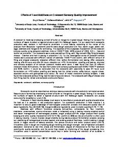
with a TCI of propofol 1.4–4.0 lg.ml 1, a continuous infusion of remifentanil 0.05–0.4 lg.kg 1.min 1 and intermittent boluses of rocuronium. The bispectral index was maintained within a range from 40 to 60, and the blood pressure and the heart rate were maintained within 20% of the baseline values. After induction of general anaesthesia, an ultrasound-guided paravertebral catheter placement was performed with an S-Nerve ultrasound machine (SonoSite Inc, Bothell, WA, USA) and a 6- to 13-MHz linear transducer (HFL 38x; SonoSite Inc), with the patient in a lateral decubitus position and the side to be blocked uppermost. The same anaesthesiologists (TY or TF), skilled in ultrasound-guided nerve blocks, performed all paravertebral catheter placements. After performing standard skin asepsis, the transducer, within a sterile sleeve, was placed on the patient in a transverse and partial oblique position to the vertebral column, parallel to the rib at the fifth intercostal space, to obtain a view of the internal intercostal membrane (IICM) and the lateral apex of the thoracic paravertebral space (TPVS) (Fig. 1a). If it was difficult to demonstrate the IICM at the fifth intercostal space, we placed the transducer at either the fourth or sixth intercostal space, for better visibility of the IICM. Thereafter, an 18-G Tuohy needle (Perican II; B. Braun AG, Melsungen, Germany) was inserted and advanced in plane with the transducer, in a lateral-tomedial direction, under ultrasound guidance through the intercostal space, as previously described [10]. After the needle tip penetrated through the IICM, 10 ml of normal saline 0.9% was injected into the TPVS to dilate it (Fig. 1b). Subsequently, a 20-G epidural catheter (Perifix Softtip Catheter; B. Braun AG) was threaded into the TPVS, 5 cm beyond the needle tip, in a medial direction. Then, the anaesthesiologist placed the transducer longitudinally to the paravertebral area to demonstrate the sagittal plane of the TPVS. Correct positioning of the catheter tip in the TPVS was ultrasonographically confirmed by injecting a mixture of 0.5 ml air and 3 ml saline through the catheter (Fig. 1c). When a hyperechoic flash induced by the injected air and saline mixture was not observed in the TPVS, the catheter was withdrawn 0.5 cm, and the air and saline mixture was re-injected, until a hyperechoic flash was observed in the TPVS. If © 2014 The Association of Anaesthetists of Great Britain and Ireland
Yoshida et al. | The spread of continuous thoracic paravertebral block
(a)
(b)
(c)
Figure 1 (a) Ultrasound cross-sectional image of the thoracic paravertebral space. (b) Ultrasound image of the needle tip placement into the thoracic paravertebral space. The thoracic paravertebral space is dilated, and the pleura is pressed ventrally by saline injected through the needle. (c) Ultrasound image of the sagittal plane of the dilated thoracic paravertebral space (*). Hyperechoic flash (Δ), produced by air injected through the catheter, was observed in the thoracic paravertebral space. TP, transverse process; TPVS, apex of thoracic paravertebral space; IICM, internal intercostal membrane; EICM, external intercostal muscle; PL, pleura; N, Tuohy needle; SCTL, superior costotransverse ligament.
© 2014 The Association of Anaesthetists of Great Britain and Ireland
Anaesthesia 2014, 69, 231–239
a hyperechoic flash was not observed, despite withdrawing the catheter more than 2 cm, the anaesthesiologist removed the catheter and reinserted it. After the catheter tip location was confirmed in the TPVS, the catheter was secured to the skin with a suture. Twenty millilitres of ropivacaine 0.5% was injected through the paravertebral catheter 15 min before the beginning of surgery. At the beginning of the surgery, patients received intravenous droperidol 1.25 mg, for prevention of postoperative nausea and vomiting (PONV) and intravenous flurbiprofen axetil 50 mg. The patients received intravenous fentanyl 5 lg.kg 1 approximately 60 min before the end of surgery. A continuous intravenous infusion of fentanyl (0.5 lg.kg 1.h 1 for 50 h) mixed with droperidol (0.1 mg.h 1 for 50 h) was initiated using a disposable pump (Linearfusor; Terumo, Tokyo, Japan) 30 min after the bolus injection of fentanyl. The patients were not administered any more fentanyl, except as described above. Twenty millilitres of ropivacaine 0.5% was injected again through the paravertebral catheter at the end of surgery. Just after the second bolus injection of ropivacaine, a continuous paravertebral infusion of the test drug (ropivacaine 0.2% or 0.5%) was started at 6 ml.h 1 using another disposable pump (Coopdech Balloonjecter 300; Daiken Medical, Osaka, Japan). After emergence from general anaesthesia, patients were administered sugammadex 4 mg.kg 1 to reverse neuromuscular blockade and then their tracheas were extubated. Patients were returned to the ward after 30 min of observation in the operating theatre. Oral administration of loxoprofen (60 mg thrice daily) was initiated on the evening of the first postoperative day. Breakthrough pain was treated with additional rectal diclofenac 25 mg or oral loxoprofen 60 mg at least 6 h apart. Patients did not receive any other analgesics. Postoperative nausea and vomiting was treated with intravenous metoclopramide 10 mg or rectal domperidone 60 mg, as needed. If the vomiting was intractable, the fentanyl infusion was discontinued or decreased by the surgeon. The primary outcome of this study was the number of anaesthetised dermatomes 24 h after surgery. The secondary outcomes were pain at rest, pain during movement, pain during coughing, additional analgesic 233
Anaesthesia 2014, 69, 231–239
Yoshida et al. | The spread of continuous thoracic paravertebral block
use, sedation level, patient satisfaction rating, incidence of PONV, and incidence of local anaesthetic toxicity. The evaluation of anaesthetised dermatomes was performed using an ice pack in a standardised fashion 1, 6, 24 and 48 h postoperatively. The blocked area was tested from the T4 dermatome between the anterior axillary line and midclavicular line, first in a cranial direction, then in a caudal direction. If required, cervical and high thoracic dermatomes were tested at the upper extremity and the neck, and lumbar dermatomes were tested at the lower extremity. The dermatome at which the patient perceived less or no sensation to the cold stimulus compared with that of the contralateral side was registered as the anaesthetised dermatome. The number of anaesthetised dermatomes was recorded on a specific chart that depicted the dermatome map. Pain was assessed using a numerical rating scale (NRS; 0, indicated no pain; 10, indicated worst pain imaginable). The sedation level was assessed using the Richmond Agitation Sedation Scale (RASS) [11]. The incidence of PONV and local anaesthetic toxicity were assessed dichotomously. These outcomes were assessed 1, 6, 24 and 48 h postoperatively by an anaesthesiologist who was blinded to the allocation and did not perform the TPVB procedure or the general anaesthesia. The patient satisfaction rating was described using a four-grade scale (dissatisfied, 1; slightly dissatisfied, 2; slightly satisfied, 3; satisfied, 4). The ward nurse obtained and recorded patient satisfaction ratings 48 h after surgery. In addition, the ward nurse recorded additional analgesic use within 48 h of surgery. According to a pilot study (n = 7, T. Yoshida and T. Fujiwara, unpublished data), the mean number (SD) of anaesthetised dermatomes 24 h after surgery produced by the continuous paravertebral infusion of 0.2% ropivacaine (at 6 ml.h 1) was 3.0 (1.2). We considered a difference of one dermatome to be clinically relevant. To demonstrate this difference using a twotailed Student’s t-test, a sample size of 22 patients per group was the minimum calculated number needed to provide a statistical power of 0.8 and a type-1 error rate of 0.05. Because of the expected dropout rate, 30 patients per group were enrolled to the study. Sample sizes were determined using StatMate 2 for Macintosh (GraphPad Software, San Diego, CA, USA). Significant 234
differences between the two groups were analysed using the two-tailed Student’s t-test (continuous data) or the Mann–Whitney U-test (non-continuous data). Proportional significant differences in the properties of the two groups were evaluated using Fisher’s exact test. Statistical analyses were conducted using R for Mac OS X version 2.15.2 (The R Foundation for Statistical Computing, Vienna, Austria). A p value < 0.05 was considered statistically significant.
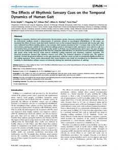
Results Sixty consecutive patients were enrolled in this study. The study was randomised, and all patients received their allocated interventions (Fig. 2). The baseline and peri-operative characteristics of the study patients were similar between the two groups (Table 1). Three patients receiving ropivacaine 0.2% group were excluded from the final analysis. The paravertebral catheter of one patient was accidentally dislodged postoperatively. Another patient’s trachea remained intubated until the second postoperative day, because of an intra-operative pulmonary artery rupture. The other patient was over-sedated and hence, we could not evaluate the outcomes until 3 h after surgery. Three patients in receiving ropivacaine 0.5% were excluded from the final analysis because of accidental paravertebral catheter dislodgement, intra-operative prolapse of the catheter into the thoracic cavity and inability to confirm the catheter tip. The fentanyl infusion was discontinued owing to intractable PONV in two patients receiving ropivacaine 0.2% (17 and 25 h after surgery, respectively) and in three patients receiving ropivacaine 0.5% (24, 25 and 19.5 h after surgery, respectively). Owing to persistent PONV, the fentanyl infusion rate was decreased to 0.25 lg.kg 1.h 1 in one patient receiving ropivacaine 0.5%, 21 h after surgery. These patients were all included in the final analysis. Finally, two groups, each containing 27 patients, were included in the analysis. Figure 3 shows the time course for the number of anaesthetised dermatomes. No statistically significant difference was found between the two groups at each evaluation time. For the primary outcome measure of this study, the median (IQR [range]) number of anaesthetised dermatomes 24 h after surgery was 4 (3–6 [1– 9]) with ropivacaine 0.2% and 4 (3–6 [2–11]) with © 2014 The Association of Anaesthetists of Great Britain and Ireland
Yoshida et al. | The spread of continuous thoracic paravertebral block
Anaesthesia 2014, 69, 231–239
Figure 2 CONSORT flow diagram. ropivacaine 0.5% (p = 0.66). The number of anaesthetised dermatomes 48 h after surgery was 4 (2–5 [1–8]) with ropivacaine 0.2% and 3 (3–6 [1–11]) with ropivacaine 0.5% (p = 0.69). A spread of the blockade over the midline was not observed. The pain scores at rest, during movement, and while coughing are shown in Fig. 4, and there were no significant differences between the two groups at each evaluation time. Other outcomes, such as RASS, incidence of PONV, additional analgesic use, and patient satisfaction rating, are shown in Table 2, and the differences were not statistically significant between the two groups. Local anaesthetic toxicity was not observed during the whole study period. There were © 2014 The Association of Anaesthetists of Great Britain and Ireland
no other study-related adverse events, such as postoperative hypotension, infection and nerve injury.
Discussion In our study, the segmental spread of sensory block produced by continuous TPVB using 0.2% ropivacaine was similar to that of 0.5% ropivacaine, when the infusion volume rate was equivalent. The analgesic efficacy was also similar between the two groups. This is the first report to investigate a factor affecting a segmental spread of sensory block produced by continuous TPVB. There have been several reports investigating a spread of single-injection TPVB [12–17]. Some investi235
Anaesthesia 2014, 69, 231–239
Yoshida et al. | The spread of continuous thoracic paravertebral block
Table 1 Baseline and peri-operative characteristics of study patients receiving continuous thoracic paravertebral block with 0.2% or 0.5% ropivacaine. Values are mean (SD) or number (proportion).
Age; years Male Height; cm Weight; kg BMI; kg.m 2 ASA physical status 1/2/3 Catheter site 4th/5th/6th intercostal space Surgical side, right Procedure, PLT/VATS Surgical time; min Anaesthesia time; min Intra-operative remifentanil dosage; lg.kg 1.min
0.2% (n = 27)
0.5% (n = 27)
67 (9) 17 (63%) 161.0 (10.0) 57.7 (9.6) 22.2 (2.9) 2/22/3
65 (9) 17 (63%) 160.5 (7.1) 55.7 (10.9) 21.5 (3.2) 4/23/0
3/23/1
1/26/0
(a)
(b) 15 (56%) 18/9
21 (78%) 16/11
219 (53) 328 (61)
228 (68) 334 (68)
0.18 (0.05)
0.17 (0.04)
1
PLT, posterolateral thoracotomy; VATS, video-assisted thoracic surgery.
(c)
Figure 3 Time course of the number of anaesthetised dermatomes after surgery in patients receiving continuous thoracic paravertebral block with 0.2% (□) or 0.5% (■) ropivacaine. Horizontal lines indicate medians; boxes indicate IQRs; and whiskers indicate ranges. No statistically significant differences (n.s.) between the groups were observed at each evaluation time.
Figure 4 Pain at rest (a), during movement (b), and during coughing (c) after surgery in patients receiving continuous thoracic paravertebral block with 0.2% (□) or 0.5% (■) ropivacaine. NRS, numerical rating scale; n.s., not significant. Horizontal lines indicate medians; boxes indicate IQRs; and whiskers indicate ranges. No statistically significant differences between the groups were observed at each evaluation time.
gators have evaluated the distribution of a single paravertebral injection using radiopaque dye and have concluded that a longitudinal distribution of the TPVB
is associated with a linear correlation between the injected volume and the number of segments covered by the dye [14, 16]. Nonetheless, it has been shown
236
© 2014 The Association of Anaesthetists of Great Britain and Ireland
Yoshida et al. | The spread of continuous thoracic paravertebral block
Table 2 Patients’ postoperative data after receiving continuous thoracic paravertebral block with 0.2% or 0.5% ropivacaine. Values are median (range) or number (proportion). 0.2% (n = 27)
0.5% (n = 27)
Richmond Agitation Sedation Scale 1 h after 2 ( 3 to 0) 2 ( 3 to 1) surgery 6 h after 1 ( 2 to 0) 1 ( 3 to 0) surgery 24 h after 0 ( 1 to 0) 0 ( 2 to 0) surgery 48 h after 0 ( 1 to 0) 0 ( 1 to 0) surgery Postoperative nausea and vomiting; % 1 h after 0 (0) 1 (4) surgery 6 h after 3 (11) 3 (11) surgery 24 h after 5 (19) 12 (44) surgery 48 h after 2 (7) 3 (11) surgery Additional 1 (0–3) 0 (0–5) analgesics use Patient satisfaction rating 1 0 0 2 3 (11%) 3 (11%) 3 9 (33%) 9 (33%) 4 15 (56%) 15 (56%)
p value 0.55 0.63 0.70 1.00
1.00 1.00 0.08 1.00 0.61
1.00
that the distribution of radiopaque dye and local anaesthetic solution do not necessarily correlate with a clinical distribution of sensory blockade [16, 17]. Cheema et al. investigated the clinical spread of sensory block produced by single-injection TPVB using bupivacaine and concluded that the clinical spreading pattern was unpredictable [12]. Although a segmental spread of sensory block produced by continuous thoracic epidural block depends on the total mass of local anaesthetic [7–9], our results suggest that the segmental spread of sensory block produced by continuous TPVB does not depend on the total mass of local anaesthetic. Compared with the TPVS, the epidural space is surrounded by the spinal column and a relatively limited area. Local anaesthetic injected into the epidural space has been considered to remain in the spinal canal and to diffuse intrathecally in accordance with its concentration gradient [18]; hence, the same total mass of local anaesthetic produces the same segmental spread of sensory block [19]. The TPVS is a © 2014 The Association of Anaesthetists of Great Britain and Ireland
Anaesthesia 2014, 69, 231–239
wedge-shaped area that contains the spinal nerve and the sympathetic trunk and is continuous with the intercostal space laterally [1]. The TPVS is larger than the epidural space, and radiopaque dye injected into the paravertebral space may spread widely [14]. Because of these anatomical properties, local anaesthetics may hardly diffuse longitudinally in the TPVS in accordance with their concentration gradients, and we suppose that the concentration and the total mass of local anaesthetic does not play an important role in the segmental spread of sensory block by continuous TPVB, as our study demonstrated. We designed this research to be a superiority study and calculated a sample size as described above, because we expected that a higher concentration of ropivacaine would provide a wider spread of sensory block. However, contrary to our expectations, we failed to find a significant difference in a sensory spread between the two groups at the end of the study. Although our study does not demonstrate equivalence in the spread of sensory block between the two different concentrations of ropivacaine, because of the study design, our results suggest that even if there was an actual difference between the two groups in sensory block spread, the difference would be miniscule and clinically inconsequential. The median number of anaesthetised dermatomes 24 h after surgery was four in both groups, and the pain score was similar between the two groups. A sensory blockade of four dermatomes can cover all incisions made during lung surgery, including videoassisted thoracic surgery. A recent meta-analysis found higher doses of paravertebral bupivacaine (890– 990 mg.24 h 1) to be associated with improved pain control after thoracotomy, compared with lower doses (325–472.5 mg.24 h 1) [4]. We determined the ropivacaine concentrations used in our study by referring to this meta-analysis [4]. Patients in the ropivacaine 0.5% group received ropivacaine 720 mg.24 h 1, whereas patients in the 0.2% group received ropivacaine 288 mg.24 h 1. However, the pain scores did not differ significantly between the two groups. There were two possible reasons for the failure to demonstrate a difference in analgesic efficacy in this study. First, the number of study patients might have been inadequate to demonstrate a difference. The primary outcome of this 237
Anaesthesia 2014, 69, 231–239
Yoshida et al. | The spread of continuous thoracic paravertebral block
study was to determine the number of anaesthetised dermatomes 24 h after surgery; hence, the sample size was not determined to detect a difference in analgesic efficacy. Second, the continuous fentanyl infusion should mask the difference in analgesic efficacy between the two groups; we planned to administer fentanyl to reduce the patients’ visceral pain induced by the phrenic and vagus nerves [20, 21]. Our study had some limitations. First, we did not inject radiopaque dye through the catheter to confirm correct catheter placement in the TPVS. Instead, we confirmed that the catheter tip was definitely located in the TPVS by ultrasound. Compared with other landmark-based techniques, our in-plane ultrasound-guided thoracic paravertebral catheter placement technique has a higher success rate [22]. Second, we did not confirm if the spreading pattern of the radiopaque dye injected into the TPVS was classified as a longitudinal or cloudlike pattern according to the previous report [14]. The segmental spread of the radiopaque dye was found to be more extensive with the longitudinal spread pattern than the cloud-like pattern [14]. However, it was suggested that the clinical segmental spread of sensory block did not depend on the radiopaque dye spreading pattern [16]. Third, there was a possibility that a difference in intensity of sensory block existed between the groups. When we evaluated the sensory block, qualitative methods, such as cold test, could hardly measure the intensity of sensory block [23]. However, even if we could detect the difference in intensity between the groups by quantitative methods, the difference would be small and clinically negligible. In other words, we believe that merely one dermatome difference is clinically relevant, if the blocked dermatome is evaluated by qualitative methods. In conclusion, contrary to our expectations, this study failed to detect a difference in the segmental spread of sensory block produced by continuous TPVB using different concentrations of ropivacaine. While our study does not prove equivalence between the two different concentrations of ropivacaine in a sensory block spread, owing to the study design limitations, our results suggest that ropivacaine concentration does not play an important role in the segmental spread of sensory block by continuous TPVB. 238
Acknowledgments The authors thank Kohei Akazawa, PhD (Department of Medical Informatics, Niigata University Medical and Dental Hospital, Niigata, Japan) for his statistical advice. The authors also thank Editage for providing editorial assistance.
Competing interests No external funding and no competing interests declared.
References 1. Karmakar MK. Thoracic paravertebral block. Anesthesiology 2001; 95: 771–80. 2. Joshi GP, Bonnet F, Shah R, et al. A systematic review of randomized trials evaluating regional techniques for postthoracotomy analgesia. Anesthesia and Analgesia 2008; 107: 1026– 40. 3. Powell ES, Cook D, Pearce AC, et al. A prospective, multicentre, observational cohort study of analgesia and outcome after pneumonectomy. British Journal of Anaesthesia 2011; 106: 364–70. 4. Kotze A, Scally A, Howell S. Efficacy and safety of different techniques of paravertebral block for analgesia after thoracotomy: a systematic review and metaregression. British Journal of Anaesthesia 2009; 103: 626–36. 5. Casati A, Alessandrini P, Nuzzi M, et al. A prospective, randomized, blinded comparison between continuous thoracic paravertebral and epidural infusion of 0.2% ropivacaine after lung resection surgery. European Journal of Anaesthesiology 2006; 23: 999–1004. 6. Catala E, Casas JI, Unzueta MC, Diaz X, Aliaga L, Villar Landeira JM. Continuous infusion is superior to bolus doses with thoracic paravertebral blocks after thoracotomies. Journal of Cardiothoracic and Vascular Anesthesia 1996; 10: 586–8. 7. Visser WA, Lee RA, Gielen MJ. Factors affecting the distribution of neural blockade by local anesthetics in epidural anesthesia and a comparison of lumbar versus thoracic epidural anesthesia. Anesthesia and Analgesia 2008; 107: 708– 21. 8. Dernedde M, Stadler M, Bardiau F, Boogaerts JG. Continuous epidural infusion of large concentration/small volume versus small concentration/large volume of levobupivacaine for postoperative analgesia. Anesthesia and Analgesia 2003; 96: 796–801. 9. Scott DA, Chamley DM, Mooney PH, Deam RK, Mark AH, Hagglof B. Epidural ropivacaine infusion for postoperative analgesia after major lower abdominal surgery – a dose finding study. Anesthesia and Analgesia 1995; 81: 982–6. 10. Shibata Y, Nishiwaki K. Ultrasound-guided intercostal approach to thoracic paravertebral block. Anesthesia and Analgesia 2009; 109: 996–7. 11. Sessler CN, Gosnell MS, Grap MJ, et al. The Richmond Agitation-Sedation Scale: validity and reliability in adult intensive care unit patients. American Journal of Respiratory and Critical Care Medicine 2002; 166: 1338–44. 12. Cheema S, Richardson J, McGurgan P. Factors affecting the spread of bupivacaine in the adult thoracic paravertebral space. Anaesthesia 2003; 58: 684–7. © 2014 The Association of Anaesthetists of Great Britain and Ireland
Yoshida et al. | The spread of continuous thoracic paravertebral block 13. Cheema SP, Ilsley D, Richardson J, Sabanathan S. A thermographic study of paravertebral analgesia. Anaesthesia 1995; 50: 118–21. 14. Naja MZ, Ziade MF, El Rajab M, El Tayara K, Lonnqvist PA. Varying anatomical injection points within the thoracic paravertebral space: effect on spread of solution and nerve blockade. Anaesthesia 2004; 59: 459–63. 15. Saito T, Den S, Cheema SP, et al. A single-injection, multi-segmental paravertebral block-extension of somatosensory and sympathetic block in volunteers. Acta Anaesthesiologica Scandinavica 2001; 45: 30–3. €nnqvist PA, Hesser U. Radiological and clinical distribution 16. Lo of thoracic paravertebral blockade in infants and children. Pediatric Anesthesia 1993; 3: 83–7. 17. Marhofer D, Marhofer P, Kettner SC, et al. Magnetic resonance imaging analysis of the spread of local anesthetic solution after ultrasound-guided lateral thoracic paravertebral blockade: a volunteer study. Anesthesiology 2013; 118: 1106– 12.
© 2014 The Association of Anaesthetists of Great Britain and Ireland
Anaesthesia 2014, 69, 231–239
18. Kamiya Y, Kikuchi T, Inagawa G, et al. Lidocaine concentration in cerebrospinal fluid after epidural administration: a comparison between epidural and combined spinal-epidural anesthesia. Anesthesiology 2009; 110: 1127–32. 19. Bromage PR. Mechanism of action of extradural analgesia. British Journal of Anaesthesia 1975; 47(suppl): 199–211. 20. Daly DJ, Myles PS. Update on the role of paravertebral blocks for thoracic surgery: are they worth it? Current Opinion in Anesthesiology 2009; 22: 38–43. 21. Scawn ND, Pennefather SH, Soorae A, Wang JY, Russell GN. Ipsilateral shoulder pain after thoracotomy with epidural analgesia: the influence of phrenic nerve infiltration with lidocaine. Anesthesia and Analgesia 2001; 93: 260–4, 1st contents page. 22. Renes SH, Bruhn J, Gielen MJ, Scheffer GJ, van Geffen GJ. Inplane ultrasound-guided thoracic paravertebral block. Regional Anesthesia and Pain Medicine 2010; 35: 212–6. 23. Curatolo M, Petersen-Felix S, Arendt-Nielsen L. Sensory assessment of regional analgesia in humans: a review of methods and applications. Anesthesiology 2000; 93: 1517–30.
239