506 Brazilian Journal of Medical and Biological Research (2009) 42: 506-514 ISSN 0100-879X
D.Q. Feng et al.
Effects of the conditioned medium of mesenchymal stem cells on mouse oocyte activation and development D.Q. Feng1, Y. Zhou1, B. Ling1,2, T. Gao2, Y.Y. Shi1, H.M. Wei3 and Z.G. Tian3 1Anhui
Province Key Laboratory of Molecular Medicine, 2Department of Obstetrics and Gynecology, Anhui Provincial Hospital, Anhui Medical University, Hefei, China 3Institution of Immunology, Hefei National Laboratory for Physical Science at Microscale and School of Life Science, University of Science and Technology of China, Hefei, China Correspondence to: B. Ling, Department of Obstetrics and Gynecology, Anhui Provincial Hospital Affiliated to Anhui Medical University, 17 Lu-Jiang Road, Hefei 230001, China Fax: +86-551-228-2121. E-mail:
[email protected] Mesenchymal stem cells (MSCs) have been reported to secrete a variety of cytokines and growth factors acting as trophic suppliers, but little is known regarding the effects of conditioned medium (CM) of MSCs isolated from femurs and tibias of mouse on the artificial activation of mouse oocytes and on the developmental competence of the parthenotes. In the current study, we investigated the effect of CM on the events of mouse oocyte activation, namely oscillations of cytosolic calcium concentration ([Ca2+]i), meiosis resumption, pronucleus formation, and parthenogenetic development. The surface markers of MSCs were identified with a fluorescence-activated cell sorter. The dynamic changes of the spindle and formation of pronuclei were examined by laser-scanning confocal microscopy. Exposure of cumulus-oocyte complexes to CM for 40 min was optimal for inducing oocyte parthenogenetic activation and evoking [Ca2+]i oscillations similar to those evoked by sperm (95 vs 100%; P > 0.05). Parthenogenetically activated oocytes immediately treated with 7.5 μg/mL cytochalasin B (CB), which inhibited spindle rotation and second polar body extrusion, were mostly diploid (93 vs 6%, P < 0.01) while CB-untreated oocytes were mostly haploid (5 vs 83%, P < 0.01). Consequently, the blastocyst rate was higher in the CB-treated than in the CB-untreated oocytes. There was no significant difference in developmental rate between oocytes activated with CM and 7% ethanol (62 vs 62%, P > 0.05), but the developmental competence of the fertilized oocytes was superior to that of the parthenotes (88 vs 62%, P < 0.05). The present results demonstrate that CM can effectively activate mouse oocytes, as judged by the generation of [Ca2+]i oscillations, completion of meiosis and parthenogenetic development. Key words: Mesenchymal stem cells; Oocyte activation; Parthenogenesis; Calcium; Cytochalasin B Research supported by grants from the China National Natural Science Foundation (#34071805) and the Science Research Foundation, Anhui Provincial Hospital.
Received September 24, 2008. Accepted March 11, 2009
Introduction Mesenchymal stem cells (MSCs), widely distributed in a variety of tissues in the adult organism, secrete many kinds of cytokines and growth factors, such as monocyte chemotactic protein-1, vascular endothelial growth factorA, epidermal growth factor (EGF), fibroblast growth factor2, interleukin-6, leukemia inhibitory factor, transforming
Braz J Med Biol Res 42(6) 2009
growth factor-β, and so on (1,2). We have demonstrated the trophic effects of these bioactive factors in supporting follicular growth and in vitro maturation of mouse oocytes (3). However, to our surprise, some MII oocytes cocultured with MSCs were activated, with the occurrence of cleavage, though none of these oocytes developed further in this culture. We later observed that the conditioned medium (CM) of MSCs, which undoubtedly contains many
www.bjournal.com.br
MSC conditioned medium and oocyte activation
kinds of cytokines and growth factors resulting from the exocrinosity of MSCs, can act equally as a stimulant. Thus, we thought it would be useful to assess the ability of CM to activate mouse oocytes parthenogenetically, and embryo development before implantation. Some of these secreted bioactive factors have been reported to improve meiotic maturation in vitro and subsequently the embryo developmental potential directly or via cumulus cells (4-6). Many studies have demonstrated a beneficial influence of leukemia inhibitory factor on embryo development in several species, such as improved cleavage of parthenogenetic embryos, higher rate of pronuclear-stage embryos with in vitro fertilization, a larger inner cell mass, and so on (7-9). The EGF receptor (EGFR) is present in cumulus cells and in oocyte at the germinal vesicle stage and following maturation. O’Donnell et al. (10) reported that activation of EGFR by EGF or transforming growth factor-α induces both auto- and trans-phosphorylation of EGFRs, which result in cytosolic calcium concentration ([Ca2+]i) elevation and subsequent membrane permeabilization in mouse cumulus-oocyte complexes (COCs), but oocytes fail to respond to the EGF stimulus. However, to our knowledge, no information is available concerning the effect of CM on oocyte activation and on the development of preimplantation parthenogenotes. A variety of artificial stimuli such as ethanol, electroporation, calcium ionophore, ionomycin, and inositol 1,4,5trisphosphate (InsP3) are used to induce the parthenogenetic activation of mammalian oocytes (11-15). Yang et al. (13) reported that combined treatment with 7% ethanol for 10 min and cycloheximide, a protein synthesis inhibitor, improved the rate of activation of the young oocytes of cattle. The histone kinase inhibitor 6-dimethylaminopurine has been reported to accelerate and enhance the formation of pronuclei in non-aged MII oocytes from mice and cattle (16). Also, cytochalasin B (CB), an inhibitor of microfilament polymerization, prevents the release of the second polar body (Pb2) after oocyte activation, which would result in diploid development (17), and may also help prevent embryo fragmentation (18,19). These artificial stimuli mimicking sperm-triggered events evoke a series of [Ca2+]i oscillations, which is critical for oocyte activation. [Ca2+]i oscillation triggers two important major events during activation: resumption of meiosis and pronuclear formation. Sperm–oocyte fusion initiates [Ca2+]i oscillations that last several hours (20). Some activators, such as ionomycin or ethanol, cause a single [Ca2+]i transient, while others such as InsP3, adenophostin A, thimerosal, and strontium evoke repetitive [Ca2+]i oscillations (21). Furthermore, some investigators have suggested that the type of [Ca2+]i change is an important factor in
www.bjournal.com.br
507
promoting subsequent embryo development (22-24). Therefore, it is important to investigate [Ca2+]i oscillations of oocytes treated with CM when judging the efficiency of CM for oocyte activation and embryo development. In the present study, we investigated the pattern of [Ca2+]i changes in mouse oocytes after treatment with CM, and the effects of time of exposure to CM and to a sequential combination of CM and CB in improving parthenogenetic activation and development of mouse oocytes.
Material and Methods Care and use of animals Kunming mice, originally derived from ICR (CD1) animals, were housed in a controlled environment (20-25°C, 40-60% humidity, and a 8:00-20:00-light cycle). Female mice of the F1 strain, 6-8 weeks old, were used. Animal care and handling were in accordance with the policies issued by the Ethics Committee of Anhui Provincial Hospital affiliated to Anhui Medical University. Isolation of mesenchymal stem cells and preparation of conditioned medium Mice were deeply anesthetized with chloral hydrate and decapitated. Bone marrow was collected by flushing femurs and tibias with DMEM-HG (Gibco™, Invitrogen Corporation, USA). Mononuclear cells were harvested by separation with 60% Percoll (Amersham Biosciences, USA) separation (density: 1.077 g/mL, 800 g for 20 min) and washed twice (270 g for 10 min) in DMEM-HG. Cells were then incubated in complete medium consisting of DMEMHG, 10% fetal calf serum (Gibco™, Invitrogen Corporation), 100 U/mL penicillin and 100 mg/mL, at a density of 5 x 10⁶ cells/mL. After 3 days, nonadherent cells were removed by two to three washes with 1X PBS and adherent cells were further cultured in complete medium to 90% confluence. At this time, adherent cells were trypsinized and seeded at a density of 3 x 10⁵ cells/mL for further expansion. After 48 h, the supernatant was collected as CM and filtered through a 0.2-μm filter for immediate use. Surface markers of MSCs, CD11b, CD34, CD44, and CD105, were analyzed with a fluorescence-activated cell sorter (FACS). FITC-anti-CD11b, FITC-anti-CD34, PE-cy5anti-CD44, and PE-anti-CD105 were purchased from eBioscience (USA). Superovulation and collection of mouse oocytes Female mice were induced to superovulate by injection of pregnant mare’s serum gonadotropin (PMSG) (7.5 IU, ip, Hangzhou Animal Medicine Factory, China), followed 48 h later by injection of human chorionic gonadotropin
Braz J Med Biol Res 42(6) 2009
508
(hCG) (7.5 IU, ip, Hangzhou Animal Medicine Factory). The superovulated mice were killed 14 h after hCG injection, and the COCs were released from the oviductal ampullae into pre-equilibrated M16 medium (Sigma, USA). The COCs and denuded MII oocytes, which were freed of cumulus cells by a brief exposure to 0.1% (w/v) hyaluronidase (Sigma), were used for in vitro activation. All manipulations were performed on a thermostatic plate (37°C). Parthenogenetic activation COCs and denuded MII oocytes were treated with freshly prepared CM for 0, 10, 40 min, 1, or 2 h. Cumulus cells in COCs were dispersed with hyaluronidase after CM treatment. Also, oocytes were activated with CZB medium containing 7% ethanol for 6 min. After activation, the oocytes were washed three times with M16 and further cultured in CZB medium containing 0.4% BSA (Sigma) in a 4-well Petri dish (Nunc, Denmark). Other batches of COC used as control were incubated with DMEM-HG containing 10% fetal calf serum for 0, 10, 40 min, 1, or 2 h followed by additional incubation in CZB medium containing 0.4% BSA after cumulus cells were removed by hyaluronidase. Oocytes that exited MII arrest and resumed meiosis were considered to have been activated. At 48 h post-activation, glutamine and glucose were directly injected into one drop of CZB to give a final concentration of 1 and 5.56 mM, respectively, which supported embryo development to the morula and blastocyst stages (25). Treatment with CB Cytochalasin B (Sigma) was dissolved as a stock solution (1 mg/mL) in dimethylsulfoxide (Sigma) and stored at -20°C. It was later diluted to a final concentration of 7.5 μg/ mL in CZB containing 0.4% BSA. The parthenogenetically activated oocytes were sequentially treated with CB for 4 h. The oocytes were removed from the medium containing CB, washed at least three times with M16, and then cultured in CB-free CZB containing 0.4% BSA. At 48 h, glutamine and glucose were directly injected into the culture medium of CZB to a final concentration of 1 and 5.56 mM, respectively. In vivo/in vitro fertilization The PMSG- and hCG-primed female mice were allowed to mate overnight with males of proven fertility. In vivo fertilized oocytes were isolated into M16 medium from the oviductal ampullae of mice 15 h after hCG administration. Cumulus cells were removed as described earlier for MII oocytes and then cultured in CZB containing 0.4% BSA. Glutamine and glucose at final concentrations of 1 and 5.56 mM, respectively, were added to the culture drop
Braz J Med Biol Res 42(6) 2009
D.Q. Feng et al.
at 48 h. For in vitro fertilization, MII oocytes were treated with 0.5% (w/v) protease E (BioShare, Germany) to remove the zona pellucida (ZP) prior to insemination. Sperm were collected from the cauda epididymis of the males, capacitated for 1-1.5 h, and diluted in HTF Xtra medium (LifeGlobal, USA) to a final concentration of 0.7-1.3 x 10⁶ sperm/mL. Insemination of ZP-free oocytes by capacitated sperm was performed in a thermostatic 4-well Petri dish, suitable for [Ca2+]i measurements, as previously described (26). Measurement of [Ca2+]i To monitor [Ca2+]i changes, COCs and denuded oocytes, either ZP-enclosed or ZP-free, were loaded with the Ca2+-sensitive dye, fura-2-AM (3 μM; Sigma), for 30 min in Ca2+/Mg2+-free M2 medium at 37°C. After dye loading, COCs and denuded oocytes were washed free of the dye and transferred to a polylysine-coated coverslip mounted on a 4-well Petri dish held at 37°C. COCs and denuded oocytes were stimulated by the addition of reagents directly to the cell dish. Free [Ca2+]i was determined by monitoring the fluorescence ratio at 340/380 nm excitation wavelengths using an inverted microscope (Zeiss, Axiovert, Germany) after appropriate background fluorescence subtraction. In all cases, data were sampled at 5-s intervals. Immunofluorescent staining and confocal microscopy At various times after activation (0, 1, 2, 4, 6, 12, 16, 20, 24 h), oocytes were fixed with 3.7% (w/v) paraformaldehyde in PBS containing 0.1% (v/v) Triton X-100 for 30 min at 37°C. Fixed oocytes were stored until processing at 4°C in a PBS blocking solution containing 1% (w/v) BSA, 2% (v/ v) normal goat serum, 0.1 mol glycine, 0.2% (w/v) sodium azide, and 0.01% (v/v) Triton X-100. A double-labeling protocol was used for the detection of microtubules and chromatin by confocal microscopy. Oocytes were first incubated for 1 h at 37°C in a mixture of mouse monoclonal anti α–tubulin and β–tubulin (Sigma) antibodies at a 1:500 final dilution in PBS blocking solution. After two washes in 0.1% (w/v) polyvinylpyrrolidone (PVP)/PBS at room temperature, oocytes were incubated at 37°C in PBS blocking solution alone for 30 min, and then in a 1:100 dilution of goat anti-mouse fluorescein-conjugated IgG (Santa Cruz, USA) for 45 min at 37°C. Oocytes were washed again twice in 0.1% PVP/PBS, and incubated at room temperature for 10 min in 10 μg/mL propidium iodide (Sigma) and mounted in 70% (v/v) glycerol/PBS containing 25 mg/mL sodium azide. Controls included nonimmune and secondary antibody alone, which did not detect spindles, microtubules or midbodies. The samples were examined with a laser-scanning confocal microscopy (Zeiss, LSM510).
www.bjournal.com.br
MSC conditioned medium and oocyte activation
509
Statistical analysis Data are reported as the means ± SEM of three replicates of each experiment. Differences between treatment groups were determined by the chi-square test, with the level of significance set at P < 0.05.
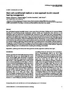
Results Characterization of mesenchymal stem cells MSCs of passage two showed typical fibroblast morphology with large flat and spindle-shaped cells, as did primary MSCs. FACS analysis demonstrated that these cells were negative for CD34 and CD11b, and positive for CD44 and CD105, consistent with the general agreement that MSCs lack typical hematopoietic antigens and markers of granulo-monocytic cells, namely, CD34 and CD11b (27). The effect of CM on [Ca2+]i changes A very early observable event occurring after spermoocyte interaction or after parthenogenetic activation is an increase in [Ca2+]i. Oocytes in COCs treated with CM exhibited an initial [Ca2+]i transient at 12-30 min, that was followed by a series of irregular [Ca2+]i oscillations of lower amplitude (0.41 ± 0.05 arbitrary units) and longer peak-topeak intervals (7.11 ± 1.59 min; Figure 1C), whereas the oscillations in fertilized oocytes were regular, with greater amplitude (0.83 ± 0.13 arbitrary units) and shorter intervals
1.6
1.0
A
(3.15 ± 0.37 min; Figure 1A). In general, [Ca2+]i increased slowly in CM-treated oocytes and all sequential changes occurred above the basal level. Oocytes treated with 7% ethanol presented a single rise in [Ca2+]i, which increased rapidly from the basal level to the peak level, and then slowly decreased but did not return to the basal level within 6 min (Figure 1B). Effects of CM on the activation of mouse oocytes in the presence or not of CB Significantly more oocytes (approximately 95%) were activated at the CM exposure times of 40 min, 1 and 2 h than at shorter exposure times as shown in Table 1. There was no difference between exposure for 40 min, 1 and 2 h, but the oocytes treated for 40 min developed well, while embryo fragmentation increased with exposure time. The optimal time for denuded MII oocyte activation (approximately 95%) was not 40 min but 2 h (data not shown). To study the effect of CB on the development of activated oocytes, COCs were treated with CM for 40 min, followed by culture for 4 h in CZB with or without 7.5 μg/mL CB after cumulus cells were removed. No significant difference in activation rate was observed in the presence or absence of CB (Table 2). However, most of the oocytes treated with CB had two pronuclei, whereas most of the activated oocytes without CB treatment contained one pronucleus or cleaved immediately. Since CB inhibited the second polar body extrusion, the activated oocytes treated
1.0
B
C
1.0 0.8 0.6
0.8 Ratio (340/380 nm)
1.2
Ratio (340/380 nm)
Ratio (340/380 nm)
1.4
0.6
0.4
0.8
0.6
0.4
0.4 0.2
0.2 0
10
30 20 Time (min)
40
0.2 0
2
4 Time (min)
6
0
10
20 Time (min)
30
40
Figure 1. Fertilization-, ethanol-, and conditioned medium (CM)-induced [Ca2+]i changes in fura-2-AM-loaded mouse oocytes. A, During in vitro fertilization, sperm triggered an initial [Ca2+]i transient that was followed by a series of regular and high [Ca2+]i oscillations. B, A single rapid [Ca2+]i rise was observed in 7% ethanol-treated oocytes, which decreased slowly. C) The type of [Ca2+]i changes in CM-treated oocytes was similar to that induced by sperm. The amplitude of the initial [Ca2+]i transient was lower than the subsequent [Ca2+]i oscillations, whereas the oscillations induced by CM were of lower amplitude and longer duration than those induced by sperm.
www.bjournal.com.br
Braz J Med Biol Res 42(6) 2009
510
D.Q. Feng et al.
with CB mostly formed diploid zygotes while those not treated with CB mostly formed haploid zygotes. A significantly higher proportion of activated oocytes in the group treated with CB developed into blastocysts than in the group not treated with CB (Table 2).
Table 1. Effect of mesenchymal stem cell conditioned medium (CM) on the activation of mouse oocytes. Duration of CM treatment
No. of oocytes examined
0 min 10 min 40 min 1h 2h
Activated oocytes (%)
59 106 104 97 102
6 46 95 95 96
± ± ± ± ±
3%a 10%b 2%c 2%c 2%c
Dynamic changes of the spindle following parthenogenetic activation Microtubules were found mainly in the well-organized spindle in MII oocytes by immunofluorescent staining. The Data are reported as means ± SD for activated oocytes. Values spindle was symmetrical, bipolar, barrel-shaped, and lowith different superscript letters differed significantly (P < 0.01, cated near the cortex of the oocyte. From 1 to 3 h postchi-square test). activation, as the oocyte exited MII and progressed to anaphase II and subsequently to telophase II, the sister chromatids migrated to opposite poles of the spindle, followed spindle rotation Table 2. Effect of cytochalasin B (CB) treatment on the activation and development of mouse oocytes. from parallel to vertical in relation to the surface of the oocyte (Figure 2A). The spindle CB No. of Activated Activated oocytes (%) with Blastocyst then elongated and formed Pb2, which was oocytes oocytes (%) (%) extruded into the perivitelline space. 1 pronucleus 2 pronuclei Fragmented Neither the resumption of meiosis nor the cytoplasm separation of daughter chromatids was dis109 97 ± 2% 5 ± 1* 93 ± 3* 0* 59 ± 2* turbed after oocyte activation with CM fol- + 94 95 ± 2% 83 ± 2 6 ± 2 7 ± 3 10 ± 3 lowed by treatment with 7.5 μg/mL CB for 4 h. However, in CB-treated parthenogenetically Data are reported as means ± SD. *P < 0.01 compared to oocytes not treated activated oocytes, spindle rotation and Pb2 with CB (chi-square test).
Figure 2. Resumption of meiosis induced by conditioned medium (CM) and effects of cytochalasin B (CB) on spindle rotation and pronucleus formation. Microtubules (green) and chromatin (red) were stained with anti-tubulin antibodies and propidium iodide, respectively. After 40-min exposure to CM, MII arrested oocytes in cumulus-oocyte complexes (COCs) resumed meiosis. A, At 1-3 h post-activation in the absence of CB, the first polar body (Pb1) extruded into the perivitelline space while the telophase spindle showed an orientation perpendicular to the plasma membrane, and daughter chromosomes separated towards the two spindle poles (SP). B, At 1 to 4 h of CB treatment, the oocytes failed to undergo spindle rotation. C and D, At 0 to 2 h post-CB treatment, because the second polar body (Pb2) extrusion was inhibited, two pronuclei (PN) containing multiple nucleoli formed in the cytoplasm connected with a midbody (MB). Scale bar = 20 μm for all panels.
SP
Pb1
SP
A
B
MB
PN
PN
MB
C
Braz J Med Biol Res 42(6) 2009
D
www.bjournal.com.br
MSC conditioned medium and oocyte activation
extrusion were inhibited. At 1 to 4 h of CB treatment, spindles were still parallel to the surface of the oocytes (Figure 2B). As no Pb2 extruded, these treated oocytes were diploid, containing two haploid female pronuclei connected at the midbody in the cytoplasm at 0-2 h after 4 h of CB treatment (Figure 2C,D). Development of activated oocytes At 4-6 h post-CM treatment, the activated oocytes completed the second meiosis and entered mitosis (Figure 3A-F). The activated oocytes without CB treatment developed faster than those with 4 h of CB treatment, a fact that may have been partly due to immediate cleavage of the parthenotes or cytokinesis post-activation in the absence of CB. At 24 h post-activation, approximately 62% of the oocytes without CB treatment developed to the 3- to 8-cell stage, which in a small proportion of cases exhibited abnormal morphology with many cleavage furrows and many nuclei within an intact membrane (Figure 3D,F),
511
while CB-treated oocytes were mostly (80%) at the 2-cell stage with normal morphology, i.e., with two nuclei containing several nucleoli (Figure 3C,E). Moreover, the CMactivated oocytes with or without the presence of CB developed faster than the oocytes activated with 7% ethanol or by fertilization, and the rate of blastocyst formation did not differ between the group treated with CM in combination with CB and the group treated with 7% ethanol but was significantly lower in both compared to the fertilized group at 96 h post-activation (Table 3).
Discussion In the present study, experiments were undertaken to investigate the effects of CM on mouse oocyte activation, the pattern of [Ca2+]i changes, and the kinetics of key nuclear events of activated oocytes, such as meiotic cell cycle resumption, spindle rotation, Pb2 extrusion, and pronucleus formation. Oocytes collected from the F1 strain
A
C
E
B
D
F
Figure 3. Development of the parthenogenetic embryos. Microtubules (green) and chromatin (red) were stained with anti-tubulin antibodies and propidium iodide, respectively. At 4 to 6 h, the activated oocytes entered mitosis. A, Oocyte at prometaphase. B, Oocyte at mitosis metaphase. C and E, At 24 h, activated oocytes formed a 2-cell embryo after 4 h of cytochalasin B treatment. D and F, At 24 h, activated oocytes without cytochalasin B treatment formed a 3-cell embryo with cytokinesis. Bar = 20 μm for all panels.
www.bjournal.com.br
Braz J Med Biol Res 42(6) 2009
512
D.Q. Feng et al.
Table 3. Development of activated mouse oocytes cultured in vitro in CZB medium. Treatment
CM CM + CB 7% ethanol + CB Fertilized control
No. of oocytes
114 119 107 68
At 24 h
At 96 h
1-cell embryos (%)
2-cell embryos (%)
3-8 cell embryos (%)
Morulae (%)
0% 8 ± 4%* 96 ± 1% 0%
26 ± 9% 80 ± 6%* 3 ± 1% 100%
62 ± 12% 9 ± 4%* 0% 0%
56 ± 7% 15 ± 3%* 9 ± 2% 0%
Blastocysts (%)
9 62 62 88
± ± ± ±
3% 2%* 3%* 8%
Data are reported as means ± SD. CM = conditioned medium of mesenchymal stem cells; CB = cytochalasin B. *P < 0.05 compared with other treatments (chi-square test).
of Kunming mice were treated with CM for 0, 10 and 40 min and for 1 and 2 h. As shown here, mouse oocytes can be effectively activated by CM treatment. The optimal time was 40 min for COCs but 2 h for denuded oocytes. Second polar body extrusion and fragmented cytoplasm increased as CM exposure exceeded the optimum time. A small fraction (6%) of mouse MII oocytes cultured in DMEM-HG or CZB without any known parthenogenetic stimulants initiated parthenogenetic activation spontaneously, which is a common phenomenon in lower vertebrate species and amphibians (28). [Ca2+]i elevation is the early observable sign of oocyte activation, and triggers meiosis resumption and pronuclear formation. Recent studies have demonstrated that [Ca2+]i mediates the degradation of cyclin B1 by increasing the activity of an E3 ubiquitin ligase (29,30). Cyclin B1 is the regulatory subunit of M-phase promoting factor and its destruction is required for meiosis resumption (31,32). In the present study, CM, as well as sperm and ethanol induced [Ca2+]i elevation. All of these activators caused mouse oocyte activation but induced different patterns of [Ca2+]i changes. CM induced a smaller first calcium transient, followed by oscillations that were lower in amplitude, more prolonged and of lower frequency than those induced by sperm. By comparison, ethanol induced only a single calcium transient. The first [Ca2+]i transient occurred at 12-30 min in CM-treated oocytes, so 10 min of exposure time was not enough to activate oocyte effectively. Exposure to CM for 40 min induced 3-4 [Ca2+]i oscillations, was optimal for oocyte activation, and exhibited a better development than 1- and 2-h treatment, possibly reflecting the toxic effects of prolonged [Ca2+]i oscillations. In this experiment, the oocyte in COCs can be activated with CM more easily than the denuded oocyte, an event that may have been partly due to the Ca2+ transients generated in the cumulus cells and/or other messengers diffused to the oocyte via gap junctions,
Braz J Med Biol Res 42(6) 2009
with a resulting increase in Ca2+ in the oocyte (33,34). Cytochalasin B, a microfilament-depolymerizing drug widely used in animal cloning (35,36) and parthenogenetic activation (14,15), can inhibit polar body extrusion and therefore chromosome expulsion; in the presence of cytochalasin B, chromosome segregation occurred, but spindle rotation and cytokinesis did not take place. This resulted in diploid zygotes with two pronuclei (14,15,17, 35,37). The present results show that CB did not affect chromosomal movement or nuclear division but did inhibit spindle rotation and, thus, cytokinesis. Ninety-three percent of CM-activated oocytes combined with 4 h of CB treatment formed diploid zygotes containing two pronuclei while 83% of activated oocytes without CB treatment extruded the second polar bodies and formed haploid zygotes. Some of them even cleaved immediately, though activation rates were similar irrespective of the presence or absence of CB. The embryos developed faster in the absence than in the presence of CB, but the rate of blastocyst was lower in the absence of CB, and the embryos with abnormal morphology, such as fragmented cytoplasm, cytokinesis, were frequently observed in this group. As far as the parthenogenetic development is concerned, it was obvious that the beneficial effect of CB treatment post-oocyte activation was due to the diploidization of parthenotes. It is well known that diploid parthenotes are less apoptotic and have greater developmental capacity than haploid parthenotes (38,39). The results of the present study demonstrate that CM can act as an effective parthenogenetic agent, fairly mimicking the important events of oocyte activation, namely, [Ca2+]i elevation, meiosis resumption, pronuclear formation, and parthenogenetic development. However, the efficient component that induced activation in CM is less clear and merits further study.
www.bjournal.com.br
MSC conditioned medium and oocyte activation
Acknowledgments We thank Dr. Hai L. Hou (Louisiana State University Health Sciences Center, New Orleans) for helpful com-
513
ments and a critical reading of the manuscript. We also thank Mr. Xu Wu (School of Life Science, University of Science and Technology of China, Hefei City) for assistance with the confocal microscopy experiments.
References 1. Caplan AI, Dennis JE. Mesenchymal stem cells as trophic mediators. J Cell Biochem 2006; 98: 1076-1084. 2. Heil M, Ziegelhoeffer T, Mees B, Schaper W. A different outlook on the role of bone marrow stem cells in vascular growth: bone marrow delivers software not hardware. Circ Res 2004; 94: 573-574. 3. Ling B, Feng DQ, Zhou Y, Gao T, Wei HM, Tian ZG. Effect of conditioned medium of mesenchymal stem cells on the in vitro maturation and subsequent development of mouse oocyte. Braz J Med Biol Res 2008; 41: 978-985. 4. Sutton ML, Gilchrist RB, Thompson JG. Effects of in-vivo and in-vitro environments on the metabolism of the cumulus-oocyte complex and its influence on oocyte developmental capacity. Hum Reprod Update 2003; 9: 35-48. 5. Chian RC, Buckett WM, Tan SL. In-vitro maturation of human oocytes. Reprod Biomed Online 2004; 8: 148-166. 6. Martins da Silva SJ, Gardner JO, Taylor JE, Springbett A, De Sousa PA, Anderson RA. Brain-derived neurotrophic factor promotes bovine oocyte cytoplasmic competence for embryo development. Reproduction 2005; 129: 423-434. 7. Tsai HD, Chang CC, Hsieh YY, Lo HY, Hsu LW, Chang SC. Recombinant human leukemia inhibitory factor enhances the development of preimplantation mouse embryo in vitro. Fertil Steril 1999; 71: 722-725. 8. Dunglison GF, Barlow DH, Sargent IL. Leukaemia inhibitory factor significantly enhances the blastocyst formation rates of human embryos cultured in serum-free medium. Hum Reprod 1996; 11: 191-196. 9. Ptak G, Lopes F, Matsukawa K, Tischner M, Loi P. Leukaemia inhibitory factor enhances sheep fertilization in vitro via an influence on the oocyte. Theriogenology 2006; 65: 1891-1899. 10. O’Donnell JB Jr, Hill JL, Gross DJ. Epidermal growth factor activates cytosolic [Ca2+] elevations and subsequent membrane permeabilization in mouse cumulus-oocyte complexes. Reproduction 2004; 127: 207-220. 11. Fissore RA, Robl JM. Intracellular Ca2+ response of rabbit oocytes to electrical stimulation. Mol Reprod Dev 1992; 32: 9-16. 12. Mitalipov SM, White KL, Farrar VR, Morrey J, Reed WA. Development of nuclear transfer and parthenogenetic rabbit embryos activated with inositol 1,4,5-trisphosphate. Biol Reprod 1999; 60: 821-827. 13. Yang X, Presicce GA, Moraghan L, Jiang SE, Foote RH. Synergistic effect of ethanol and cycloheximide on activation of freshly matured bovine oocytes. Theriogenology 1994; 41: 395-403. 14. Ma SF, Liu XY, Miao DQ, Han ZB, Zhang X, Miao YL, et al. Parthenogenetic activation of mouse oocytes by strontium
www.bjournal.com.br
15.
16.
17.
18.
19.
20.
21.
22.
23.
24.
25.
26.
27.
28.
29.
chloride: a search for the best conditions. Theriogenology 2005; 64: 1142-1157. Yi YJ, Park CS. Parthenogenetic development of porcine oocytes treated by ethanol, cycloheximide, cytochalasin B and 6-dimethylaminopurine. Anim Reprod Sci 2005; 86: 297-304. Leal CL, Liu L. Differential effects of kinase inhibitor and electrical stimulus on activation and histone H1 kinase activity in pig oocytes. Anim Reprod Sci 1998; 52: 51-61. Fukui Y, Sawai K, Furudate M, Sato N, Iwazumi Y, Ohsaki K. Parthenogenetic development of bovine oocytes treated with ethanol and cytochalasin B after in vitro maturation. Mol Reprod Dev 1992; 33: 357-362. Collas P, Robl JM. Factors affecting the efficiency of nuclear transplantation in the rabbit embryo. Biol Reprod 1990; 43: 877-884. Yang X, Jiang S, Kovacs A, Foote RH. Nuclear totipotency of cultured rabbit morulae to support full-term development following nuclear transfer. Biol Reprod 1992; 47: 636-643. Tomashov-Matar R, Tchetchik D, Eldar A, Kaplan-Kraicer R, Oron Y, Shalgi R. Strontium-induced rat egg activation. Reproduction 2005; 130: 467-474. Jellerette T, He CL, Wu H, Parys JB, Fissore RA. Downregulation of the inositol 1,4,5-trisphosphate receptor in mouse eggs following fertilization or parthenogenetic activation. Dev Biol 2000; 223: 238-250. McDougall A, Sardet C. Function and characteristics of repetitive calcium waves associated with meiosis. Curr Biol 1995; 5: 318-328. Vincent C, Cheek TR, Johnson MH. Cell cycle progression of parthenogenetically activated mouse oocytes to interphase is dependent on the level of internal calcium. J Cell Sci 1992; 103 (Part 2): 389-396. Ozil JP, Swann K. Stimulation of repetitive calcium transients in mouse eggs. J Physiol 1995; 483 (Part 2): 331346. Chatot CL, Lewis JL, Torres I, Ziomek CA. Development of 1-cell embryos from different strains of mice in CZB medium. Biol Reprod 1990; 42: 432-440. Ben-Yosef D, Oron Y, Shalgi R. Intracellular pH of rat eggs is not affected by fertilization and the resulting calcium oscillations. Biol Reprod 1996; 55: 461-468. Pittenger MF, Mackay AM, Beck SC, Jaiswal RK, Douglas R, Mosca JD, et al. Multilineage potential of adult human mesenchymal stem cells. Science 1999; 284: 143-147. Krivokharchenko A, Popova E, Zaitseva I, Vil’ianovich L, Ganten D, Bader M. Development of parthenogenetic rat embryos. Biol Reprod 2003; 68: 829-836. Hyslop LA, Nixon VL, Levasseur M, Chapman F, Chiba K,
Braz J Med Biol Res 42(6) 2009
514
30.
31.
32.
33.
34.
D.Q. Feng et al.
McDougall A, et al. Ca(2+)-promoted cyclin B1 degradation in mouse oocytes requires the establishment of a metaphase arrest. Dev Biol 2004; 269: 206-219. Jones KT. Turning it on and off: M-phase promoting factor during meiotic maturation and fertilization. Mol Hum Reprod 2004; 10: 1-5. Nixon VL, McDougall A, Jones KT. Ca2+ oscillations and the cell cycle at fertilisation of mammalian and ascidian eggs. Biol Cell 2000; 92: 187-196. Carroll J. The initiation and regulation of Ca2+ signalling at fertilization in mammals. Semin Cell Dev Biol 2001; 12: 3743. Webb RJ, Bains H, Cruttwell C, Carroll J. Gap-junctional communication in mouse cumulus-oocyte complexes: implications for the mechanism of meiotic maturation. Reproduction 2002; 123: 41-52. Homa ST, Carroll J, Swann K. The role of calcium in mammalian oocyte maturation and egg activation. Hum Reprod 1993; 8: 1274-1281.
Braz J Med Biol Res 42(6) 2009
35. Wakayama T, Perry AC, Zuccotti M, Johnson KR, Yanagimachi R. Full-term development of mice from enucleated oocytes injected with cumulus cell nuclei. Nature 1998; 394: 369-374. 36. Ogura A, Inoue K, Takano K, Wakayama T, Yanagimachi R. Birth of mice after nuclear transfer by electrofusion using tail tip cells. Mol Reprod Dev 2000; 57: 55-59. 37. Liu L, Ju JC, Yang X. Differential inactivation of maturationpromoting factor and mitogen-activated protein kinase following parthenogenetic activation of bovine oocytes. Biol Reprod 1998; 59: 537-545. 38. Liu L, Trimarchi JR, Keefe DL. Haploidy but not parthenogenetic activation leads to increased incidence of apoptosis in mouse embryos. Biol Reprod 2002; 66: 204-210. 39. Latham KE, Akutsu H, Patel B, Yanagimachi R. Comparison of gene expression during preimplantation development between diploid and haploid mouse embryos. Biol Reprod 2002; 67: 386-392.
www.bjournal.com.br