Electrocardiograph Changes in Acute Ischemic Cerebral Stroke
Recommend Documents
Neurocardiology. An interdisciplin- ary area for the 80's. Arch Neurol 1985; 42:180-4. 14. Kocan MJ. The brain heart connection: cardiac effects of acute ischemic ...
Jun 25, 2009 - Their evolution allowed us to classify them in fourth stages. Phase one changes (1â2 days after onset) (n=2 patients) included red hypoxic and ...
Jun 17, 2008 - ischemic stroke after unsuccessful rtPA thrombolysis. It is bi- laterally preserved in .... cal signs of major anterior circulation stroke on admission [Na- tional Institute of Health ... End-tidal CO 2 partial pressure (P ETCO2) was .
Jan 17, 2004 - with low molecular weight heparin (LMWH) and aspirin.2. Therefore, anticoagulation with ... withdrawal of nadroparin) as compared to none in the aspirin group. ... low doses.2 Atypical antipsychotics (clozapine, olanzapine).
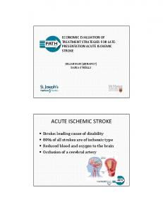
PRESENTATION ACUTE ISCHEMIC. STROKE. JULIAN NAM (@JNAM27).
DARIA O'REILLY. ACUTE ISCHEMIC STROKE. ▫ Stroke: leading cause of
disability.
[28] Hansen AT, Nedergaard M. Brain ion homeostasis in cerebral ischemia. ...... Neurovascular, Freemont, CA), and Enterprise (Codman, Raynham, MA).
Menu of Radiological Tests. • Cerebral Angiogram. • CT: ... transformation.
Adapted from Osborn, 1994. O ften. 4-6 h ... Osborn, Diagnostic Neuroradiology,
1994 ...
Aug 9, 2007 - life-years in high-income countries and as a cause of death ... the rate of residual blood flow and the du
in particular: sickle cell disease (SCD), cardiac disorders, trauma, and major ...... Epub 2011 Sep 10. 46. Fleisher GR, Ludwig .... antithrombotic therapy and prevention of thrombosis, 9th ed: American. College of Chest ... Becker K. Intensive care
Aug 9, 2007 - the rate of residual blood flow and the duration of ischemia, the ..... clinical course and prognostic sig
SYNTHESIS and Mechanical Retrieval and. Recanalization of Stroke Clots Using Embolectomy. (MR Rescue)] have shown that endovascular therapy is.
Bernard SA, Gray TW, Buist MD, Jones BM, Silvester W, Guta teridge G, et al: Treatment of ... Guluma KZ, Hemmen TM, Olsen SE, Rapp KS, Lyden PD: A trial of ...
5 : 153-159. ONAL M. Z., LI F., TATLISUMAK T., LOCKE K. W.,. SANDAGE B. W., JR., et al. Synergistic effects of citicoline and MK-801 in temporary experimental.
Jun 7, 2016 - volume (CBV), mean transit time (MTT), time-to-peak (TTP), and max, are computed using a bolus tracking method based on.
to apply this into routine clinical practice to improve the out- ... Seaman Family MR Research Centre, Foothills Medical Centre, .... of stent retrievers), enrolling proximal and distal vascular oc- clusions ... vestigators targeted a stroke onset to
Hunt SA, Abraham WT, Chin MH, et al. 2009 focused update incorporated into ... He J, Ogden LG, Bazzano LA, et al. Risk factors for congestive heart failure in ...
mRS 0-3, good neurological outcome (variable criteria on the. National Institutes of Health Stroke Scale [NIHSS] score across trials), good activity of daily living ...
Stryker and Codman. Turgut Tatlisumak has no conflicts of interest. Wim van Zwam was one of the principal investigators of MR. CLEAN. Nils Wahlgren is ...
Jun 29, 2015 - bral perfusion in acute IS has been the use of various throm- bolytic agent. These agents ... enzyme plasmin and thrombolysis is achieved by conversion of plasminogen to ... Streptokinase) or proteolytic tissue plasminogen activators.
Nov 18, 2013 - thrombolysis achieved by a stroke center in a centralized organizational .... total effect, 911 calls 0.9%, EMS transport 2.8%, and high priority.
stroke outcome in relation to treatment of hypertension in the management of acute ischemic stroke and its .... edema, hypertensive encephalopathy) received.
Apr 1, 2014 - followed by acetylsalicylic acid (Trombyl; Pfizer AB,. Sollentuna, Sweden; bolus of 300 mg and then 75 mg daily for a minimum of 6 months) ...
Mar 19, 2008 - Universitat Autònoma de Barcelona, Spain. J. Bruguera, MD ..... vere acute ischemic stroke. Neuroepi- .... Gaasch, William H (2004) Diastolic.
Dec 5, 2007 - In- travenous ancrod for treatment of acute ischemic stroke: the STAT study: a randomized controlled trial. Stroke Treatment with Ancrod Trial.
Electrocardiograph Changes in Acute Ischemic Cerebral Stroke
diac abnormalities during cerebral injury. The electrocardiogram changes and cardiac arrhythmias frequently encountered after stroke are not solely explicable ...
Electrocardiograph Changes in Acute Ischemic Cerebral Stroke Koochaki Ebrahim1 Arami Mohamadali2 Mazoochi Majid1 Alizargar Javad3 Assistant Professor of Kashan University of Medical Sciences, Beheshti Hospital 2 Neurologist, Milad Hospital, Tehran, Iran 3 Kashan University of medical Science, Esfahan, Iran 1
KEY WORDS: Stroke, Electrocardiography, Ischemic heart disease, ST segment changes ABSTRACT Introduction: Electrocardiography (ECG) changes are observed in patients with acute stroke and may cause diagnostic and management dilemmas. The aim of this study is to determine the frequency of ECG changes in patients with acute ischemic stroke. Material and Methods: In a case-control hospital-based study 262 patients with cerebral infarction were observed for ECG changes during their hospitalization. The ECG changes were compared with those of the matched control group consisting of 102 individuals. Results: Of the 262 patients of the study group, 112 (42.8%) were females and 150 (57.2%) were males. The mean age was 67.5 ± 11.9 (range 34–91 years). The control group consisted of 48 (47%) females and 54 (53%) males. The mean age was 64.5 ± 11.9 (range 31–87 yrs). The frequency of the The Journal of Applied Research • Vol.12, No. 1, 2012.
ECG changes observed in 179 (68.3%) of patients with cerebral infarct and 30 (29.4%) of the control group (P