Mar 27, 1980 - able by benzidine staining and radioimmunoassay, and we (5) have demonstrated that K562 cells exposed to this agent during growth in ...
Proc. Natl. Acad. Sci. USA Vol. 77, No. 6, pp. 3509-3513, June 1980 Cell Biology
Embryonic-fetal erythroid characteristics of a human leukemic cell line (K562 cell line/human globin mRNA and cDNA/molecular hybridization/i cell surface antigen/lactate dehydrogenase isoenzymes)
EDWARD J. BENZ, JR.*, MARY J. MURNANE*, BARRY L. TONKONOW*, BRIAN W. BERMANt, ERIC M. MAZUR*, CESIRA CAVALLESCO*, TRINA JENKOt, EDWIN L. SNYDER*, BERNARD G. FORGET*, AND RONALD HOFFMAN* *Departments of Internal Medicine, Pediatrics, and Laboratory Medicine, Yale University School of Medicine, New Haven, Connecticut 06510; and tThe
Connecticut Red Cross Blood Center, Farmington, Connecticut 06032
Communicated by Helen M. Ranney, March 27,1980
ABSTRACT We have studied a number of cell surface, enzyme, and protein markers in the human leukemic K562 cell line. We have confirmed previous observations that these cells accumulate human embryonic hemoglobins after exposure to hemin. In addition, our results demonstrated that these cells possess in their "uninduced" state i surface antigen, lactate dehydrogenase isoenzymes characteristic of embryonic or fetal erythroid cells, fetal and embryonic globin chains, and globin mRNAs. The levels of i antigen, embryonic globin chains, and embryonic globin mRNA increased substantially after exposure of the cells to hemin in suspension culture. In contrast, K562 cells lacked several surface, enzymatic, and functional prop erties typical of granulocytes, Iymphocytes, monocytes, or adult erythroblasts, including HLA surface antigens, surface immunoglobulins, sheep erythrocyte rosetting, phagocytosis, terminal deoxynucleotidyl transferase, carbonic anhydrase, ABO and Rh blood groups, and adult hemoglobins. The K562 cell, line therefore exhibits phenotypic properties of embonic erythroid progenitor cells and a quantitative increase in the expression of some of these properties can be achieved by exposure of the cells to hemin.
The K562 cell line is a Philadelphia chromosome-positive line derived from a pleural effusion of a patient with chronic granulocyte leukemia in terminal blast crisis (1). A number of recent observations suggest that suspension cultures of K562 cells may contain cells that can exhibit erythroid properties when exposed to appropriate in uitro culture conditions. The K562 line has been shown to produce glycophorin, the major sialoglycoprotein of the erythroid cell surface (2). At least two agents (hemin and sodium butyrate) known to "induce" the development of erythroid phenotypic features by mouse erythroleukemia cells (3) also promote hemoglobin accumulation in K562 cells: Andersson et al. (4) have shown that exposure to sodium butyrate leads to accumulation of hemoglobin detectable by benzidine staining and radioimmunoassay, and we (5) have demonstrated that K562 cells exposed to this agent during growth in semisolid culture systems give rise to benzidinepositive colonies resembling erythroid colonies derived from normal human erythroid stem cells (BFU-E). Rutherford et al. (6) have rigorously established that embryonic hemoglobins (Hb Portland and Hb Gower), as well as small quantities of fetal hemoglobin (Hb F) are produced by the K562 line after exposure to hemin in suspension culture. K562 cells thus appear to possess the capacity for at least partial erythroid differentiation. These cells are potentially valuable for studies of the dynamics of erythropoiesis and hemoglobin switching, and for analysis of expression of specific genes in neoplastic cells. Lozzio et al. (7) have recently challenged the assumption that The publication costs of this article were defrayed in part by page charge payment. This article must therefore be hereby marked "ad-
vertisement" in accordance with 18 U. S. C. §1734 solely to indicate this fact.
K562 cells are erythroid-like stem cells, on the basis of their observation that a K562 line exposed to hemin failed to exhibit morphologic characteristics of erythroid differentiation or to accumulate adult globins (a and ,3 globin chains). In contrast, Andersson et al. (8) have reported that the same clone of K562 cells contains glycophorin. In order to characterize further the erythroid potential of K562 cells, we have examined uninduced and hemin-induced K562 cells for the presence of surface and enzymatic characteristics, as well as for the accumulation of globin and globin mRNA production. Our findings suggest that uninduced K562 cells possess detectable erythroid features whose phenotypic expression is enhanced by exposure to the inducing agent hemin. Moreover, the phenotype expressed is more characteristic of early embryonic or fetal, as opposed to adult, hematopoietic cells. MATERIALS AND METHODS Cell Line. K562 cells, clone 6, were a kind gift of C. G. Gahmberg; this culture has been shown to contain glycophorin on its surface (2), and was kindly provided to us by H. Furthmayr, Dept. of Pathology, Yale University School of Medicine. A second culture of K562 cells, designated in this report as "clone R," was the kind gift of T. R. Rutherford and J. B. Clegg; these cells have been shown to produce embryonic hemoglobin in response to hemin (6). Cells were maintained at 37°C in a humidified atmosphere in RPMI 1640 medium supplemented with 10% fetal calf serum. "Induction" was accomplished by adding a 10-fold or 100-fold concentrated stock of hemin to a final concentration of 0.05 mM. Hemin was prepared as described (9) and sterilized by filtration prior to addition to the cultures. Cell growth was continued for 4 or 6 days after the addition of hemin. Cells were then harvested by centrifugation, washed with 0.9% saline, and resuspended in the appropriate buffer for analysis. Exposure to hemin reduced cell viability, as assessed by trypan blue exclusion, by 10-35%. Surface, Functional, and Enzymatic Assays. K562 cells were examined before and after hemin induction for HLA surface antigens, lymphocyte surface and enzymatic markers, phagocytosis, erythrocyte surface antigens, and lactate dehydrogenase (LDH), using established methods described in Results and table legends (cf. refs. 9-14). Terminal deoxyribonucleotidyl transferase assays were kindly performed by H. S. Allaudeen, Dept. of Pharmacology, Yale University School of Medicine, using published methods (15). Analysis of Globin and Hemoglobin. Cells were harvested by centrifugation, washed, and lysed in a solution of 150 mM NaCl/50 mM Tris-HCl, pH 8.0/3 mM MgCI2/0.5% Triton X-100. In some cases, protein was concentrated by pressure Abbreviation: LDH, lactate dehydrogenase.
3509
3510
Cell Biology: Benz et al.
Proc. Natl. Acad. Sci. USA 77 (1980)
Table 1. Surface and functional markers absent from K562 cells Principal cell type Result associated with marker Induced Uninduced Assay Surface immunoglobulin B lymphocytes Absent Absent T lymphocytes "E-rosetting" Absent Absent Terminal transferase Pre-T lymphoblasts Absent Absent Latex particle phagocytosis Monocytes, macrophages Absent Absent HLA antigens Multiple cell types Absent Absent Assays for surface immunoglobulins, E-rosetting with sheep erythrocytes, and phagocytosis were performed exactly as described by Dwyer (13). Terminal transferase screening was performed exactly as described by Shaw et al. (15). K562 cells (clone 6) were analyzed before and after 4 days of induction with 0.05 mM hemin. For each assay, appropriate positive and negative control cells were analyzed simultaneously with K562 cells and yielded the appropriate positive or negative results. HLA antigen activity was assessed by a sensitive complement-mediated micro-cytotoxicity assay (11, 12). Appropriate control cells gave positive results (data not shown).
dialysis prior to analysis. Hemoglobin was detected by electrophoresis on acrylamide gels in Tris/borate buffer at pH 8.6 and staining with benzidine in nitroprusside solution, using methods described in detail previously (6, 16). Globin chains were analyzed on acetic acid/urea/Triton X-100/acrylamide gels, using the method of Rovera et al. (17), as modified by Alter (18). Protein bands were detected by staining with 1% Coomassie blue; radioactive protein from mRNA translation assays was detected by autoradiography of dried gels. Isolation and Analysis of mRNA. Purification of mRNA, translation of mRNA in a wheat germ mRNA-dependent cell-free protein-synthesizing system, synthesis of cDNAs by reverse transcriptase from the mRNA templates, and molecular hybridization analysis of the cDNAs and mRNAs were performed, using the methods we have described in detail elsewhere (19, 20). Globin mRNA species were detected by gel blotting, using the methods described by Wahl et al. (21) and Alwine et al. (22). Electrophoretic separation of RNAs was performed on 2.5% agarose gels in 10 mM sodium phosphate buffer at pH 7.0 prior to transfer of the RNA bands to activated cellulose paper and hybridization to cloned human, a, /, and y globin cDNA probes (23), which had been rendered radioactive by nicktranslation in the presence of [32P]dCTP, using established methods (24). After hybridization overnight at 37-420C in the buffer described by Alwine et al. (22), the strips were washed initially in a solution of 50% (wt/vol) formamide/0.75 M NaCl/0.075 M sodium citrate/50 mM sodium phosphate, pH 7.0, then washed a final time in 0.30 M NaCl/0.030 M sodium citrate/0. 1% sodium dodecyl sulfate prior to drying and autoradiography. RESULTS The results of a survey of clone 6 cells before and after induction with hemin for surface, functional, and enzymatic properties are shown in Table 1. Surface characteristics of B and T lymphoid cells were absent, as was latex particle phagocytosis, a property of monocytes and macrophages. These characteristics remained undetectable after induction with hemin. As shown in Table 2, clone 6 cells also lack ABO and Rh blood group antigens, but possess i antigen activity, a feature characteristic of fetal erythrocytes. Uninduced K562 cells were weakly reactive with anti-i antiserum but, after exposure to 0.05 mM hemin for 4 days, the cells exhibited a strong increase in i antigen activity. The level of i antigen observed was comparable to that obtained in parallel assays with cord blood erythrocytes and the same antiserum. As shown in Table 2, our anti-i antiserum reacted only with cord, and not with adult human, erythrocytes. LDH isoenzymes were analyzed by electrophoresis of con-
centrated cell lysates on Cellogel, followed by specific enzymatic staining, as described by Ruddle and Nichols (14); Human fetal liver erythroblasts, from a 16-week-old abortus, yielded a symmetrical pattern with five LDH isoenzyme bands indicating approximately equal production of LDH A and LDH B subunits, whereas adult erythrocytes showed three rapidly migrating anodal bands demonstrating a greater proportion of LDH B than LDH A subunits. Both uninduced and hemin-induced K562 cells were observed to give a five-banded symmetrical pattern virtually identical to that of fetal liver erythroblasts (data not shown). Hemin induction had no effect on the intensity or relative expression of the LDH isoenzymes.
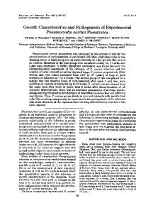
Analysis of uninduced and induced K562 cells for the presof hemoglobin was accomplished by benzidine staining of proteins separated by acrylamide gel electrophoresis in a Tris/borate/EDTA system. As shown in Fig. 1, the positions of authentic hemoglobin markers were analyzed in parallel lanes; the positions of Hb A (aA232), Hb F (a2y2), and Hb A2 (a262) are shown in lanes 1 and 2. The positions of the embryonic hemoglobins, Hb Barts (Q4) and Hb Portland (M2Y2), were verified by analysis of an erythrocyte lysate from a fetus with homozygous a-thalassemia (hydrops fetalis with Hb Barts) (lane 7); the position shown for Hbs Gower (t2E2 and a2E2) is similar to that obtained by Rutherford et al. (6). Both clones of uninduced K562 cells contained low levels of hemoglobin, consisting predominantly of Hb Gower and Hb Portland (lane 3) or of Hb Gower alone (lane 4). A considerable increase in hemoglobin accumulation was observed after hemin induction with both clone 6 and clone R cells (lanes 5 and 6). Moreover, distinct differences in the types of hemoglobin accumulating were observed: clone 6 cells (lane 5) contained Hb Gower, Hb Portence
Table 2. i erythrocyte surface antigen on K562 cells i antigen titer Cell type 1:4 Uninduced K562 Hemin-induced K562 1:256 Adult group 0 erythrocytes Absent undiluted 1:256 Cord blood group 0 erythrocytes A 3-5% suspension of K562 cells, clone 6, before and after 4 days of hemin induction, was suspended in 0.9% NaCl after packed cells had been washed twice in 0.9% NaCl. Anti-i antiserum was kindly provided by Marie Crookston. Serial dilutions of anti-i in 0.9% NaCl were incubated with the cells for 30 min at 4VC. The mixtures were centrifuged in a Dade Immunofuge for 15 sec, and titration scores were determined macroscopically. Standard blood bank techniques (10) were used to screen for the following antigens, all of which were absent from induced and uninduced clone 6 cells but present in high titer on appropriate test erythrocytes: A, B, C, c, E, e, Fya, Jka, M, N, and P1.
Cell Biology: Benz et al. 2
3
Proc. Natl. Acad. Sci. USA 77 (1980) 4
5
7
6
orA
aolo
Hb F Hb A
3511
-p