Apr 12, 1977 - 16-day mouse embryos (appearance of vaginal plug. = day 0) using a Wild ..... evidence in support of this and it seems more likely that the H-2 ...
Immunology 1978 34 69
Embryonic
mouse
thymus development: stem cell entry and differentiation
MARY A. RITTER Zoology Department, South Parks Road, Oxford
Received 28 March 1977;
accepted for
Summary. The entry and subsequent division and differentiation of blood-borne stem cells within the embryonic thymus has been studied in a system where the inflowing cells were either chromosomally or antigenically marked. H-2 positive stem cells enter the thymus within 5 days of grafting but may wait for up to 14 days before proliferating to give rise to Thy-i positive thymocytes.
publication
12
April
1977
host, and their growth, repopulation and differentiation studied by weight, histological appearance, the T6 chromosome marker method and cytotoxicity testing using anti-H-2k and anti-Thy-i.1 antisera. MATERIALS AND METHODS
Embryonic thymus grafts Thymic rudiments were removed from 12-, 14- and 16-day mouse embryos (appearance of vaginal plug = day 0) using a Wild stereomicroscope and fine cataract knives. Rudiments were grafted under the kidney capsule (four to six lobes per kidney) of 2 to 4 month old male syngeneic or F1 (one parental strain in common with the graft) hosts.
INTRODUCTION Thymus lymphoid development is dependent upon an inflow of stem cells from the blood stream (Owen & Ritter, 1969; Ford & Micklem, 1963; Moore & Owen, 1967). Although mouse thymocytes are known to be characterized by certain differentiation antigens (cortical thymocytes: low H-2; high Ly-1, -2, -3, and Thy-i; TL positive; medullary thymocytes: high H-2; low Ly-1, -2, -3 and Thy-i; TL negative in appropriate strains (for review see Cantor & Weissman, 1976)); little is known of the sequence of events leading from stem cell to thymus lymphocyte. Experiments described in this paper were designed to follow the entry and subsequent division and differentiation of the blood-borne precursor cells within the thymus, in a system where the inflowing cells were either chromosomally or antigenically marked. Embryonic mouse thymic rudiments were grafted to a perirenal site in an F1
Weighing 12-, 14- and 16-day embryonic CBA/H and CBA/HT6T6 donor tissue was grafted to syngeneic hosts and removed for weighing after 7, 12, 14, 17, 19, 21, 24 and 28 days. Tissues were placed in medium 199 immediately after removal to prevent excessive drying, before blotting and weighing on a Sartorius microbalance.
Histology CBA/H and CBA/H-T6T6 mice were used for both donors and recipients. Thymuses were sampled at 1, 2, 5, 7, 12, 14, 17, 21 and 28 days after grafting. Tissues were fixed in Bouin's, embedded in Polyester wax (British Drug Houses), sectioned at 7 gm and stained in Giemsa R66 (G. T. Gurr).
Correspondence: Dr M. A. Ritter, Pathology Department, University of Connecticut Health Center, Farmington, Connecticut 06032, U.S.A.
69
70
Mary A. Ritter
Chromosome marker technique 12-, 14- and 16-day embryonic thymic rudiments from CBA/H and CBA/H-T6T6 donors were grafted into CBA/H-T6T6 and CBA/H recipients respectively. Grafts were sampled after 7, 12, 14, 17, 19, 21, 24 and 28 days. Three hours before sampling, the host was injected i.p. with 0-4 ml of 0 5 mg/ml colcemid (CIBA) in medium 199. Chromosomes were prepared by a modified hypotonic citrate air-drying technique (Ford, 1966), stained in lactic-aceticorcein and mounted in Euparol. Metaphase plates were examined under phase contrast microscopy and scored under high power for the presence or absence of two T6 chromosomes. The total number of chromosomes in each metaphase plate was checked before scoring. Only those that were complete (40 chromosomes) were scored. Preparation of anti-Thy-i.1 antisera Two batches of anti-Thy-i.1 antisera were raised in CBA/H females using AKR thymocytes (Reif & Allen, 1966). Batch I was titrated against 4 week old CBA/H and F. (CBA/H x AKR) thymocytes (for use in experimental situation (b) below), showing a plateau of kill against the F, cells out to a final dilution of 1/1024 (mean background-corrected kill on plateau= 91 %). Batch II was titrated against 4 week old BALB/c and F, (BALB/c x AKR) thymocytes (for use in experimental situation (c) below) showing a plateau of kill against F, cells out to a final dilution of 1/256 (mean background-corrected kill on plateau = 90 %). To avoid slight anti-BALB/c autoantibody activity (Boyse, Bressler, Iritani & Lardis, 1970) a final dilution of 1/32 and 1/64 was used in all experiments involving batches I and II respectively. Preparation of anti-H-2" antisera Anti-H-2k antisera were prepared by the same method (Reif & Allen, 1966) using CBA/H spleen cells and BALB/c female recipients. The antiserum was titrated against BALB/c, F, (BALB/c x CBA/HT6T6) and F, (BALB/c x AKR) thymocytes from 4 week old animals. To avoid anti-BALB/c autoantibodies a final dilution of 1/8 was used in experimental situations (a) and (c) below. This dilution was still on the plateau of the titration curve against F1 (BALB/c x CBA/H-T6T6) thymocytes (mean kill on plateau = 94%). No such plateau was obtained for F, (BALB/c x AKR) thymus cells. The corrected cell kill at 1/8 final dilution was 73%. These low
percentage kills were compensated for in repopulation studies by the inclusion of a 'maximum kill control'. The greater activity of the anti-H-2k antiserum against F1 (BALB/c x CBA/H-T6T6 thymocytes may have been due to the presence of anti-Ly-1.1 antibodies in the serum. However, since Ly antigens are not present on thymocyte precursors in the bone marrow (Boyse, Miyazawa, Aoki & Old, 1968), this should not affect the accuracy of the serum in detecting stem cell entry into the thymus.
Cytotoxicity testing ofgrafted tissue Three immunogenetically compatible combinations of host and donor tissue were used. Grafts were removed for testing after 5, 7, 12, 14, 17, 19, 21 and 24 days. The 4 to 6 lobes per host were pooled to provide sufficient cells for analysis. (a) 12-, 14- and 16-day embryonic BALB/c rudiments grafted to F. (BALB/c x CBA/H-T6T6) were sampled for the proportion of host-derived cells using anti-H-2k antiserum. (b) 14-day CBA/H embryonic thymus was grafted to F1 (AKR x CBA/H) hosts and the tissue analysed for host type cells using anti-Thy-1.l antiserum. (c) 14-day BALB/c thymic rudiments grafted to F. (BALB/c x AKR) hosts were tested for host derived cells using anti-Thy-1.1 and anti-H-2k antisera. In each experiment the cell suspension was divided for testing with both antisera. The cytotoxic assay used was that of Boyse et al. (1964) as modified in Schlesinger's two step system (1965) to minimize anticomplementary effects. Guinea pig serum, absorbed with agar (Difco Noble Agar) to remove anti-thymocyte activity, was used as a source of complement (Cohen &
Schlesinger, 1970). Results were corrected for nonspecific background cell death: Corrected _ observed % kill -background % kill 100- background % kill % kill An additional correction was made for all data obtained using anti-H-2k antiserum since the antiserum was a relatively weak one giving little or no plateau against F1 thymocytes. (This may be due to there being less H-2 than Thy-i on thymocytes (Aoki, Hammerling, De Harven, Boyse & Old, 1969), and a reduced amount of H-2 in the F1). Thus slight variations in experimental conditions (temperature, complement activity, complement and antiserum dilution) could affect the percentage of
Stem cell differentiation in embryonic thymus
cells killed. To overcome this, a 'maximum kill control' was included in each test. Thymocytes from 2 to 3 week old normal animals of the same genotype as the host (and therefore the inflowing cells) were processed for cytotoxicity testing in parallel with experimental cells. Both experimental values and 'maximum kill control' were corrected for nonspecific background cell death. Experimental values were then expressed as a percentage of the 'maximum kill control' for that experiment.
71
Table 1. Embryonic mouse thymus grafts: weights
Weights (g) ± s.e. Days after grafting 7 12 14 17 19 21 24 28
12 day
14 day
16 day
n.t. 1-6 ±0-2 16±0-2 2-7 ±03 3-3 ±07 4-3 ±07
0-9 0-1 2-8 ±03
2-4 0-2 5 6 0-7
65±07 n.t.
3-6±04
6-2±0-5
4-5 ±0-6 3-9 0-3 5-8 ±08 7-2±05 10-7 ±0-5
8-2* 7-0 0-6 10-2 1-2 8.5±0-9 11 1 1-2
RESULTS
Histology A vascular supply to grafted rudiments became established within 2 to 3 days. No necrosis was seen in 12-day thymic grafts. In 16-day, and to a much lesser extent in 14-day thymic grafts, a small necrotic area could be seen at the centre of the rudiment during the first 2 days after grafting. This cell damage presumably resulted from an inadequate gaseous and nutrient supply to the central portion of the larger rudiments during the period before a vascular supply became established (2-3 days). At day 2 after transplantation 12- and 14-day thymic rudiments were composed of epithelial cells and large basophilic cells, 16-day grafts contained in addition some lymphoid cells. By 5 days both younger stages also contained lymphocytes. During the rest of the culture period (23 days) all grafts increased in size and became progressively more lymphoid.
Weights All rudiments increased steadily in weight during the grafting period. Sixteen-day grafts were the heaviest and 12-day grafts the lightest at every stage. The average rate of weight gain in grafts of all ages was approximately 05 mg/day. Results are shown in Table 1. Chromosome marker repopulation The results of chromosome marker studies on CBA/H and CBA/H-T6T6 thymus graft repopulation by marked syngeneic host cells are summarized in Table 2. In 12-day rudiments very few host derived cells (< 3 %) were seen at 7 and 12 days after grafting. At day 14 the proportion had risen to 30 %,
12-, 14- and 16-day CBA/H and CBA/H-T6T6 thymic rudiments were grafted perirenally in syngeneic hosts. Rudiments were removed and weighed after grafting. Between 6 to 8 grafts were sampled at each stage. * Six grafts fused together. n.t., not tested. Table 2. Embryonic mouse thymus grafts: chromosome marker repopulation % Repopulation Days after grafting
12 day
14 day
16 day
7 12 14 17 19 21 24 28
2 3 31 83 95 99 100 n.t.
2 1 0 33 68 92 98 100
0 1 1 2 12 50 98 100
12-, 14- and 16-day embryonic mouse thymic rudiments from CBA/H and CBA/H-T6T6 donors were grafted perirenally in syngeneic hosts, CBA/H-T6T6 and CBA/H respectively (repopulating cells distinguished chromosomally from donor graft-type cells). Grafts from 2 to 3 hosts (8-18 thymic lobes) were sampled at each stage. 100-200 metaphases were scored at each stage. n.t., not tested.
and by day 24 the grafts were completely repopulated (as measured by the dividing cell population). Few host-type cells were found in the 14-day grafted rudiments until 17 days after grafting. At this stage 33 % were of host origin. These grafts were fully repopulated by 28 days. Cell entry was delayed yet further in 16-day grafted thymus. Appreciable numbers of host-derived cells appeared only after
72
Mary A. Ritter
Table 3. Embryonic mouse thymus grafts: H-2k repopulation
Table 4. Embryonic mouse thymus grafts: Thy-I .1 repopulation
% Repopulation Days after grafting
12 day
14 day
16 day
5 7 12 14 17 19 21 24
36 45 54 89 76 94 102 105
44 52 49 44 66 92 96 102
28 35 46 54 64 66 95 100
Days after grafting
% Repopulation
5 7 12 14 17 19 21 24
2 2 0 1 43 51 85 88
Fourteen-day CHA/H mouse thymic rudiments were grafted perirenally in F1 (CBA/H x AKR) hosts. Rudiments were removed at various intervals after grafting and the percentage of host-derived cells was assessed in cytotoxicity tests using anti-Thy-1.1 antiserum and guinea-pig complement. Experimental values were corrected for background cell death. Grafts from 2 to 3 hosts (8-18 thymic lobes) were sampled at each stage.
12-, 14- and 16-day BALB/c embryonic mouse thymic rudiments were grafted perirenally in F1 (BALB/c x CBA/H) hosts. Rudiments were removed at various intervals after grafting and the percentage host-derived cells was assessed in cytotoxicity tests using anti-H-2k antiserum and guinea-pig complement. Experimental values were corrected for background cell death and for the 'maximum control' for each experiment. Grafts from 2 to 3 hosts (8-18 thymic lobes) were sampled at each stage.
19 days (12 %). Again, repopulation was complete within 28 days.
loor c
H-2 repopulation Table 3 gives the results of repopulation studies of BALB/c grafts by F1 (BALB/c x CBA/H-T6T6) host cells using anti-H-2k antiserum. Five days after grafting, rudiments of all ages contained a considerable proportion of H-2k positive cells (12-day: 36%; 14-day: 44 %; 16-day: 28 %). The percentage of host cells rose until by 21 days (12-day thymus) and 24 days (14- and 16-day thymus) all rudiments were
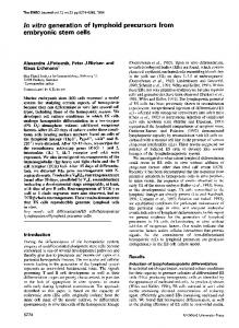
completely repopulated. Thy-i repopulation Repopulation of 14-day CBA/H embryonic thymus grafts by F1 (CBA/H x AKR) host cells, measured using anti-Thy-1.1 antiserum, is given in Table 4. During the first 14 days of grafting the proportion of Thy-1.1 positive cells was between 0 and 2%. By 17 days this had risen steeply to 43 %. At 24 days, 88 % of cells were of host origin. Combined H-2 and Thy-i repopulation Fig. 1 gives the data for repopulation of 14-day BALB/c embryonic mouse thymus grafts by F1 (BALB/c x AKR) host cells, measured by both
0 Q 0 0. 4)
50
A-A
I,1
,
0 0
20 10 Duration of grafting (days)
30
Figure 1. Fourteen-day embryonic BALB/c thymic rudiments grafted perirenally in adult F1 (BALB/c x AKR) hosts. Rudiments were removed at intervals after grafting and sampled for both Thy-l.l positive and H-2k positive host derived cells in cytotoxicity tests. Grafts from 2 to 3 hosts (8-18 lobes) were sampled at each stage. (0) Percentage H-2k positive cells; (A) percentage Thy-l.l positive cells.
anti-H-2k and anti-Thy-1.1 antisera. The two values given at each sampling stage are the proportions of H-2k and Thy-1.1 positive cells within a single sample of grafted tissue. The results are comparable to those obtained in independent H-2k and Thy-1.1 grafting experiments. By 12 days grafts contained 33% H-2k positive cells. Thy-i .1 positive cells first appeared at day 17. After 24 days, 99% H-2k and
Stem cell differentiation in embryonic thymus Table 5. Embryonic mouse thymus grafts: H-2k and Thy-I.1 repopulation
% Repopulation Days after grafting
H-2k
Thy- 1.1
12 14 17 19 21 24
33 64 79 78 93 99
0 0 44 62 91 94
Fourteen-day BALB/c embryonic mouse thymic rudiments were grafted perirenally in F1 (BALB/c x AKR) hosts. Rudiments were removed at various intervals after grafting and the percentage of host-derived cells was assessed in cytotoxicity tests using anti-H-2k and anti-Thy-i.1 antisera and guinea-pig complement. H-2k and Thy-i .1 experimental values were corrected for background cell death. H-2k values were also corrected for the 'maximum control' in each experiment. The two values given at each sampling stage are the percentages of H-2k and Thy-l.l positive cells within a single sample of grafted tissue (8-18 thymic lobes).
94% Thy-i.1 positive cells were found within the grafted thymic tissue. DISCUSSION The work described in this paper was designed to study the migration of stem cells into and their subsequent development within the embryonic mouse thymus, in a system where the inflowing cells were either chromosomally or antigenically (H-2k, Thy-I .1) marked. All grafts increased steadily in weight at a rate of approximately 0 5 mg/day. At all stages after grafting the weight differential between 12-, 14- and 16day rudiments was maintained. This supports the concept that thymic development is autonomous (independent of the host), and that it is the thymic epithelium rather than the supply of blood-borne stem cells that controls thymus growth (Metcalf, 1963, 1965). Repopulation of embryonic mouse thymic grafts was followed in three ways. Firstly, since most cells express H-2 antigens (Klein, 1975) anti-H-2" anti-
73
serum was used to trace inflowing stem cells. Secondly, anti-Thy-I.1 antiserum was used to detect host-derived thymocytes within the grafts (Reif & Allen, 1964). Finally, using the T6 chromosome marker method the proportion of host-type dividing cells was determined (Ford, 1966). Analysis of 12-day grafts (Tables 2 and 3) shows that although some host cells (36%) had entered the rudiment by 5 days, not until 14 days after grafting were many host-derived cells found in division (31 %). By 21 to 24 days all cells, whether in division or not, were derived from the host. For 14-day grafted rudiments (Fig. 2) again at 5 days some host cells (44%) had entered the graft. However, not until 17 days after grafting were there appreciable numbers of hostderived dividing and Thy-i.1 bearing cells (33 and 43 % respectively). At this stage 66 % of all cells were of host H-2k type. Grafts were fully repopulated by 24 to 28 days. In 16-day thymic grafts (Tables 2 and 3), 28% host cells were found after 5 days. Only after 19 days were many host-derived cells found in division (12 %.). Rudiments were completely repopu-
lated 24-28 days after grafting. In order to discount the possibility that the differences in the rates of repopulation as measured by anti-H-2k and anti-Thy-i .1 antiserum simply reflected genetically controlled differences in cell kinetics in the two genotype combinations used, a second series of experiments was performed. In these the repopulation of each 14-day BALB/c thymic graft by F1 (BALB/c x AKR) host cells was studied with both anti-H-2k and anti-Thy-i .1 antisera. The results corresponded closely to those obtained in the separate 14-day graft experiments (Fig. 1). Host-derived cells bearing Thy-I.1 were first found 17 days after grafting; by 24 days grafts were almost fully repopulated. Hence genetic disparity cannot be responsible for the differences between H-2k and Thy-i.1 results. Unfortunately, it was not possible to study all three markers (T6, H-2 and Thy-i) in the same thymus graft since both the T6 (CBA/H strain) and Thy-i.i (AKR and RF strains available) were on the same H-2k genetic background. Another possible explanation of the results is that two separate cell populations colonize the thymus: the first, non-dividing H-2 positive and Thy-i negative; the second, rapidly dividing, H-2 negative and Thy-i negative that gives rise to H-2 positive, Thy-i positive thymocytes. However, there is no evidence in support of this and it seems more likely that the H-2 positive, Thy-l negative stem cells have
74
Mary A. Ritter
to undergo some maturational steps prior to proliferation within the thymus. Thus, when embryonic thymus rudiments are grafted into a syngeneic or semiallogeneic host, stem cells bearing H-2 antigens migrate into the graft. At least some host cells enter grafts within the first 5 days of grafting; cell entry probably starts as soon as a vascular supply is established (2-3 days). The rate at which stem cells enter the graft is determined by its age: the older the rudiment at the start of grafting the lower the rate of inflow, and so the slower the rate of repopulation. This may reflect the fact that the older rudiments already contain a considerable number of stem cells at the time of grafting. Ultimately all grafts become completely repopulated by host cells, and fully lymphoid. However, there was a considerable lag (9, 12 and 14 days in 12-, 14- and 16-day grafted rudiments respectively) between the time when H-2k positive host stem cells enter the grafted rudiment and when appreciable numbers of these cells start to divide and to acquire Thy-i .1 antigen. Other studies of lymphoid development in either thymus grafts in a genetically distinct host (Metcalf & Wakonig-Vaartaja, 1964; Schlesinger & Hurvitz, 1968; Owen & Raff, 1970) or in the thymus in vivo in an irradiated host given a genetically marked bone marrow cell inocculum (Kadish & Basch, 1976), also demonstrate the first appearance of host-derived dividing cells and lymphocytes 2 to 3 weeks after the start of the experiment, although in none of these studies was the actual entry of stem cells followed from the start of grafting. In contrast, a shorter period of 'waiting' is suggested by histological studies of the normal embryonic thymus (Owen & Ritter, 1969). Large basophilic stem cells are first seen at day 11 while the first lymphocytes appear at day 15, after a lag of only 4 days. In addition, at least 60% of cells in the embryonic thymus bear Thy-1 by day 16 (Owen & Raff, 1970). The extension of this 'waiting' period in grafted or irradiated thymuses could be an artifact of the experimental situations used. Alternatively, it may be due to differences in the proliferative potential of embryonic and adult stem cell, since grafted rudiments, although themselves embryonic, are repopulated by cells from the adult host. In the light of these observations, the failure to detect hostderived dividing Thy-1 positive cells in the thymuses of irradiated, bone marrow reconstituted mice before day 12 (Kadish & Basch, 1976; Basch & Kadish, 1977) may not reflect diminutive numbers of these
100 _
0 0
, 50
a_ 0Il
0
20 10 Duration of grafting (days)
30
Figure 2. Fourteen-day embryonic thymus grafted perirenally in adult hosts. Rudiments were removed at intervals after grafting and analysed for host-derived cells using T6, H-2k and Thy-1.1 markers. CBA/H thymus grafted to CBA/H-T6T6 hosts: analysed by T6 chromosome marker. BALB/c thymus grafted to F, (BALB/c x CBA/H) hosts: analysed by anti-H-2k antiserum. CBA/H thymus grafted to F, (AKR x CBA/H) hosts: analysed by anti-Thy-1.1 antiserum. Grafts from 2 to 3 hosts (8-18 lobes) were sampled at each stage. (0) Percentage H-2k positive cells; (A) percentage T6 positive dividing cells; (0) percentage Thy-1.1 positive cells.
cells, but rather the fact that during this time host cells comprise a Thy-i negative, temporarily nondividing ('waiting') population. The marked parallel between the T6 chromosome marker and Thy-i.1 repopulation in 14-day grafts (Figs 1 and 2) points to a close temporal association between these two components of thymus lymphocyte development. Fourteen-day thymic rudiments examined after a 14 day grafting period contain little or no (2%) host-derived dividing or Thy-1.1 positive cells. By 17 days, 33 % of dividing cells are of host type and 43% of all cells bear the Thy-1.1 antigen. Therefore within 3 days host cells have left the 'waiting' phase, undergone cell division and developed the Thy-i.1 surface antigen. With an average cell division time of 7 to 8 h (Metcalf 1969) a maximum of 9 divisions would be possible during this period to give rise to the Thy-i .1 positive lymphocytes. Whether these lymphocytes that have developed within the grafts represent both cortical (high Thy-1) and medullary (low Thy-1) thymocytes is not known. Thus, the results indicate that the thymus is seeded by blood-borne stem cells bearing H-2 but lacking Thy-1 surface antigens. Under the inductive influence of the thymus these cells divide and differen-
Stem cell differentiation in embryonic thymus
tiate to give Thy-1 positive thymocytes; a process that may occur more rapidly for the stem cell of the embryo than for that of the adult. The complete nature of the thymus inductive environment is unknown, although clearly thymic humoral factors produced by the epithelium play a part (Komuro & Boyse. 1973; Mandel, 1970; Mandi & Glant, 1973). A CKNOWLEDGMENTS
The author is indebted to Professor J. J. T. Owen for many helpful discussions during the course of this work. This research was supported by the Medical Research Council. REFERENCES AOKI T., HAMMERLING U., DE HARVEN E., BOYSE E.A. & OLD L.J. (1969) Antigenic structure on cell surfaces. An immunoferritin study of the occurrence and topography of H-2, 0 and TL alloantigens on mouse cells. J. exp. Med. 130, 979. BASCH R.S. & KADISH J.L. (1977) Hematopoietic thymocyte precursors. II. Properties of the precursors. J. exp. Med. 145, 405. BOYSE E.A., BRESSLER E., IRITANI C.H. & LARDIS M. (1970) Cytotoxic yM autoantibody in mouse alloantiserum. Transplantation, 9, 339. BOYSE E.A., MIYAZAWA M., AOKI T. & OLD L.J. (1968) Ly-A and Ly-B: two systems of lymphocyte isoantigens in the mouse. Proc. R. Soc. Lond. B. Biol. Sci. 170, 175. BOYSE E.A., OLD L.J. & CHOUROULINKOV I. (1964) Cytotoxic test for demonstration of antibody. Methods Med. Res. 10, 39. CANTOR H. & WEISSMAN I. (1976) Development and function of subpopulations of thymocytes and T lymphocytes. Progr. Allergy, 20, 1. COHEN A. & SCHLESINGER M. (1970) Adsorption of guineapig serum with agar. Transplantation, 10, 130.
75
FORD C.E. (1966) In: Tissue grafting and radiation (ed. by H. S. Micklem and J. F. Loutit). Academic Press, New York. FORD C.E. & MICKLEM H.S. (1963) The thymus and lymph nodes in radiation chimaeras. Lancet, i, 359. KADISH J.L. & BASCH R.S. (1976) Hematopoietic thymocyte progenitors. I. Assay and kinetics of the appearance of progeny. J. exp. Med. 143, 1082. KLEIN J. (1975) Biology of the mouse histocompatibility-2 complex. Springer, New York. KoMURo K. & BOYSE E.A. (1973) Induction of T lymphocytes from precursor cells in vitro by a product of the thymus. J. exp. Med. 138, 479. MANDEL T. (1970) Differentiation of epithelial cells in the mouse thymus. Z. Zellforsch. mikrosk. Anat. 106, 498. MANDI B. & GLANT T. (1973) Thymosin-producing cells of the thymus. Nature New Biol. 246, 25. METCALF D. (1963) The antonomous behaviour of normal thymic grafts. Aust. J. exp. Biol. med. Sci. 41, 437. METCALF D. (1965) Multiple thymus grafts in aged mice. Nature, 208, 87. METCALF D. (1969) Lymphocyte kinetics in the thymus. In: The lymphocyte in immunobiology and haemopoiesis. (Ed. by J.M. Yoffey), p.333 Edward Arnold, London. METCALF D. & WAKONIG-VAARTAJA R. (1964) Stem cell replacement in normal thymus grafts. Proc. Soc. exp. Biol. Med. 115, 731. MOORE M.A.S. & OWEN J.J.T. (1967) Experimental studies on the development of the thymus. J. exp. Med. 126, 715. OWEN J.J.T. & RAFF M.C. (1970) Studies on the differentiation of thymus-derived lymphocytes. J. exp. Med. 132, 1216. OWEN J.J.T. & RIrrER M.A. (1969) Tissue interaction in the development of thymus lymphocytes. J. exp. Med. 129, 431. REIF A.E. & ALLEN J.M.V. (1964) The AKR thymic antigen and its distribution in leukaemias and nervous tissue. J. exp. Med. 120, 413. REIF A.E. & ALLEN J.M.V. (1966) Mouse thymic iso-antigens. Nature, 209, 521. SCHLESINGER M. (1965) Immune lysis of thymus and spleen cells of embryonic and neonatal mice. J. Immunol. 94, 359. SCHLESINGER M. & HURVITZ D. (1968) Serological analysis of thymus and spleen grafts. J. exp. Med. 127, 1127.