J OURNAL OF FOREST PRODUCTS & INDUSTRIES, 2014, 3(6), 248-256 ISSN:2325–4513( PRINT ) ISSN 2325 - 453X (ONLINE )
Research Article
Endophytic Fungal Communities Associated with Ethno-medicinal Plants from Sudan and their Antimicrobial and Antioxidant Prospective Tawasol Mahdi1, Ietidal Mohamed2, Sakina Yagi*3 1-3
248
Botany Department, Faculty of Science, University of Khartoum, P.O. Box 321, Khartoum, Sudan *Corresponding author e-mail:
[email protected] (Received: September 14, 2014; Accepted: November 21, 2014)
Abstract- The investigation was carried out to isolate and characterize the endophytic fungi associated to three selected medicinal plants growing in Sudan and to evaluate their bioactive potential. The endophytic fungal populations were explored in leaves and stems of Datura stramonium L (family Solanaceae), Moringa oleifera Lam. (family Moringaceae) and Prosopis chilensis (Molina) Stuntz (family Fabaceae). Seventeen isolates were recovered where, 10 isolates were identified to species level and 7 to the genus level. The endophytic fungal communities of D. stromonium comprised of fungi belonging to genera Aspergillus and Curvularia. The tissue of M. oleifera was found to be colonized with endophytic fungi of the genera Emericella, Aspergillus and Curvularia while those of P. chilensis were colonized with Emericella, Aspergillus and Chaetomium. The ethyl acetate extracts obtained from the isolated fungal endophytes were screened for evaluation of their antimicrobial activity and antioxidant capacity. The antimicrobial activity of extracts was determined using the disc diffusion method. Endophytic fungi crude extracts isolated from of P. chilensis showed the highest antibacterial activity compared to other isolates. They all inhibited Escherichia coli, Pseudomonas aeruginosa, Klebsiella pneumonia, Salmonella Typhi and and Staphylococcus aureus with minimum inhibitory concentration value of 25 µg/mL. The antioxidant activity was determined using the 1, 1-diphenyl-2-picrylhydrazyl (DPPH) assay. A remarkable scavenging activity was obtained from endophytic fungi Aspergillus sp. (D-4) and Emericella bicolor (M-6) isolated from D. stramonium and M. oleifera respectively where they possessed activity higher than that obtained from the standard control ascorbic acid. Moreover, a positive correlation between the phenolic content of the extracts with their antioxidant activity was also observed. In conclusion, the endophytic flora associated within these widely used medicinal plants could be a potential source of novel products of great importance in medicine and industry.
endophyte, they have been isolated from various organs of different plant species, tissues of liverworts, hornworts, mosses, lycophytes, equisetopsids, ferns, and spermatophytes from the tropics to the arctic, and from the wild to agricultural ecosystems [4, 5]. Endophytic fungi have received considerable investigation for their biodiversity and their potential to produce bioactive metabolites that may have applicability in medicine, agriculture and industry [6, 7]. However, information of the endophytic biodiversity of plants from the Sudan and their potential to produce bioactive secondary metabolites are scanty. The aim of this study was to isolate and characterize fungal endophytes from three medicinal plants, namely; Datura stramonium L (family Solanaceae), Moringa oleifera Lam. (family Moringaceae) and Prosopis chilensis (Molina) Stuntz (family Fabaceae) and to determine the antimicrobial and antioxidant activities of crude extracts of the isolated endophytic fungi.
Index terms: Endophytes, Datura stramonium, Moringa oleifera, Prosopis chilensis, Antibacterial activity, Antioxidant activity.
Screening, identification and preservation of endophytes
I.
Today, the
INTRODUCTION
future of many medicinal plants is being
endangered with extinction through over exploitation by herbalists, medicinal plants traders and also through deforestation and habitat loss. This has prompted industries, as well as scientists to consider the possibilities of investigation into endophytic microorganisms as an alternative supply for the production of plant pharmaceuticals [1]. The existence of fungi inside the organs of asymptomatic plants has been known since the end of the 19th century [2], and the term ‘‘endophyte’’ was first proposed in 1866 [3]. To date, all plant species studied have been found to harbor at least one
II. MATERIAL AND METHODS Sample collections Healthy plant samples of D. stramonium, M. oleifera and P. chilensis were collected from around the University of Khartoum, Khartoum State (Central Sudan). Voucher specimens have been deposited in the herbarium of Botany Department, Faculty of Science, University of Khartoum. Plant samples were brought to the laboratory within 24 hours after samples collection following guidelines of Monnanda et al. [8]. Fresh leaves and stems of the selected plants were separated for further isolation of endophytes.
All selected parts were washed properly under running tap water followed by double distilled water before processing. The samples were cut into small pieces. Stem samples were cut into 1.0 × 1.0 cm pieces and leaves were cut into small discs using sterile cork borer. To eliminate epiphytic microorganisms, all samples were initially surface sterilized. The samples were immersed in 70% ethanol for 1-3 min. then sterilized with aqueous sodium hypochlorite (4% available chlorine) for 3-5 min and then rinsed in 70% ethanol for nearly 2-5 s, before a final rinse in sterilized double distilled water. Each sample was then dried under aseptic conditions. Segments (a total of 30 at three to six segments per Petri plate) were placed on the surface of potato dextrose agar (PDA) medium in Petri plates. The parafilm sealed Petri dishes were then incubated at 27° C, checked on alternate days and hyphal
JOURNAL OF FOREST PRODUCTS & INDUSTRIES, 2014, 3(6), 248-256 ISSN:2325–4513(PRINT) ISSN 2325 - 453X (ONLINE)
tips of actively growing fungi were then subcultured. Appropriate controls were also set up in which no plant tissues were inoculated. The purified endophytic isolates were transferred separately to PDA slants and accessioned [9]. Finally all the purified endophytes were maintained at 4º C till further used. The isolated endophytic fungi were identified according to their macroscopic and microscopic characteristics such as the morphology of the hyphae, fruiting structures and spores using the lactophenol and cotton blue staining methods [10, 11, 12]. Preparation of extracts The most dominant fungal strains from all parts were selected for large scale cultivation. Each fungal strain was inoculated in 15 Petri dishes containing PDA media. Fungal biomass, including the medium were cut into small pieces and soaked into 500 mL ethyl acetate in 1L conical flasks for 6 days and the flasks were shacken on alternate days and then filtered using Whatman No. 1 filter paper. The filtrates were evaporated to dryness using a rotatory evaporator. Antimicrobial activity Standard strains of microorganism, obtained from Medicinal and Aromatic Institute of Research, National Research Center, Khartoum, were used in this study. The bacterial species used were the Gram-negative Escherichia coli (ATCC 25922), Pseudomonas aeruginosa (ATCC27853), Klebsiella pneumoniae ATCC (700603) and Salmonella typhi (ATCC 6539) and the Gram-positive Staphylococcus aureus (ATCC 25923). Fungal laboratory isolates use were Candida albicans and Aspergillus niger. Antibacterial assay Antibacterial activity of endophytic fungi crude ethyl acetate extracts was evaluated by the agar disc diffusion method [13]. The concentration of bacterial suspensions was adjusted to 10 6 – 108 colony forming units per millilitre (CFU/mL). Twenty five mL/plate of pre-autoclaved Mueller-Hinton agar (MHA) medium were poured into each of 90 mm diameter pre sterilized Petri plates. After the plates were solidified the freshly prepared microbial broth culture suspension (about 0.1 mL) was spread over the MHA medium using L-shaped sterilized glass spreader under aseptic condition in a laminar flow cabinet. Blank paper discs (Schleicher & Schuell, Spain) with a 6.0 mm diameter were impregnated with 20 μL of 10 mg/mL crude extracts and left to dry. After 5 min, antibacterial discs were dispensed onto the surface of the inoculated agar plates then the plates were incubated for 24 h at 37º C. The solvent dimethyl sulfoxide (DMSO) was used as a negative control, while ciprofloxacin was used as a positive control. Three replicates were carried out for each extract against each of the test organisms. After incubation, the diameters of clear zone of inhibition produced around the discs were measured in mm. Antifungal assay Antifungal activity was also evaluated by the disc diffusion method [13]. The inoculum was evenly spread on the surface of 90 mm Petri dishes containing Sabouraud Dextrose Agar
249 (SDA) medium and exposed to dry. Then, the paper discs were impregnated with 20 μL of 10 mg/mL crude extracts. After 5 min, antifungal discs were dispensed onto the surface of the inoculated agar plates, after which the plates were incubated at 25° C for 24 h. Zones of inhibition around the discs, were measured and recorded. DMSO was used as a negative control, while nystatin was used as a positive control. Three replicates were carried out for each extract against each of the test organisms. Minimum inhibitory concentration assay The two-fold serial microdilution method described by Eloff [14] was used to determine the minimum inhibitory concentration (MIC) values for the endophytic fungi extracts against bacteria. All dilutions were prepared under aseptic conditions. A volume of 100 μL of the crude extract (1 mg/mL) dissolved in DMSO in duplicate was serially diluted two-fold with sterile distilled water and 100 μL of bacterial culture in MH broth was added to each tube. Ciprofloxacin and DMSO were used as positive and negative controls, respectively. Tubes were sealed in plastic bags to avoid contamination in the laboratory and were incubated overnight at 37° C. Afterwards, 40 μl of 0.2 mg/mL of piodonitrotetrazolium violet (INT) was added to each test tube to indicate microbial growth. The colourless salt of tetrazolium acts as an electron acceptor and is reduced to a red coloured formazan product by biologically active organisms. The solution in the test tubes remains clear or shows a marked decrease in intensity of colour after incubation with INT at the concentration where bacterial growth is inhibited. Test tubes were further incubated at 37° C for 2 h and the MIC was determined as the lowest concentration inhibiting microbial growth, indicated by a decrease in the intensity of the red colour of the formazan. The experiment was performed in triplicate. DPPH radical scavenging activity Antioxidant activity of the extracts was estimated using 1, 1-diphenyl-2-picrylhydrazyl (DPPH) in vitro method [15]. Test samples were dissolved separately in methanol to get test solution of 1 mg/mL. Series of extract solutions of different concentrations (1, 5, 10, 20, 40, 60, 80 and 100 µg/mL) were prepared by diluting with methanol. Assays were performed in 96-well, microtiter plates. 140 µL of 0.6 x 10⁻⁶ mol/L DPPH were added to each well containing 70 µL of sample. The mixture was shaken gently and left to stand for 30 min in dark at room temperature. The absorbance was measured spectrophotometrically at 517 nm using a microtiter plate reader (Synergy HT Biotek®, logiciel GEN5). Ascorbic acid was used as a reference antioxidant compound. Analysis was done in triplicate. The ability to scavenge DPPH radical was calculated by the following equation: I% = [(Ablank - Asample) / Ablank] × 100, Where Ablank is the absorbance of the control reaction (containing all reagents except the test sample), and A sample is the absorbance of the extracts/reference.
JOURNAL OF FOREST PRODUCTS & INDUSTRIES, 2014, 3(6), 248-256 ISSN:2325–4513(PRINT) ISSN 2325 - 453X (ONLINE)
Determination of total phenolics Extracts were resuspended separately in ethanol to make 50 mg/mL stock solutions. Total phenol contents in the extracts were determined using modified Folin-Ciocalteu method [16]. An aliquot of the extract was mixed with 5 mL FolinCiocalteu reagent (previously diluted with water at 1:10 v/v) and 4 mL (75 g/L) of sodium carbonate. The tubes were vortexed for 15 sec and allowed to stand for 30 min at 40° C for color development. Absorbance was then measured at 765 nm. Sample extracts were evaluated at a final concentration of 0.1 mg/mL. Total phenolic contents were expressed as gallic acid equivalents (mg/L) using the following equation based on the calibration curve: y = 0.0011x + 0.1341, R² = 0.9994 where x was the absorbance and was the gallic acid equivalent (mg/L). Determination of total flavonoids Estimation of the total flavonoids in the plant extracts was carried out using the method of Zhishen, et al. [17]. To 0.5 mL of sample, a volume of 0.5 mL of 2% AlCl 3 ethanol solution was added. After one hour at room temperature, the absorbance was measured at 420 nm. A yellow colour indicated the presence of flavonoids. Extract samples were evaluated at a final concentration of 0.1 mg/mL. Total flavonoids content was calculated as quercetin (mg/g) using the following equation based on the calibration curve: y =3E05x + 0.0098, R² = 0.955, where y was the absorbance and was the quercetin equivalent (mg/L). III. RESULTS AND DISCUSSION Identification of the isolated endophytic fungi Endophytic organisms have received considerable attention as they are found to protect their host against pest, pathogens and even domestic herbivorous [18]. The wide colonization of an endophyte in plant tissue provides protection by creating a barrier effect against the attack of the pathogens to the host [19]. The mechanism of host tissue recognition and colonization within the tissue is thought to be common in the closely related fungi [20]. Hawksworth and Rossman [21] and Dreyfuss and Chappela [22] estimated that there may be as many as one million different fungal species, yet only about 100,000 have been described. Several studies have been conducted about the endophytic biodiversity, taxonomy, reproduction, host ecology and their effects on host [23]. In this study, endophytic fungi were collected from D. stramonium, M. oleifera and P. chilensis trees located in Khartoum. The isolated fungi were identified on the basis of their colony characterization on PDA medium and microscopical characters. A total of 17 isolates, obtained from the stems and leaves of the three plant samples, were recorded where, 10
250 isolates were identified to species level and 7 to the genus level (Table 1). The five endophytic fungi isolated from D. stramonium were belonged to class Deuteromycetes. Six endophytic fungi were isolated from M. oleifera, four belonged to the fungal class Deuteromycetes and two to class Ascomycetes. Six endophytic fungi were isolated from P. chilensis, four belonged to the fungal class Ascomycetes and two to Deuteromycetes. Petrini [24] reported that, Mitosporic fungi (Hyphomycetes and Coelomycetes), Ascomycetes and sterile forms were present as endophytes in the green leafy plants, whereas, Basidiomycetes and Oomycetes were not encountered as they are rarely occur as endophytes. The endophytic flora, both numbers and types, differ in their host and depends on host geographical position [25]. The endophytic fungal communities of D. stramonium comprised of fungi belonging to genera Aspergillus and Curvularia. The tissue of M. oleifera was found to be colonized with endophytic fungi of the genera Emericella, Aspergillus and Curvularia while, those of P. chilensis were colonized with Emericella, Aspergillus and Chaetomium (Table 1). The dominant genus associated with the three plant species in this study was Aspergillus which consisted of 4 different species in D. stramonium and 3 different species in each of M. oleifera and P. chilensis. Bills and Polishook [26] considered this genus as one of ‘almost exclusive’ endophytes. In fact, this genus of endophytes is isolated frequently from tropical hosts and now its host range is widened. The rich diversity of Aspergillus species as endophytes in different tissues of plants may be due germination of more number of spores of this fungi due to favourable environmental condition. Seasonal variation plays a major role in endophyte harvesting where environmental conditions pave the way for the symbiotic microbes to survive and explore; precipitation may be one of the major factors that influences the infection of endophytes [27, 28]. Several endophytic fungi were previously isolated from the medicinal plant D. stramonium grew in India [30] where Alternaria spp were reported to be dominant endophytic fungi. Kumaresan et al. [31] isolated a total of 23 endophytic isolates from M. oleifera belonging to 12 species where Hyphomycetes dominated the endophyte assemblage of this plant and Nigrospora sp. [32] was identified as the dominant endophyte species. In this study Nigrospora sp. was not detected. Furthermore, endophytic fungi isolated from Indian M. oliefera revealed the presence of similar genera to those obtained in this study like Curvularia sp and Aspergillus sp [33]. Curvularia species which was isolated from M. oleifera and D. stramonium, is common phylloplane fungi known to occur as endophytes [34]. Several workers have reported the occurrence of phylloplane fungi as endophytes from diverse groups of plants [35]. O’Donnell and Dickinson [36] suggested that phylloplane fungi might resort to an endophytic mode of life to overcome adverse environmental conditions.
JOURNAL OF FOREST PRODUCTS & INDUSTRIES, 2014, 3(6), 248-256 ISSN:2325–4513(PRINT) ISSN 2325 - 453X (ONLINE)
251 Table 1. Endophytic fungi isolated from Datura stramonium, Moringa oleifera and Prosopis chilensis. Isolate No. Plant host/organ Endophytes Colony colour D. straomonium D-1
Stem
Aspergillus pulvinus
Grey
D-2
Stem
A. terreus
Beige to brown
D-3
Stem
A. flavus
Yellowish- green
D-4
Stem
Aspergillus sp
Grey
D-5
Stem
Curvularia sp
Black
M. oleifera M-1
Stem & leaf
Emericella sp
Dark green
M-2
Stem & leaf
Aspergillus parasiticus
Yellowish- green
M-3
Stem & leaf
A. tamari
Beige to brown
M-4
Stem & leaf
Aspergillus sp
Beig to greenish
M-5
Stem
Curvularia sp
Black
M-6
Stem
E. bicolor
Beige to yellow
P. chilensis P-1
Stem
Emericella rugulosa
Yellowish- green
P-2
Stem
E. nidulans
Dark green
P-3
Stem
Aspergillus niger
Black
P-4
Stem & leaf
A. pulvinus
Grey
P-5
Stem & leaf
Chaetomium sp
Light grey
P-6
Stem & leaf
Aspergillus sp
Beige to brown
Antimicrobial Activity The antimicrobial activity at concentration of 10 mg/mL of ethyl acetate extracts of endophytic fungi isolated from D. stramonium, M. oleifera and P. chilensis was determined against the Gram negative Escherichia coli, Klebsiella pneumoniae, Pseudomonas aeruginosa and Salmonella Typhi and the Gram positive Staphylococccus aureus and two fungi; Aspergillus niger and Candida albicans using the disc diffusion method. Minimum inhibitory concentration (MIC) was determined for the most active extracts. Results are presented in Tables 2 and 3. From the 17 endophytic fungi strains, 16 (94%) were able to exhibit antimicrobial activity, with inhibition zones diameters ranging from 11 to 37 mm. Extracts of Emericella rugulosa (P-1), E. nidulans (P-2), Aspergillus niger (P-3), A. pulvinus (P-4), Chaetomium sp (P-5) and Aspergillus sp. (P6) isolated from P. chilensis presented the best
results. They all inhibited the tested bacteria with diameters of inhibition zones ranging from 23 to 37 mm, against K. pneumoniae, P. aeruginosa, S. Typhi and S. aureus and MIC value of 25 µg/mL. Out of 6 isolates from M. oleifera, only Aspergillus tamari (M-3) showed potential antibacterial activity against all tested bacteria, exception was E. coli, with inhibition zones ranging from 22 to 29 mm and MIC value of 50 µg/mL, while out of 5 isolates from D. stramonium, only Aspergillus sp. (D-4) showed potential inhibition (20 mm) against E. coli. All tested bacteria were resistant to ethyl acetate extract of endophyte Aspergillus flavus (D-3) isolated from D. stramonium. Thus, it was clear that, ethyl acetate extracts of endophytic fungi obtained from the same plant showed different antibacterial activity. The same was true for similar endophytic fungi species obtained from different host plants. These differences in susceptibility could be attributed to the type of isolates and nature and level of the antimicrobial agents present in their extracts as well as their mode of action on different test microorganisms [37].
JOURNAL OF FOREST PRODUCTS & INDUSTRIES, 2014, 3(6), 248-256 ISSN:2325–4513(PRINT) ISSN 2325 - 453X (ONLINE)
252 Table 2. Antimicrobial activity of ethyl acetate extracts derived from PDA of endophytic fungi cultures. Zone of inhibition in mm Samples D-1
E. coli 6
K. pneumoniae 9
P. aeruginosa 14
D-2
9
14
13
14
6
D-3
6
6
8
8
6
D-4
20
6
8
10
6
D-5
17
6
9
8
6
M-1
11
6
13
13
6
M-2
16
12
11
9
6
M-3
13
26
29
22
23
M-4
6
6
11
7
7
M-5
15
19
14
11
8
M-6
7
7
12
14
6
P-1
33
35
37
27
33
P-2
18
32
35
24
27
P-3
17
32
35
23
25
P-4
20
32
32
24
30
P-5
19
35
30
24
27
P-6
25
30
34
31
28
Std*
33
27
35
22
10
Std**
S. typhi 10
S. aureus 6
A. niger
C. albicans
na
na
na
na
na
na
na
na
na
na
na
na
na
na
na
na
na
na
na
na
na
na
na
na
na
na
na
na
na
na
na
na
na
na
26
30
* Ciprofloxacin (5 mcg/disc); **nystatin (5 mcg/disc); na, not active; D-1→D-5 isolates from D. stramonium; M-1→ M-6 isolates from M. oleifera; P-1 →P-6 isolates from P. chilensis.
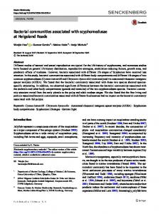
Antioxidant activity and phenolic content One mechanism by which antioxidants inhibit oxidation is by quenching reactive species through hydrogen or electron donation [38]. The DPPH assay measures this capacity by monitoring the decrease in absorbance of DPPH radicals as they react with the antioxidant, marked by the colour change from purple to yellow in DPPH assay [39]. The antioxidant activity, measured as percentage inhibition of DPPH free radicals scavenging activity, of ethyl acetate extracts of endophytic fungi isolated from D. stramonium, M. oleifera and P. chilensis was determined and results are listed in Figure (1). Generally, typical compounds that possess antioxidant activity have been characterized as phenolic compounds [40].
Therefore, it was reasonable to investigate their total levels (total polyphenols and flavonoids) in the tested extracts. Antioxidant activity of endophytic fungi extracts from D. stramonium ranged from 45 to 118%. The highest activity was obtained from Aspergillus sp. (D-4) followed by A. pulvinus (D-1) and A. terreus (D-2) with inhibition of DPPH free radicals of 118, 77 and 67% respectively. A. flavus (D-3) and Curvularia sp. (D-5) displayed weak scavenging activity. The total polyphenolic content of the extracts ranged from 146 to 830 mg gallic acid equivalent/g whereas the flavonoids content ranged from 549 to 920 mg quercetin equivalent/g. Aspergillus sp. (D-4) revealed the highest phenolic contents.
JOURNAL OF FOREST PRODUCTS & INDUSTRIES, 2014, 3(6), 248-256 ISSN:2325–4513(PRINT) ISSN 2325 - 453X (ONLINE)
253
Table 3.Minimum inhibitory concentration (MIC) of the endophytic fungi ethyl acetate extracts against bacteria. Samples
MIC value (µg/mL) E. coli
K. pneumoniae
P. aeruginosa
S. typhi
S. aureus
D-1
> 100
> 100
> 100
> 100
> 100
D-2
> 100
> 100
> 100
> 100
> 100
D-3
> 100
> 100
> 100
> 100
> 100
D-4
> 100
> 100
> 100
> 100
> 100
D-5
> 100
> 100
> 100
> 100
> 100
M-1
> 100
> 100
> 100
> 100
> 100
M-2
> 100
> 100
> 100
> 100
> 100
M-3
50
50
50
50
50
M-4
> 100
> 100
> 100
> 100
> 100
M-5
> 100
> 100
> 100
> 100
> 100
M-6
> 100
> 100
> 100
> 100
> 100
P-1
25
25
25
25
25
P-2
25
25
25
25
25
P-3
25
25
25
25
25
P-4
25
25
25
25
25
P-5
25
25
25
25
25
P-6
25
25
25
25
25
D-1→D-5 isolates from D. stramonium; M-1→ M-6 isolates from M. oleifera; P-1 →P-6 isolates from P. chilensis.
Antioxidant activity of endophytic fungi extracts from M. oleifera ranged from 24 to 113%. Strong DPPH radical scavenging activity was displayed by Emericella bicolor (M-6) with inhibition of DPPH free radicals of 113%. Aspergillus sp. (M-4) and Curvularia sp. (M-5) showed moderate scavenging activity whereas extracts of other endophytic isolates revealed weak activity. The total polyphenolic content of extracts ranged from 241 to 821 mg gallic acid equivalent/g where E. bicolor (M-6) contained the highest content. All isolates extracts of endophytic fungi isolated from M. oleifera contained high flavonoids content that ranged from 735 to 869 mg quercetin equivalent/g. Antioxidant activity of endophytic fungi extracts from P. chilensis ranged from 44 to 79%. The highest activity was obtained from Chaetomium sp. (P-5) followed by Emericella rugulosa (P-2), A. pulvinus (P-4) and E. rugulosa (P-1) with inhibition of DPPH free radicals of 79,
69, 67 and 66% respectively. A. niger (P-3) and Aspergillus sp (P-6) displayed weak activity. The total polyphenolic content of extracts ranged from 159 to 488 mg gallic acid equivalent/g whereas the flavonoids content ranged from 524 to 837 mg quercetin equivalent/g. Chaetomium sp. (P-5) exhibited the highest total polyphenolic content whereas, E. rugulosa (P-2) possessed the highest favonoids level. In summary, it was observed that DPPH radical scavenging activity was varied in all tested endophytic fungi. A remarkable scavenging activity was obtained from endophytic fungi Aspergillus sp. (D-4) and E. bicolor (M-6) isolated from D. stramonium and M. oleifera respectively where they possessed antioxidant activity higher than that obtained from the standard control ascorbic acid. The results of our study indicated that endophytic fungi from these selected Sudan’s medicinal plants may serve as potential source of antioxidants. Moreover, mutualistic associations of endophytes
JOURNAL OF FOREST PRODUCTS & INDUSTRIES, 2014, 3(6), 248-256 ISSN:2325–4513(PRINT) ISSN 2325 - 453X (ONLINE)
also enhance host plant defense responses against various biotic and abiotic stresses. It was observed that, increased endophytic fungal biomasses have been associated with increased host tolerance of a range of stresses and low percentages of occurrence of endophytes were correlated with poor total antioxidants and antioxidant activity [41].
254 antioxidant activity was also observed. These results suggested that polyphenols contributed largely for the antioxidant activities of endophytic fungi extracts isolated from D. stramonium M. oleifera and P. chilensis. Many researchers have reported a positive relation between the phenolic contents to antioxidant activity [42]. According to Huang et al., [43], phenolic content were the major antioxidant constituents of the endophytes
Moreover, a positive correlation between the phenolic content of the extracts with their
inhibition of DPPH (%)
. 140 120 100 80 60 40 20 0 D1 D2 D3 D4 D5 M1 M2 M3 M4 M5 M6 P1 P2 P3 P4 P5 P6 Std Fig. 1. Antioxidant activity of ethyl acetate extracts derived from PDA of endophytic fungi cultures. D-1→D-5 isolates from D. stramonium; M-1→ M-6 isolates from M. oleifera; P-1 →P-6 isolates from P. chilensis; Std, Standard control (Ascorbic acid).
Table 4. Total polyphenols and flavonoids of the endophytic fungi. Samples Total polyphenolsa Total flavonoidsb D1
448
549.5
D2
297
844.5
D3
1146
639
D4
830
920
D5
151
678
M1
255
785
M2
741
735
M3
622
869.5
M4
449
837.5
M5
59
737.5
M6
821
849.5
P1
352
524.5
P2
657
837.5
P3
292
765
P4
135
689.5
P5
288 807.5 P6 159 599.5 a Expressed as mg g-1 extract as gallic acid equivalent. b Expressed as mg g-1 extract as quercetin equivalent. D-1→D-5 isolates from D. stramonium; M-1→ M-6 isolates from M. oleifera; P-1 →P-6 isolates from P. chilensis.
JOURNAL OF FOREST PRODUCTS & INDUSTRIES, 2014, 3(6), 248-256 ISSN:2325–4513(PRINT) ISSN 2325 - 453X (ONLINE)
It is also important to note that extracts of endophytic fungi strains that did not possess antimicrobial or antioxidant activities do not necessarily means they devoid of agents responsible of these activities. Scherlach and Hertweck [44] and Kusari and Spiteller [45] reported that, bioprospecting endophytes capable of producing desired bioactive secondary metabolites traditionally involves screening of a plethora of different endophytes isolated from a single host plant for identifying the ‘‘competent’’ endophyte with the desired trait. When employing the classical approach, often, only a few or even none of the endophytes is capable of possessing the desired potential. The rest so-called ‘‘incompetent’’ endophytes are discarded without further investigation leading to the loss of the entire suite of natural products that they might produce under suitable conditions mimicking their natural habitat. However, recent whole-genome sequencing strategies have revealed that the number of genes encoding the biosynthetic enzymes in various fungi and bacteria undoubtedly is greater than the known secondary metabolites of these microorganisms [44, 26]. Therefore, these researchers stated that the discarded endophytes might actually express only a subset of their biosynthetic genes under in vitro standard laboratory conditions such that only a minor portion of their actual biosynthetic potential is harnessed. The large reservoir of ‘‘cryptic’’ natural metabolites is, thus, yet to be exploited. It is even possible that they produce the desired target compounds in quantities below the limit of detection, sometimes coupled with a large ‘‘metabolic background’’ and discrete culture conditions. Hence, it is necessary to understand and unravel the chemical ecological interaction of endophytes to fully exploit their inexhaustible potential of natural product biosynthesis [47]. In conclusion, these endophytic fungi associated with medicinal plants grow in Sudan could be effective alternative sources of antimicrobial and antioxidant agents and with high potential in producing novel metabolites for the development of drugs. ACKNOWLEDGEMENTS Authors would like to acknowledge Prof. Maha Kordofani (Botany Department, Faculty of Science, University of Khartoum) for the identification of the plants. REFERENCES [1] Shukla, S. T., Habbu, P. V., Kulkarni, V. H., Jagadish, K., Aprajita, S., Pandey, R., Sutariya, V. N. 2014. Endophytic microbes: A novel source for biologically/pharmacologically active secondary metabolites. Asian J. Pharmacol. Toxicol. 2 (03): 01-16. [2] Guerin, P. 1898. Sur la presence d’un champignon dans l’ivraie. J. Botanique. 12: 230–238. [3] de Bary, A. 1866. Morphologie und Physiologie der Pilze, Flechten, und Myxomyceten. Hofmeister’s Handbook of Physiological Botany, Volume II (Leipzig, Germany: Engelmann).
255 [4] Arnold, A. E. 2007. Understanding the diversity of foliar endophytic fungi: progress, challenges, and frontiers. Fungal Biol. Rev. 21: 51-66. [5] Tan, R. X. and Zou, W. X. (2001). Endophytes: a rich source of functional metabolites. Nat. Prod. Rep. 18: 448-459. [6] Strobel, G. A. 2002. Microbial gifts from rain forests. Can. J. Plant Pathol. 24: 14-20. [7] Chen, Y., Mao, W., Tao, H., Zhu, W., Qi, X., Chen, Y., Li, H., Zhao, C.; Yang, Y., Hou, Y., Wang, C. and Li, N. 2011. Structural characterization and antioxidant properties of an exopolysaccharide produced by the mangrove endophytic fungus Aspergillus sp. Y16. Bioresource Technol. 102: 8179– 8184. [8] Monnanda, S. N., Basavanna, M., Mysore, V. T., Harischandra, S. P., Ven, S., Kukkundur, R. K. and Huntrike, S. S. 2005. Fungal endophytes from the three-leaved caper, Crataeva magna (Lour.) DC. (Capparidaceae). Mycopathologia.159: 245-249. [9] Petrini, O., Sieber, T. N., Toti, L. and Viret. O. 1992. Ecology, metabolite production, and substrate utilization in endophytic fungi. Nat. Toxins. 1: 185-196. [10] Webster, J. 1970. Coprophilous fungi. Trans. Br. Mycol. Soc. 54:161-180. [11] Webster, J. 1980. Introduction to Fungi. 2 nd ed. Cambridge University Press. Cambridge. [12] Watanabe, T. 2002. Pictorial atlas of soil and seed fungi: morphologies of cultured fungi and key to species .2 nd ed. CRC Press LLC. Washington. DC. [13] Mothana, R. A. A. and Lindequist, U. 2005. Antimicrobial activity of some medicinal plants of the island Soqotra. J. Ethnopharmacol. 96: 177-181. [14] Eloff, J. N. 1998. A sensitive and quick microplate method to determine the minimal inhibitory concentration of plant extracts for bacteria. Planta Medica. 64:711-713. [15] Yamaguchi, T., Takamura, H., Matoba, T. and Teroa, J. 1998. HPLC method for evaluation of the free radical scavenging activity of foods by using 1, 1-diphenyl-2picrylhydrazyl. Biosci., Biotechnol., Biochem. 62: 1201-1204. [16] Iqbal, S., Bhanger, M. I. and Anwar, F. 2005. Antioxidant properties and components of some commercially available varieties of rice bran in Pakistan. Food Chem. 93:265–272. [17] Zhishen, J., Mengcheng, T. and Jianming, W. 1999. The determination of flavonoid contents in mulberry and their scavenging effects on superoxide radicals. Food Chem. 64: 555–9. [18] Weber, J. 1981. A natural control of Dutchelm disease. Nature, London. 292: 449-451. [19] Stierle, A.; Strobel, G.A., and Stierle, D. (1993). Taxol and taxane production by Taxomyces andreanae, an endophytic fungus of Pacific yew. Science. 260:214–216. [20] Fisher, P. J. and Petrini, O. 1994. A comparative study of fungal endophytes in xylem and bark of Alnus species in England and Switzerland. Mycol. Res. 94: 313-319. [21] Hawksworth, D. C. and Rossman, A. Y. 1987. Where are the undescribed fungi? Phytopathol. 87:888-891. [22] Dreyfuss, M. M. and Chapela, I. H. 1994. Potential of Fungi in the discovery of novel, low-molecular weight
JOURNAL OF FOREST PRODUCTS & INDUSTRIES, 2014, 3(6), 248-256 ISSN:2325–4513(PRINT) ISSN 2325 - 453X (ONLINE)
pharmaceuticals. In: The discovery of natural products with therapeutic potential. (Ed.): V. P. Gullo. Butter-worthHeinemann, London, United Kingdom. P: 49-80. [23] Faeth, S. H., Helander, M. L. and Saikkonen, K. J. T. 2004. Asexual Neotyphodium endophytes in a native grass reduce competitive abilities. Ecol. Lett. 7: 304-313. [24] Petrini, O. 1996. Ecological and physiological aspects of host-specificity in endophytic fungi. In: Endophytic fungi in grasses and woody plants: systematics, ecology and evolution. Ed. by Redlin, S. C.; Carris, L. M. St. Paul, MN: APS Press, P: 87–100. [25] Gange, A. C., Dey, S., Currie, A. F. and Sutton, B. V. C. 2007. Site and species-species differences in endophyte occurrence in two herbaceous plants. J. Ecol. 95: 614-622. [27] Bills, G. F. and Polishook, J. D. 1992. Recovery of endophytic fungi from Chamaecyparis thyoides. Sydowia. 44: 1-12. [28] Sahashi, N., Miyasawa, Y., Kubono, T. and Ito, S. 2000. Colonization of beech leaves by two endophytic fungi in northern Japan. Forest Pathol. 30: 77–86. [29] Jena, S. K. and Tayung, K. 2013. Endophytic fungal communities associated with two ethno-medicinal plants of Similipal Biosphere Reserve, India and their antimicrobial prospective. Journal of Applied Pharmaceutical Science. 3: 712. [30] Sun, J., Awakawa, T., Noguchi, H. and Abe, I. 2012. Induced production of mycotoxins in an endophytic fungus from the medicinal plant Datura stramonium L. Bioorg. Med. Chem. Lett. 15: 6397-400. [31] Kumaresan,V., Veeramohan, R., Shrivastava, P., Deepika, R., Suganya, M., Vennila, S., Datchayani, A. and Kalpana, P. 2013. Fungal Endophytes of Some Green Leafy Vegetables. World Journal of Agricultural Sciences. 9: 415420. [32] Zhao, J. H., Zhang, Y. L., Wang, L. W., Wang, Y. and Zhang, C. L. 2012. Bioactive secondary metabolites from Nigrospora sp. LLGLM003, an endophytic fungus of the medicinal plant Moringa oleifera Lam. World J. Microb. Biot. 28:107-12. [33] Barnabas, J., Sushma, S., Murthy, G. and Jagdeesh, S. 2013. Antimicrobial properties of endophytic fungi isolated from Cynodondactylon and Moringa oliefera. IJBPR. 2:98104. [34] Verhoeff, K. 1974. Latent infections by fungi. Ann Rev Plant Physiol. 12: 99-110. [35] Peláez, F., Collado, J., Arenal, F., Basilio, A., Cabello, A., Díez Matas, M. T., García, J. B., González Del Val, A., González, J., Gorrochategui, J., Hernández, P., Martín, I., Platas, G., and Vicente, F. 1998. Endophytic fungi from plants living on gypsum soils as a source of secondary metabolites with antimicrobial activity. Mycol. Res.102: 755761. [36] O’Donnell, J. and Dickinson, C. H. 1980. Pathogenicity of Alternaria and Cladosporium isolates on Phaseolus. Trans. Br. Mycol. Soc. 74: 335-342. [37] Barbour, E. K., Sharif, M. A., Sagherian, V. K., Habre, A. N., Talhouk, R. S. and Talhouk, S. N. 2004. Screening of selected indigenous plants of Lebanon for antimicrobial activity. J. Ethnopharmacol. 93:1-7.
256 [38] Singh, N. and Rajini, P. S. 2004. Free radical scavenging activity of an aqueous extract of potato peel. Food Chem. 85: 611-616. [39] Elmastas, M. and Gulcin, O. I., Kufrevioglu, O. I., Ibaoglu, K. and Aboul-Enein, H. Y. 2006. Radical scavenging capacity and antioxidant activity of bay leaf extracts. J. Iran. Chem. Soc. 3:258-266 [40] Jerez, M., Selga, A., Sineiro, J. L., Torres, L. J. and Nunez, M. J. 2007. A comparison between bark extracts from Pinuspinaster and Pinusradiata: antioxidant activity and procyanidin composition. Food Chem. 100: 439–444. [41] Swarthout, D., Harper, E., Judd, S., Gonthier, D., Shyne, R., Stowe, T. and Bultman, T. 2009. Measures of leaf-level water-use efficiency in drought stressed endophyte infected and non-infected tall fescue grasses. Environ Exp Bot. 66: 88– 93. [42] Saboo, S., Tapadiya, R., Khadabadi, S. S. and Deo, U. A. 2010. In vitro antioxidant activity and total phenolic, flavonoid contents of the crude extracts of Pterospermum acerifolium wild leaves (Sterculiaceae) J. Chem. Pharm. Res. 2: 417-423. [43] Huang, D., Ou, V. and Proir, R. L. 2005. The chemistry behind antioxidant capacity assays. J. Agr. Food Chem. 53:1841-1856. [44] Scherlach, K. and Hertweck, C. 2009. Triggering cryptic natural product biosynthesis in microorganisms. Org. Biomol. Chem. 7:1753–1760. [45] Kusari, S., Zu¨ hlke, S. and Spiteller, M. 2011. Effect of artificial reconstitution of the interaction between the plant Camptotheca acuminata and the fungal endophyte Fusarium solani on camptothecin biosynthesis. J. Nat. Prod. 74: 764– 775. [46] Winter, J. M., Behnken, S. and Hertweck, C. 2011. Genomics-inspired discovery of natural products. Curr. Opin. Chem. Biol. 15: 22–31. [47] Kusari, S., Verma, V. C., Lamsho¨ ft, M. and Spiteller, M. 2012. An endophytic fungus from Azadirachta indica A. Juss. That produces azadirachtin. World J. Microb. Biot. 28: 1287–1294.