Pathophysiology/Complications O R I G I N A L
A R T I C L E
Endothelial Perturbation in Children and Adolescents With Type 1 Diabetes Association with markers of the inflammatory reaction MARIO ROMANO, MD1 MARIAPINA POMILIO, MD2 SERGIO VIGNERI, MD3 ANGELA FALCO, MD4
PIERLUIGI LELLI CHIESA, MD5 FRANCESCO CHIARELLI, MD2 GIOVANNI DAV`I, MD4
OBJECTIVE — The progression of diabetic angiopathy is, in most cases, unpredictable. The aim of this study was to investigate early events that could influence the development of diabetic angiopathy. RESEARCH DESIGN AND METHODS — Circulating levels of von Willebrand factor (vWF) and tissue-plasminogen activator (tPA), defining endothelial perturbation, were measured in 40 young patients with type 1 diabetes. Patients were divided into two groups according to the duration of diabetes (group A, ⬍1 year; group B, ⬎1 year) and compared with a control group of age- and sex-matched healthy individuals. Prothrombin fragment 1 and 2 (F1⫹2), tumor necrosis factor-␣ (TNF-␣), and C-reactive protein (CRP) levels were also determined as markers of a prothrombotic state and inflammatory response. A total of 16 of the 20 children in group A were re-examined after 12 months. RESULTS — Compared with either normal subjects or patients in group B, children in group A showed increased levels of vWF, tPA, F1⫹2, TNF-␣, and CRP. Significant direct correlations between TNF-␣ or CRP and either vWF, tPA, or F1⫹2 were observed. Endothelial perturbation was shown in 70% of group A and 20% of group B. After 1 year, 16 of the 20 patients in group A showed a significant reduction in vWF, tPA, F1⫹2, TNF-␣, and CRP levels, whereas endothelial perturbation was reversed in 5 of these patients. CONCLUSIONS — Endothelial perturbation represents an early and, in some cases, reversible event in the chronology of type 1 diabetes in children. A correlation might exist between the initial inflammatory reaction and the appearance of endothelial perturbation. Diabetes Care 24:1674 –1678, 2001
T
ype 1 diabetes is associated with the development of vascular complications involving both the micro- and macrocirculation. Nephropathy, retinopathy, and atherosclerosis are frequently observed in patients with type 1 diabetes, and the progression of these conditions
has a considerable impact on the prognosis of this disease (1). Vascular endothelium is a primary target of the unbalanced glycemic metabolism in type 1 diabetes. Evidence indicates that high glucose levels may alter the anti-atherogenic, anti-thrombotic prop-
erties of vascular endothelium, thus favoring the development of diabetic angiopathy (2). On the other hand, inflammatory reactions taking place in the early phase of diabetes determine an increase in serum levels of a wide array of cytokines, including TNF-␣, interleukin-1 (IL-1), and interleukin-6 (IL-6), which may impair endothelial cell functions (3). The initial phase of vascular endothelium damage is defined by the term “endothelial perturbation,” which refers to a reversible dysfunction of the endothelial cell. In vivo, endothelial perturbation is shown by a simultaneous increase in circulating vWF and tPA to levels 2 SD higher than the control values (4,5). Endothelial perturbation is associated with an ongoing prothrombotic state and an increased risk of thrombotic events (6), and its persistence could represent a negative prognostic index. Because the risk of developing vascular disease is not distributed homogeneously among patients with type 1 diabetes and the pathogenesis of vascular complications still is not completely defined, we examined the presence of endothelial perturbation in two populations of type 1 diabetic children and adolescents at different stages of the disease. We also investigated the possible relationship between inflammatory reaction and endothelial dysfunction. In this report, we show that endothelial perturbation represents an early and, in some cases, reversible event in children with type 1 diabetes.
● ● ● ● ● ● ● ● ● ● ● ● ● ● ● ● ● ● ● ● ● ● ● ● ● ● ● ● ● ● ● ● ● ● ● ● ● ● ● ● ● ● ● ● ● ● ● ● ●
From the 1Department of Human Pathology, University of Messina, Messina; the 2Department of Medicine and Aging, Division of Pediatrics, University of Chieti, Chieti; the 3Department of Medicine, University of Palermo, Palermo; the 4Department of Medicine and Aging, Division of Hematology, University of Chieti, Chieti; and 5the Department of Medicine and Aging, Division of Pediatric Surgery, University of Chieti, Chieti, Italy. Address correspondence and reprint requests to Giovanni Davı`, MD, Cattedra di Ematologia, Universita` di Chieti, Via dei Vestini 31, 66013 Chieti, Italy. E-mail:
[email protected]. Received for publication 11 January 2001 and accepted in revised form 31 May 2001. Abbreviations: CRP, C-reactive protein; ELISA, enzyme-linked immunosorbent assay; F1⫹2, prothrombin fragment 1 and 2; ICA-IgG, anti-islet cell antibodies; IL-1, interleukin-1; IL-6, interleukin-6; TNF-␣, tumor necrosis factor-␣; tPA, tissue-plasminogen activator; vWF, von Willebrand factor. A table elsewhere in this issue shows conventional and Syste`me International (SI) units and conversion factors for many substances.
1674
RESEARCH DESIGN AND METHODS — A total of 40 young patients with diabetes (18 girls and 22 boys, 12.5 ⫾ 4.6 years of age) attending the Diabetes Clinic at the Division of Pediatrics, University of Chieti, Italy, were examined several times between December 1996 and November 1998. All patients had type 1 diabetes, as defined in accordance with the criteria of the American Diabetes Association (7). None of the
DIABETES CARE, VOLUME 24, NUMBER 9, SEPTEMBER 2001
Romano and Associates
Table 1—Clinical characteristics of patients with diabetes and healthy subjects Patients with type 1 diabetes Variable
Healthy subjects
Group A
Group B
10 4/6 9.6 ⫾ 2.6
20 9/11 10.6 ⫾ 3.4
20 9/11 11.4 ⫾ 3.2
— — ND* 4.5 ⫾ 0.7
4.2 ⫾ 3.8 7–10 174 ⫾ 88 8.9 ⫾ 2.5
58 ⫾ 35 14–132 182 ⫾ 82 7.8 ⫾ 1.5
n Sex (F/M) Age (years) Duration of diabetes Months Range Fasting blood glucose (mg/dl) HbA1c (%) Data are n or (mean ⫾ SD). *Not determined.
children had clinical evidence of retinopathy, as assessed by routine ophthalmologic examinations and stereoscopic fundus photography, or nephropathy (absence of microalbuminuria or macroalbuminuria). Patients were subdivided into two groups according to the duration of diabetes (defined as time since first injection of insulin): Group A (n ⫽ 20), duration of diabetes ⬍1 year (4.2 ⫾ 3.8 months); and group B (n ⫽ 20), duration of diabetes ⬎1 year (58 ⫾ 35 months). All patients were being treated with insulin (0.8 –1.2 units 䡠 kg⫺1 䡠 day⫺1). A total of 10 age- and sex-matched healthy children were also studied. Of the 20 children in group A, 16 were re-examined after 12 months. Informed consent was obtained from each subject (or from his/her parents) participating in the study. The protocol was approved by the local ethical committee. The baseline characteristics of patients and control subjects are detailed in Table 1. Venous blood samples were collected in tubes containing 3.8% sodium citrate. Plasma was prepared by centrifugation (2000g for 20 min), aliquoted, and stored at ⫺20°C. vWF and tPA antigens were determined by enzyme-linked immunosorbent assay (ELISA) (American Diagnostica, Greenwich, CT). The intra- and interassay variation coefficients were 9 and 13% and 7 and 9%, respectively. Patients were considered to have endothelial perturbation when both vWF:Ag and tPA:Ag were more than 2 SD higher than the control mean value (4). Plasma F1⫹2 levels were measured using an ELISA kit (Behringwerke, Marburg, Germany). Intra- and interassay variation coefficients were 8 and 9%, respectively. TNF-␣ plasma levels were measured using an ELISA kit (R&D Systems Europe, Abing-
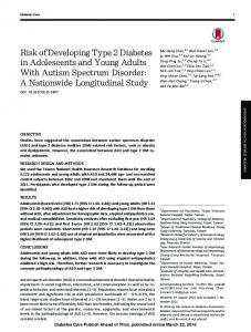
don, Oxford, U.K.). Intra- and interassay coefficients of variation were ⬍9%. Fasting plasma glucose levels were measured by the glucose oxidase method. HbA1c was determined by automated highperformance liquid chromatography (8). CRP was measured using a highly sensitive nephelometric assay (BN-II Nephelometer; Dade Behring, Milan, Italy). Conventional anti-islet cell antibodies (ICA-IgG) were detected by means of indirect immunofluorescence on cryostat sections of blood type O human pancreas (9). Positive samples were then titered by dilutions in phosphate-buffered saline. End-point titers were converted to Juvenile Diabetes Foundation units. The threshold of detection was 5–10 Juvenile Diabetes Foundation units. Statistical analyses were performed by 2 statistics or Fisher’s exact test (if n ⬍5) for independence and by appropriate Student’s t test. Correlations were analyzed by the Spearman rank correlation test. Data are presented as the mean ⫾ SD and 95% CI. TNF-␣ values are expressed as median and range because they showed an appreciably skewed distribution. Only two-tailed probabilities were used for testing statistical significance (P ⬍0.05). All calculations were made with the computer program Stat-View II (Abacus Concepts, Berkeley, CA). RESULTS — Circulating levels of vWF:Ag were significantly higher in patients in group A compared with agematched healthy subjects or patients in group B (1,536 ⫾ 438 vs. 671 ⫾ 146 units/l, P ⫽ 0.0001, and vs. 979 ⫾ 252 units/l, P ⫽ 0.0008, respectively) (Fig. 1A). Patients in group A also had increased plasma tPA:Ag levels in comparison with healthy subjects or patients in
DIABETES CARE, VOLUME 24, NUMBER 9, SEPTEMBER 2001
Figure 1—vWF (A), tPA (B), and F1⫹2 (C) levels in patients with type 1 diabetes and ageand sex-matched healthy subjects. Dots depict individual measurements; horizontal bars indicate average values.
group B (11.2 ⫾ 4.4 vs. 5.3 ⫾ 1.3 ng/ml, P ⫽ 0.0036, and vs. 7.4 ⫾ 3.0 ng/ml, P ⫽ 0.0087, respectively) (Fig. 1B). Of the 20 patients in group A, 14 (70%) had an increase in both vWF and tPA ⬎2 SD higher than control values (endothelial perturbation). In contrast, only 20% of patients in group B had signs of endothelial perturbation. Higher F1⫹2 levels compared with normal subjects or patients in group B (1.05 ⫾ 0.42 vs. 0.419 ⫾ 0.144 nmol/l, P ⫽ 0.0016, and vs. 0.498 ⫾ 0.295 nmol/l, P ⫽ 0.002, respectively) were found in patients in group A (Fig. 1C). Direct correlations were found between F1⫹2 and either vWF ( ⫽ 0.809, P ⫽ 0.0001) or tPA levels ( ⫽ 0.811, P ⫽ 0.0001) (results not shown). Patients in group A also had elevated circulating TNF-␣ levels compared with normal subjects or patients in group B (21.9 [6.5– 44.4] vs. 4.4 [1.5– 8.2] pg/ml, 1675
Endothelial perturbation in diabetic children
Table 2—Laboratory findings of Group A patients reexamined after 1 year Variable Glycemia (mg/dl) HbA1c (%) vWF (units/l) tPA (ng/ml) F1⫹2 (nmol/l) TNF-␣ (pg/ml) Endothelial perturbation
Baseline
After 1 year
P
195 ⫾ 94 8.6 ⫾ 2.4 1,459 ⫾ 381 10.8 ⫾ 4.2 1.0 ⫾ 0.39 21.3 (6.5–44.4) 11 (16)
142 ⫾ 56 7.5 ⫾ 1.3 1,135 ⫾ 276 8.3 ⫾ 2.5 0.70 ⫾ 0.26 4.1 (1.2–10.1) 6 (16)
NS NS 0.0008 0.0007 0.0004 0.0001
Data are means ⫾ SD, median (range), or n (%). NS, not significant.
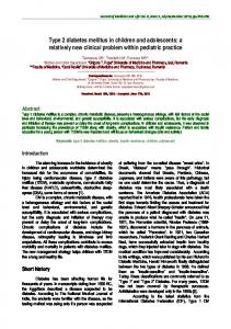
Figure 2—Measurements of TNF-␣ (A) and CRP (B) levels in patients with type 1 diabetes and sex- and age-matched healthy subjects. Data points are from individual measurements; horizontal bars indicate average values.
P ⫽ 0.0003, and vs. 2.45 [1.4 – 4.1] pg/ml, P ⫽ 0.0001, respectively) (Fig. 2A). This was paralleled by an increase in CRP levels (1.67 ⫾ 0.99 vs. 0.38 ⫾ 0.1 mg/l, P ⫽ 0.0051, and vs. 0.77 ⫾ 0.5 mg/l, P ⫽ 0.004) (Fig. 2B). Interestingly, patients with endothelial perturbation displayed the highest levels of F 1⫹2 (1.13 ⫾ 0.37 vs. 0.45 ⫾ 0.19 nmol/l), TNF-␣ (20.8 [2.6 – 44.4] vs. 6.9 [1.5– 36.4] pg/ml), and CRP (2.31 ⫾ 1.1 vs. 1.1 ⫾ 0.65 mg/l). In addition, statistically significant correlations were found between TNF-␣ and CRP ( ⫽ 0.71, P ⫽ 0.0001); between TNF-␣ and vWF ( ⫽ 0.685, P ⫽ 0.0001), tPA ( ⫽ 0.469, P ⫽ 0.0034), or F1⫹2 levels ( ⫽ 0.61, P ⫽ 0.0001); and between CRP and vWF ( ⫽ 0.838, P ⫽ 0.0001), tPA ( ⫽ 0.694, P ⫽ 0.0001), or F 1⫹2 ( ⫽ 0.718, P ⫽ 0.0001). In contrast, neither blood/ urinary glucose, nor HbA1c influenced vWF, tPA, and F1⫹2 levels (results not shown). A total of 16 patients in group A were reviewed after 1 year. The results of the follow-up are summarized in Table 2. Endothelial perturbation was still appreciable in 6 of the 11 patients in whom it was initially displayed, whereas in five cases, it was reversed. Interestingly, when patients 1676
were divided into groups according to changes in endothelial perturbation, their TNF-␣ levels showed significant differences. In fact, the patients who still had endothelial perturbation after 1 year were those who had the highest TNF-␣ levels at the beginning of the study, whereas patients in whom endothelial perturbation was reversed after 1 year were those who had significantly lower initial TNF-␣ levels. Patients who did not have endothelial perturbation from the beginning were those with the lowest initial TNF-␣ values (Fig. 3). A similar pattern was observed with CRP (results not shown). In these patients, statistically significant correlations were observed between ICA and TNF-␣ ( ⫽ 0.44, P ⫽ 0.014), vWF ( ⫽
0.54, P ⫽ 0.0025), tPA ( ⫽ 0.45, P ⫽ 0.0012), and F1⫹2 levels ( ⫽ 0.56, P ⫽ 0.0018) levels. CONCLUSIONS — This study shows for the first time that the occurrence of endothelial perturbation represents an early and, in some cases, reversible event in the chronology of type 1 diabetes in children and adolescents. We observed that patients with duration of diabetes shorter than 1 year had a significant increase in both vWF and tPA levels and that endothelial perturbation was reversed in ⬃45% of these patients after 1 year (Fig. 1). Therefore, it seems that signs of a vascular involvement can be appreciated as early as during the first decade of life of patients with diabetes.
Figure 3—Relationship between TNF-␣ levels and the evolution of endothelial perturbation. Measurements are relative to a 1-year follow-up in a selected number of patients in group A. Horizontal bars indicate average values.
DIABETES CARE, VOLUME 24, NUMBER 9, SEPTEMBER 2001
Romano and Associates
Considering the central role played by the endothelium in the regulation of vascular homeostasis (10), the question arises regarding the pathophysiological relevance of an early and, in most cases, transient endothelial perturbation in children and adolescents with diabetes. Should it be regarded as a transient epiphenomenon of the initial inflammatory autoimmune events taking place in diabetes? Or should it be considered an indicator of the risk for developing diabetic angiopathy in children and adolescents with type 1 diabetes? Long-term studies will hopefully answer this question. Consistent with previous observations (11), the present results also indicate that measurements of vWF and tPA may be useful to detect initial endothelium involvement in type 1 diabetes. In this regard, it must be noted that vWF is considered the best available noninvasive marker for endothelial dysfunction (12,13). Indeed, an elevation in vWF levels may be observed in diabetes (14) (in which it can precede the occurrence of microalbuminuria by ⬃3 years [2]), as well as in hypertension (15), smoking (16), hypercholesterolemia (17), and ischemic heart disease (18). However, because vWF may also originate from platelets (19), the combined determination of vWF and tPA, which is also secreted by endothelial cells (20), may be a more specific and sensitive index of endothelial cell alteration. In our patients, endothelial perturbation was associated with an increment in F1⫹2 levels (Fig. 1). This finding, previously unappreciated in children with diabetes, indicates that at the beginning of the disease, these patients may have a subclinical prothrombotic disorder. In fact, F1⫹2 generates thrombin and is formed when active tissue factor and activated coagulation factor X accumulate (21). Increased F1⫹2 levels can be observed during coagulative disorders and are associated with a prothrombotic condition (4). Therefore, an increase in F1⫹2 levels is both a marker of an unbalanced tissue factor/tissue factor inhibitor pathway and an indicator of augmented thrombin generation. However, in approximately half of our children with type 1 diabetes, endothelial perturbation was a transient event; therefore, it would be difficult to draw a conclusion on the validity of endothelial perturbation and F1⫹2 determinations as predictive markers of diabetic angiopathy. However, it is striking that signs of endothelial perturbation and coagulative disorders can be already detected during
childhood. Future studies will determine whether children showing persistent endothelial perturbation are at higher risk for developing vascular complications. How is endothelial perturbation established in children with type 1 diabetes? Although previous studies have shown that poor glycemic control is related to the occurrence of vascular complications in juvenile diabetes (22,23), we were unable to observe a correlation between endothelial perturbation and HbA1c levels. On the other hand, an inflammatory response is frequently observed in the initial phase of type 1 diabetes. Therefore, taking into account that among our patients, those with the shorter duration of disease had the higher incidence of endothelial perturbation, we hypothesized that a relationship might exist between the appearance of immune-inflammatory markers and the occurrence of endothelial perturbation. Indeed, we observed a strong increment in the levels of the inflammatory cytokine TNF-␣ and of the inflammation marker CRP, which correlated with the levels of endothelial perturbation indexes (Figs. 2 and 3). Moreover, ICA, which appeared at a very early stage in type 1 diabetes, positively correlated with TNF-␣ as well as with vWF, tPA, and F1⫹2 levels. Considered together, these findings seem to be consistent with the possibility that the early immune-inflammatory reaction may influence the appearance of endothelial perturbation in children with type 1 diabetes. These results are consistent with a recent observation showing a significant correlation between CRP and markers of endothelial dysfunction in patients with type 1 diabetes (24) and suggest that TNF-␣ may represent at least one of the agents that link the inflammatory reaction to the appearance of endothelial perturbation in children with type 1 diabetes. Indeed, TNF-␣ induces vWF release and transient and reversible endothelial dysfunction in humans (25,26). However, this does not exclude the possibility that some other factors may intervene in this process to determine whether endothelial perturbation becomes persistent. In this regard, the observation that patients in group A with the highest initial TNF-␣ levels (Fig. 3) corresponded to those with persistent endothelial perturbation after 1 year is intriguing. It might indicate that the degree of the initial inflammatory reaction could influence the chance of developing permanent endothelial dys-
DIABETES CARE, VOLUME 24, NUMBER 9, SEPTEMBER 2001
function. However, due to the relatively small number of patients observed, this hypothesis must be investigated further. In conclusion, in this study, we have shown that endothelial perturbation is an early event that correlates with the appearance of inflammatory markers in children and adolescents with type 1 diabetes. Although the persistence of endothelial perturbation could represent a potential index of the risk for developing vascular complications, the relationship between the occurrence of endothelial perturbation and the development of diabetic angiopathy in young patients still remains to be established by future prospective studies. Acknowledgments — This work was supported, in part (60%), by a grant from the Italian Ministry of University. Financial support was also provided by the local Juvenile Diabetes Foundation (AGDA). We thank Antonella Bascelli for secretarial help and the patients and their families for their participation in this study.
References 1. Danne T, Kordonouri O, Ho¨vener C, Weber B: Diabetic angiopathy in children. Diabet Med 14:1012–1025, 1997 2. Stehouwer CDA, Lambert J, Donker AJM, van Hinsberg VWM: Endothelial dysfunction and pathogenesis of diabetic angiopathy. Cardiovasc Res 34:55– 68, 1997 3. Hussain MJ, Peakman M, Gallati H, Lo SS, Hawa M, Viberti GC, Watkins PJ, Leslie RD, Vergani D: Elevated serum levels of macrophage-derived cytokines precede and accompany the onset of IDDM. Diabetologia 39:60 – 69, 1996 4. Ferro D, Pittoni V, Quintarelli C, Basili S, Saliola M, Caroselli C, Valesini G, Violi F: Coexistence of anti-phospholipid antibodies and endothelial perturbation in systemic lupus erythematosus patients with ongoing prothrombotic state. Circulation 95:1425–1432, 1997 5. Ferro D, Basili S, Roccaforte S, Di Franco M, Cipollone F, Ciabattoni G, Davı` G: Determinants of enhanced thromboxane biosynthesis in patients with in systemic lupus erythematosus. Arthritis Rheum 42: 2689 –2697, 1999 6. Rodgers GM: Hemostatic properties of normal and perturbed vascular cells. FASEB J 2:116 –123, 1998 7. National Diabetes Data Group: Classification and diagnosis of diabetes mellitus and other categories of glucose intolerance. Diabetes 28:1039 –1057, 1979 8. Khuu HM, Robinson CA, Goolsby K,
1677
Endothelial perturbation in diabetic children
9.
10.
11. 12. 13. 14.
15.
Hardy RW, Konrad RJ: Evaluation of a fully automated high-performance liquid chromatography assay for hemoglobin A1c. Arch Pathol Lab Med 123:763–767, 1999 Bottazzo GF, Florin-Christensen A, Doniach D: Isle-cell antibodies in diabetes mellitus with autoimmune polyendocrine deficiencies. Lancet 2:1279 –1283, 1974 Cines DB, Pollak ES, Buck CA, Loscalzo J, Zimmerman GA, McEver RP, Pober JS, Wick TM, Kondle BA, Schwarts BS, Barnathan ES, McCrae KR, Hug BA, Schmidt AM, Stern DM: Endothelial cells in physiology and in the pathophysiology of vascular disorders. Blood 91:3527–3561, 1998 Blann AD: Von Willebrand factor and the endothelium in vascular disease. Br J Biomed Sci 50:125–134, 1993 Mannucci PM: Von Willebrand factor: a marker of endothelial damage? Arterioscl Thromb Vasc Biol 18:1359 –1362, 1998 Blann AD, Taberner DA: A reliable marker of endothelial dysfunction: does it exist? Br J Haematol 90:244 –248, 1995 Parving HH, Nielsen FS, Bang LE, Smidt UM, Svendsen TL, Chen JW, Gall MA, Rossing P: Macro-microangiopathy and endothelial dysfunction in NIDDM patients with and without diabetic nephropathy. Diabetologia 39:1590 –1597, 1996 Pedrinelli R, Giampietro O, Carmassi F,
1678
16. 17.
18.
19.
20.
21.
22.
Melillo E, Dell’Olmo G, Catapano G, Matteucci E, Talarico L, Morale M, De Negri F, Di Bello V: Microalbuminuria and endothelial dysfunction in essential hypertension. Lancet 344:14 –18, 1994 Blann AD, McCollum CN: Adverse influence of cigarette smoking on the endothelium. Thromb Haemost 70:707–711, 1993 Blann AD, Maxwell SR, Burrows G, Miller JP: Antioxidants, von Willebrand factor and endothelial cell injury in hypercholesterolemia and vascular disease. Atherosclerosis 116:191–198, 1991 Jansson JH, Nilsson TK, Johnson O: von Willebrand factor in plasma: a novel risk factor for recurrent myocardial infarction and death. Br Heart J 66:351–355, 1991 Sporn LA, Chavin SI, Marder VJ, Wagner DD: Biosynthesis of von Willebrand protein by human megakaryocytes. J Clin Invest 76:1102–1106, 1985 Schleef RR, Bevilacqua MP, Sawdey M, Gimbrone MA Jr, Loskutoff DJ: Cytokine activation of vascular endothelium: effects on tissue-type plasminogen activator and type 1 plasminogen activator inhibitor. J Biol Chem 263:5797–5803, 1988 Bauer KA, Rosenberg RD: The pathophysiology of the prothrombotic state in humans: insight gained from studies using markers of hemostatic system activation. Blood 70:343–350, 1987 Lestradet H, Papoz L, Hellouin de Meni-
23.
24.
25.
26.
bus C, Levavasseur F, Besse J, Billaud L, Battistelli F, Tric P, Lestradet F: Longterm study of mortality and vascular complications in juvenile-onset (type 1) diabetes. Diabetes 30:175–179, 1981 The Diabetes Control and Complications Trial Research Group: The effect of intensive treatment of diabetes on the development and progression of long-term complications in insulin-dependent diabetes mellitus. N Engl J Med 329:977–986, 1993 Schalkwijk CG, Poland DC, van Dijk W, Kok A, Emeis JJ, Drager AM, Doni A, van Hinsbergh VW, Stehouwer CDA: Plasma concentration of C-reactive protein is increased in type 1 diabetic patients without clinical macroangiopathy and correlates with markers of endothelial dysfunction: evidence for chronic inflammation. Diabetologia 42:351–357, 1999 Kahaleh MB: The role of vascular endothelium in the pathogenesis of connective tissue disease: endothelial injury, activation, participation and response. Clin Exp Rheumatol 8:595– 601, 1990 van der Poll T, van Deventer SJ, Pasterkamp G, van Mourik JA, Buller HR, ten Cate JW: Tumor necrosis factor induces von Willebrand factor release in healthy humans. Thromb Haemost 67:623– 626, 1992
DIABETES CARE, VOLUME 24, NUMBER 9, SEPTEMBER 2001