18th Annual International Conference of the TEEE Engineering in Medicine and Biology Society, Amsterdam 1996
4.4.5: Image Enhancement I - Multiscale Methods
ENHANCEMENT OF MICROCALCIFICATIONS O N MAMMOGRAMS USING A FRACTAL MO.DELING APPROACH Huai Li'12, K. J. Ray Liu', and ShihChung B. Lo2 'Electrical Engineering Department and Institute for Systems Research University of Maryland at College Park, College Park, Maryland 20742 21SIS Center, Georgetown University Medical Center 2115 Wisconsin Avenue, NW, #603, Washington, D.C. 20007 huaili0eng.umd.edu and
[email protected]
Abstract- The objective of this research is to model the mammographic parenchymal, ductal patterns and enhance microcalcifications using a fractal approach. According to the theory of deterministic fractal geometry, images can be modeled by deterministic fractal objects which are attract o r s of sets of two dimensional afflne transformations. In this paper, a methodology based on fractal image modeling is developed to analyze and extract various mammographic textures. We show that general mammographic parenchymal and ductal patterns can be well modeled by the proposed approach. Therefore, microcalcifications can be enhanced by taking the difference between the original mammogram and modeled mammogram. Our results are compared with those of the partial wavelet reconstruction and morphological operation approaches. The results demonstrate that the fractal modeling method is an effective way to enhance microcalciflcations, and thereby facilitate the radiologists' diagnosis. It is also able to improve the detection and classification of microcalciflcations in a computer system.
I. INTRODUCTION The abnormal mammogram is usually composed of the normal breast tissues (mammographic parenchymal, ductal patterns), the disease patterns(masses, microcalcifications), and the film defects (noises). Microcalcifications are one of the disease patterns. They are early signs of breast cancer. In mammogram, Microcalcifications are visible as small bright spots and have less structure. Some microcalcifications are very subtle and are easy to be missed by radiologists [l].On the other hand, the breast tissues possess structures with high self-similarity. If the tissue patterns can be modeled and be taken out from the original image, the microcalcification information will be enhenced. It is very helpful for doctors to detect the microcalcifications. Recently, fractal-based techniques have been applied in many areas of images processing, such as image segmentation, image analysis [2], [3]. In fractal theory, images could be modeled by deterministic fractal objects which are attractors of scts of two-dimensional affine transformations. We use this approach to model breast tissue structures and enhance microcalcifications by taking the difference between the original mammogram and modeled image. The results are very encouraging. This work was supported in part by a NSF Grant MIP-9457397and an U.S. Army Grant DAMD17-93-J-3007.
rr.
METHODS
The mathematical theories of Iterated Function Systems and the Collage Theorem are the foundations of fractal image modeling [4]. Let ( M , d ) denote a metric space of digital images, where d is a given distortion niea.sure, and let f be an original image which to be modeled. We want ) 6 to find a contractive mapping r such that: d ( f , ~ ( f ) < where 6 is a tolerance. The Collage Theorem shows that, if T is known, then from any given image fo, after a number of iterations, the constructed image fn = ~ " ( f o ) will close visually to original image f . The key point of fractal modeling is to explore the self-similar property of images. Real world images are almost never self-similar, but almost all real images have a local self similarity. The image can be divided into n small blocks, and for each block, find a corresponding ri. Then, we can define r = uy=l~i. Let Nl = [0,1,...,M ] , N 2 = [ O , l , ..., N ] , N 3 = [0,1,...,L ] , then for any digital gray-level image f ( z , y), we have ( k , Z , j ( k , Z ) ) E NI x N2 x N3. Let D1,...,D, and RI,...,Rn be subsets of N I x N z , such that UF=lRi = N I x NZ and Ri n Rj = q5,i # j. We define a set of mixing functions mi : N I x N2 + NI x Nz, such that mi(Ri) = Di using a affine mapping. ri can be defined as: ri(f(k,Z))= sif(mi(k,Z))oi, where si is a scaling factor and oi is an offset factor. Also, let us denote f l ~ , be the restriction of the function f to the set R,. In practice, we use d(.,.) as the mean square root metric and f ( m i ( k ,I)) = f(k,I), where
+
The goal is for each R,, a Di C NI x N Z where a constraint Ri nDi = is given and ri : N I x Nz x N3 + N3 are sought such that
Ilf(k".l)ERi
(.if(k W ( k J ) C D i + Oi)1122
(2) is minimized. Once all ri are found, the modeled image can be got by the iterations from any starting image of the same size according to D, and ~ i . The microcalcifications can be enhanced by taking the difference between the original mammogram and modeled mammogram. -
111. RESULTS Ten real mammograms with clustered and single microcalcifications were chosen as testing images. The areas of
0-7803-381 1-1/97/$10.00 QIEEE
1111
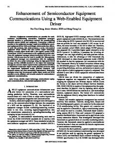
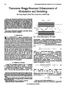
18th Annual International Conference of the IEEE Engineering in Medicine and Biology Society, Amsterdam 1996 4.4.5: Image Enhancement I - Multiscale Methods suspicious microcalcifications were identified by a highly experienced radiologist. The selected mammograms were digitized with an image resolution of 100pm x 100pm per pixel by the Lumisys DIS-1000. All images are 512 x 512 x 12 bpp. We found that the general mammographic parenchymal and ductal patterns could be well modeled and most microcalcifications were enhanced successfully. Fig. 1 shows the modeled and enhanced results of one of the real mammograms. As we can see in Fig. 1 (b), the general mammographic parenchymal and ductal patterns in mammograms were well modeled. In Fig. 1 (c), we can see that all less-structured spots, which include clusters of microcalcifications, single microcalcifications, and film detects, were clearly enhanced. For the purpose of evaluating the performance of our proposed fractal enhancement method, we chose for comparison two similar enhancement techniques of background removal: the morphological and partial wavelet reconstruction methods [ 5 ] , [ 6 ] . A global thresholding was applied to reduce unreliable noise in the fractal, morphological and wavelet approaches. Fig. 2 shows the enhancement results of clustered and single microcalcifications in the mammograms. The first, second, third, and fourth rows in Fig. 2 correspond to original ROIs, fractal enhancement, wavelet enhancement, and morphological enhancement, respectively.
IV.
of the wavelet and morphological approaches. All results obtained in this study are very encouraging, and indicate that the fractal modeling and segmentation method is an effective technique to enhance microcalcifications embedded in inhomogeneous breast tissues.
V. CONCLUSIONS In this research, we proposed a microcalcification enhancement algorithm based on the fractal modeling scheme. We compared the enhancement results with those based on morphological operations and partial wavelet reconstruction methods. The results indicated that the proposed method can facilitate the radiologists’ diagnosis of breast cancer. We expect that the proposed fractal method can also be used for improving the detection and classification of microcalcifications in a computer system.
REFERENCES J. G . Elmore, C. K. Wells, C. H. Lee, D. H. Howard, and A. R. Feinstein, “Variability in Radiologists’ Interpretations of Mammograms,” The New England Journal of Medicine, Vol. 331, pp. 1493-1499, 1994. R. Rinaldo and A. Zakhor, “Fractal Approximation of Images,” Pwc. Data Compression Conf., pp. 451-455, 1993. E. W. Jacobs, Y.Fisher and R. D. Boss, “Image Compression: A Study of The Iterated Transform Method,” Signal Processing, Vol. 29, No. 3, pp. 251-263, 1992. M. F. Barnsley, Fractals Euerywhere. New York: Academic Press, 1988. S. C. Lo, H. P. Chan, J. S. Lin, H. Li, M. T. Freedman, and S. K. Mun, “Artificial Convolution Neural Network for Medical Image Pattern Recognition,” Neural Networks, Vol. 8, No. 7/8, pp. 1201-1214, 1995. J. Dengler, S. Behrens, and J. F. Desaga, “Segmentation of Microcalcifications in Mammograms,” IEEE Bans. on Med. Imaging, Vol. 12, No. 4, pp. 634-642, December 1993.
DISCUSSION
The size of Ri and the value of 6 can affect the modeling process. Large block size would result in visible artificial edge effects on the modeled image, which are not good for segmenting the microcalcifications. On the other hand, the small Ri would have less structure information which causes it to be difficult to search for correct Di. A similar situation occurs when we choose 6. Large 6 would introduce more noise and wrong structure on the modeled image. But small 6 would result in no solution of the search process. In our experiment, we found that the suitable block size of Ri is from 32 x 32 to 8 x 8, and the range of S is 10.0 20.0.
-
The results in Fig. 2 indicated that all three approaches removed the background, and in turn enhanced less structured spots, including microcalcifications. We noted that even for the spots embedded in the bright background (such as dense tissues), the enhancement results were still very promising. firthermore, we observed that the fractal and morphological approaches can remove more background structures than the wavelet approach does, especially for those R.OIs with very low contrast compared with the surrounding background. But the wavelet approach can preserve the overall shape of spots better than the other two approaches. In order to quantitatively measure the enhancement performance with different approaches, we computed the contrast, the noise level, the peak signal to noise ratio. Among these three approaches, the noise level of the fractal approach was the lowest. The contrast and the peak signal to noise ratio of the fractal approach were better than those
Fig. 1. The modeling and enhancement results of one real mammogram using the fractal modeling approach. (a) Original mammogram; (b) Modelcd mammogram; (c) Enhanced result.
Fig. 2 . The enhancement results of clustered microcalcifications on selected ROIs on mammogram using the fractal, wavelet, and morphological approaches.
1112
’