Enterococcus faecalis-mediated biomineralized biofilm formation on root canal dentine in vitro A. Kishen,1 S. George,1 R. Kumar2 1 Department of Restorative Dentistry, Faculty of Dentistry, National University of Singapore, Singapore 2 Department of Materials Science and Engineering, College of Engineering, University of Florida, Gainesville, FL 32608, USA Received 5 July 2005; revised 8 September 2005; accepted 8 September 2005 Published online 27 January 2006 in Wiley InterScience (www.interscience.wiley.com). DOI: 10.1002/jbm.a.30622 Abstract: Enterococcus faecalis is the most predominant bacteria in teeth with failed root canal therapy and is found to survive harsh conditions prevailing in the root canals of endodontically treated teeth. This study aims to investigate the interaction between E. faecalis and root canal dentine substrate. Towards this end, tooth specimens were prepared and divided into two groups. The tooth specimens in group 1 were incubated with E. faecalis for periods of 2-, 4-, and 6-week intervals and the chemical composition of the biofilm was determined using X-ray diffraction and Fourier transform infrared (FTIR) spectroscopy. The tooth specimens in group 2 were incubated with E. faecalis for a period of 6 weeks and the topography and ultrastructure of the biofilm were examined using scanning electron microscopy
(SEM), light microscopy, and laser confocal scanning microscopy. The sediments formed from the bacterial interaction on the dentine (in group 1) were also examined by SEM and FTIR. These experiments highlighted different stages in the interaction of E. faecalis with root canal dentine. Further, a bacterial-induced apatite reprecipitation on mature biofilm was also observed. This ability of E. faecalis to form such calcified biofilm on root canal dentine may be a factor that contributes to their persistence after endodontic treatment. © 2006 Wiley Periodicals, Inc. J Biomed Mater Res 77A: 406 – 415, 2006
INTRODUCTION
in pH.7 Further, it has been suggested that microbial growth as biofilm is an adaptive process that enables the microorganisms to survive harsh growth conditions. In a biofilm, the microbes get adsorbed onto a solid nonshedding surface, and are embedded in a common self-produced extra-cellular matrix.8 The structural features of biofilm allow efficient transfer of nutrients, removal of waste materials, and circulation of secondary metabolites and pheromones.9,10 The altered microbial genetic and metabolic processes, along with the extra-cellular matrix, are thought to resist the actions of antimicrobials.11 Dentine is a composite material made up of an organic fraction (⬃30 wt %), which is mainly collagen, and an interpenetrant inorganic fraction (⬃60 wt %). The latter is composed primarily of a poorly crystalline carbonated-hydroxyapatite with needle and/or plate like morphology (10 ⫻ 50 nm2), which exist both within the collagen fibrils (intrafibrillarly mineralized) and between fibers (interfibrillarly mineralized) on a nanometric scale.12 It is established that the biofilm forming capacity and its structural organization are influenced by the chemical nature of the substrate.13 Biofilm experiments conducted on polycarbonate or
Enterococci are part of the normal human flora, and are usually found in relatively small concentrations in the mouth. They are the most frequently isolated species in tooth with persistent infection after root canal treatment (RCT).1 Different bacteriological examinations have shown that Enterococcus faecalis is present in 30 – 48% of teeth with posttreatment infection, mostly as monoculture.2,3 These findings highlight the ability of E. faecalis to survive harsh environmental conditions such as postendodontic root canal environment.4,5,6 It has been documented that E. faecalis survive endodontic treatment procedures by resisting high concentrations of intracanal medicaments and wide variations Correspondence to: A. Kishen MDS, Ph.D., Department of Restorative Dentistry, Faculty of Dentistry, National University of Singapore, National University Hospital, 5 Lower Kent Ridge Road, Singapore 119074; e-mail:
[email protected] Contract grant sponsor: National University of Singapore (ARF); contract grant numbers: Fy 03 R-224 – 000-021–101, R-224 – 000-020 –112 © 2006 Wiley Periodicals, Inc.
Key words: bacteria; dentine; biofilm; calcification; apatite
ENTEROCOCCUS FAECALIS-MEDIATED BIOMINERALIZED BIOFILM FORMATION
glass substrate will not provide a true indication of the bacteria–substrate interaction. Clinical examination of root tips of teeth associated with refractory periapical periodontitis has suggested the presence of bacterial biofilm at the apical portion of the root canal and also at the extraradicular region.14,15 Our former experiments have highlighted the ability of E. faecalis to develop biofilm under different growth conditions such as aerobic, anaerobic, nutrient-rich, and nutrient-deprived environment.16 The purpose of this study was to examine the interaction between E. faecalis biofilm and root canal dentine substrate (1) by examining the shift in the chemical composition of the biofilm structure with time and (2) by studying the topography and ultrastructure of the biofilm and the dentine substrate. This information could aid in understanding the interaction of E. faecalis with dentine and subsequently facilitate in the designing of treatment-strategies that prevents bacterial persistence in root canal.
MATERIALS AND METHODS The Institutional Review Board of the National University of Singapore approved the collection and use of extracted teeth for this study. Forty intact noncarious human maxillary molars were used in this study. The tooth specimens were sectioned below the cemento– enamel junction (CEJ) to obtain a standard tooth length of 8 mm. The canals were cleaned and shaped to Mean-Apical-File size of 40. The tooth specimens were then randomly divided into two equal groups. The tooth specimens in group-1 were vertically sectioned along the mid-sagittal plane into two halves, and both halves (mesial and distal) were ground minimally on the root canal surfaces with wet emery paper of grit size 800 and 1000, to obtain a uniformly flat plane. The tooth specimens in group-2 were used without sectioning. The smear layer on the dentine surface of specimens in both group-1 and group-2 were removed with 17% ethylenediaminetetraacetic acid followed by 5.25% sodium hypochlorite (NaOCl). The final flush was done using phosphate-buffered saline (PBS) solution. The specimens were subsequently sterilized by ␥-irradiation,17 and later placed in each wells of the multiwell plate, and were inoculated with 2 mL of E. faecalis (ATCC 29212) cultured overnight (37°C) in All Culture medium (Sigma Aldrich, USA), adjusted to an optical density of 1 at 600 nm having 108 cells/mL (UV–VISIBLE Spectrophotometer, Shimadzu Japan). To ensure a constant supply of medium and to remove excess bacterial cells, the culture medium was replaced every alternative day. At the end of 2-, 4-, and 6-week intervals, six specimens from group 1 were removed from the wells and were examined using X-ray diffraction (XRD) analysis, and Fourier transform infrared (FTIR) spectroscopy. Interestingly, it was observed that sediments were formed in microwells containing E. faecalis and dentine specimens (group 1). These sediments were collected at 4- and 8-week interval, and examined by
407
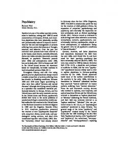
scanning electron microscopy (SEM) and FTIR spectroscopy. From group 2, three specimens were removed at 1-, 3-, and 6-week intervals, sectioned further, and examined using SEM to study the surface topography of the dentine surface and biofilm (Fig. 1). Six specimens from this group were removed after 6 weeks and examined using light microscopy (LM) and laser confocal scanning microscopy (LCSM) (Fig. 1). This was carried out to study the structure of the mature biofilm. The LM provides information on the structure of the biofilm, while the LCSM coupled with viable staining provides information on the distribution of the viable and dead cells in the biofilm. Control specimens under group 1 and group 2 were subjected to all treatments except bacterial inoculation.
Group-1: chemical composition of the biofilm structure FTIR spectroscopy FTIR spectrometer (Magna-IR 560, Nicolet, Madison, WI) with an attenuated total reflectance (ATR) accessory (Golden Gate Single Reflection Diamond ATR, GA) was used to study the biofilm. This method provided a qualitative analysis of chemical groups within 2 m of the surfaces of monolithic specimens. An incident infrared beam lost energy at characteristic wavelengths due to interaction with the biofilm. The absorbed wavelengths were taken to correspond to the harmonic frequencies of molecular structures present on the biofilm surfaces. The transmission range of the ATR accessory employed was 4000 to 500 cm⫺1. The spectra were obtained at 4 cm⫺1 resolution by averaging 20 scans per specimen group.
X-ray diffraction-Rietveld procedure Grazing angle XRD was carried out using Shimadzu 6000 Lab X diffractometer. Cu K␣ radiation at 40 kV/30 mA, a beam divergence slit of 1°, receiving slit width of 0.1 mm, scan rate of 1°/min, and step size of 0.02 were utilized for the experiments. The 2 range considered was from 24° to 50°, as all the major signature peaks of the hydroxyapatite structure [from (002) to (310)] fell within this range. Since a thin film on the dentine substrate was being studied, a grazing angle of 5° was used to obtain information from the biofilm surface. The raw data from the XRD scans were then input into the “Rietquan” quantitative analysis software 2.3, for Rietveld refinement purposes.18,19 The HA crystal model was built using the information from the International Crystal Structure Database. The details of the crystallographic model are listed in Table I.20 Peak shapes were modeled using the pseudo-Voigt function and two asymmetry parameters were refined. In each case, four background parameters, a scale factor, five peak shape parameters, 2 offset (zero point correction), sample displacement, cell parameters and atomic positions were refined. The occupancy of the oxygen and hydrogen atoms associated with the hydroxyl (OH⫺) group was refined as a group (i.e.
408
KISHEN, GEORGE, AND KUMAR
Figure 1. Schematic diagram showing the preparation of tooth specimens in (A) group 1 and (B) group 2.
OH⫺ occupancy) using the same reasoning as Knowles et al.21 The program was also tested for its accuracy using a three-phase mixture of known composition of aluminum (20%), ␣-alumina (50%), and monoclinic-zirconia (30%). Results of the analyses showed 21.4% aluminum, 49.4% Al2O3, and 29.2% ZrO2, confirming the competence of the software.
Group 2: topography and ultrastructure of E. faecalis biofilm on dentine LM, SEM, and LCSM analysis Tooth specimens for the microscopic examination were prepared by cross-sectioning (200 m thick) the root portion,
TABLE I The HA Crystallographic Model Information Used in this Study Phase
Crystal System
Space Group
Lattice Parameters
Atomic Coordinates
HA
Hexagonal
P63/m
a ⫽ 9.422 c ⫽ 6.88
Atom Ca1: x ⫽ 0.333, y ⫽ 0.667, z ⫽ 0.001; Atom Ca2: x ⫽ 0.246, y ⫽ 0.993, z ⫽ 0.25; Atom P: x ⫽ 0.4, y ⫽ 0.369, z ⫽ 0.25; Atom O1: x ⫽ 0.329, y ⫽ 0.484, z ⫽ 0.25; Atom O2: x ⫽ 0.589, y ⫽ 0.466, z ⫽ 0.25; Atom O3: x ⫽ 0.348, y ⫽ 0.259, z ⫽ 0.073; Ion OH: x ⫽ 0, y ⫽0, z ⫽ 0.25
ENTEROCOCCUS FAECALIS-MEDIATED BIOMINERALIZED BIOFILM FORMATION
409
Figure 2. The FTIR spectra of the E. faecalis biofilms formed on root canal dentine at different time intervals. 3 mm apical to the CEJ using microtome (Microslice-2, England) (Fig. 1). The cross-sections prepared were subjected to a modified Gram staining procedure and were examined under an oil immersion microscope for LM. Since the microbial cells in a biofilm were attached to the dentine surface, the heat fixation step was excluded from the original Gram staining protocol. The specimens for SEM were washed with PBS, and immersed in a fixative solution containing 4% Gluteraldehyde in sodium cacodylate buffer at 4°C for 3 h. The specimens were later subjected to increasing concentrations of ethanol for serial dehydration. The dehydrated specimens were critical point dried in Critical-Point-Drier (BALTEC CPD030, Liechtenstein), sputter coated with gold (BALTEC-SCD005, Liechtenstein), and were examined using SEM (JEOL JSM5500LV, Japan) to evaluate the surface topography of the biofilm structure formed on root canal wall. The specimens for the LCSM were first washed with PBS. The root canal surfaces were then stained with LIVE/DEAD BacLight bacterial viability stain (Invitrogen, Molecular probes), and were observed with a laser scanning confocal microscope (Olympus FV500, Japan) fitted with 60⫻ and 100⫻ oil immersion lenses. The LCSM analysis aided in understanding the three-dimensional ultrastructure of the biofilm and to learn the distribution of the viable and dead cells within the biofilm structure.
Sediments from E. faecalis– dentine interaction FTIR spectroscopy and SEM analysis The sediment collected at 4- and 8-week intervals were centrifuged, washed, and allowed to settle. The sediment
portion were removed and divided into two portions. One portion was mixed with KBr and pressed into pellets for FTIR analysis. The remaining powder was dehydrated at room conditions, sputter coated with gold, and was examined using SEM.
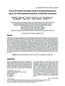
RESULTS Group-1: chemical composition of the biofilm structure ATR-FTIR analysis Figure 2 shows a comparison of the FTIR reflectance spectra for E. faecalis biofilm on dentine. The dentine surface in the medium without bacterial cells (control) did not show any characteristic peaks corresponding to phosphate or carbonate bands. This is expected in the ATR mode, as the reflected signal is coming from the top most layers and the apatite in dentine is shielded or incoherently scattered by the liquid-conditioning layer. However, there was a systematic increase in peak intensity and definition at around 1530, 1450, 1400, and 980 cm⫺1 with incubation period (2, 3, and 6 weeks). This increase corresponded to the increase in carbonate (both A and B type substitution in apatite) and phosphate groups in apatite on the biofilm surface. The higher definition of the peaks indicated that the apatite structure was improving and gaining higher regularity with time. The details of
410
Figure 3. The XRD spectra of the E. faecalis biofilms formed on the root canal dentine at different time intervals.
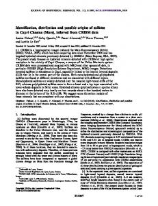
carbonate substitution in the apatite lattice have been provided by many others.22–25 It has been shown that the two different site substitutions (1) for OH⫺ (Atype substitution) and (2) CO32⫺PO43⫺ (B-type substitution) corresponded to infrared bands around 1450 and 1535 cm⫺1, and 1430 and 1405 cm⫺1, respectively.26,27 A prominent hump was also noticed in our spectrum below 877 cm⫺1, which extended beyond 600 cm⫺1. This increase is attributed to, POO (865 cm⫺1) and PO4 (620 cm⫺1, 600 cm⫺1). X-ray diffraction-Rietveld procedure The XRD spectra of the E. faecalis biofilm grown on root canal dentine displayed changes evident with time. However, the control specimens did not show any change even with 6-week incubation. Figure 3 shows the XRD spectra of E. faecalis biofilm formed on the dentine at different time interval. The characteristic peaks for hydroxyl-carbonated apatite as seen by the (002), (211), (112), and (300) peaks were present. Also there was a broad hump around 2 ⫽ 22° due to the scattering from the collagen in dentine.20 With increasing incubation time, the hump due to the matrix diminished, as does the peak intensity of apatite (evident at 3 weeks). After 6 weeks, the broad hump (at 22°) due to the collageneous matrix is hardly seen, while the apatite peaks again became more intense. Such a trend under grazing angle conditions implicates the precipitation and growth of a fresh apatite layer on the dentine surface. Another feature of the spectra was the peak shift to lower 2 angles, indicating subtle but definite differences in the structure of the reprecipitated apatite as compared to the one present initially as part of the dentine substrate. When the XRD spectra were digitized and refined
KISHEN, GEORGE, AND KUMAR
by the Rietveld method, it was found that the a-axis of the apatite unit-cell of the control sample (without bacteria) was smaller than the synthetic hydroxyapatite. This apatite was also found to be similar to carbonated-flor-apatite present in dentine (with B-type carbonate substitution) and corresponded to the JCPDS card number 31– 0267. Apatite with this composition has been observed in teeth and bone by McClellan28 and documented in detail by Elliot.25,29 The structure of the reprecipitated apatite was closer to synthetic HA with possible minor A- and B-type carbonate substitution (JCPDS card number 09 – 432) and complimented by FTIR spectroscopy results. With increasing incubation time in the culture medium (especially ⬃6 weeks), the results of the Rietveld analysis clearly indicated that there were crystallographic differences between the original apatite in dentine (dahllite—where the B-type substitution was prominent) and the latter (hydroxyapatite—where the B-type substitution was not), which was formed on the biofilm, confirming the formation of the new layer. The details of the changes to a- and c-axes of the apatite structure obtained from Rietveld refinement are given in Table II.
Group 2: topography and ultrastructure of E. faecalis biofilm on dentine LM, SEM, and LCSM analysis The SEM examinations showed that after 1-week incubation, bacterial cells aggregated on the dentine surface and characteristic interconnecting network of polymer strands were also observed on the biofilm [Fig. 4(A)]. Manifestations of dentine surface dissolution were also observed [Fig. 4(B)]. After 2 weeks, bacterial aggregates were found to coalesce together to form bigger aggregate over the surface of the root canal dentine [Fig. 4(C,D)]. The dentine surface below the biofilm at this stage displayed typical corroded appearance [Figs. 4(B,C)]. After 3 weeks, there were thick bacterial growth on the surface of dentine [Fig. 4(E)] and beyond 4-week incubation, elevated clumps TABLE II The a-Axis and the c-Axis Parameters of the Apatite Structure Formed by the E. faecalis-Denfine Interaction from the Rietveld Analysis Immersion Time (weeks)
a-Axis (Å)
c-Axis (Å)
0 2 3 6
9.353 9.349 9.409 9.449
6.821 6.824 6.864 6.893
ENTEROCOCCUS FAECALIS-MEDIATED BIOMINERALIZED BIOFILM FORMATION
411
Figure 4. SEM showing the stages in the development of E. faecalis biofilm on root canal dentine. (A and B) The biofilm and the dentine surface topography after 1 week, (C and D) the biofilm and the dentine surface topography after 2 weeks, (E) the biofilm surface topography after 3 weeks, and (F) after 4-week incubation. Arrows indicates region of dentine dissolution (B, D).
of bacterial cells that covered the entire dentine surface was observed [Fig. 4(F)]. The matured biofilm after 6-week incubation showed a highly organized honey-comb like structures [Fig. 5(A,D)]. It was found from the light microscopic examination that E. faecalis formed distinct raised and uneven biofilm structures on the surface of the root canal dentine. The LM examination of the mature biofilm structure also showed signs of calcification and crystal growth that extended from the dentine substrate to the surface within the biofilm matrix [Fig. 5(B)]. Figure 5(C) shows the typical image of the internal architecture of the mature biofilm obtained from the tooth
cross-section. The LSCM examination after staining with LIVE/DEAD BacLight bacterial viability stain showed that the majority of the cells present in the mature biofilm were dead (red color cells). However, pockets of live bacterial cells (green color cells) were found inside the biofilm structure. The interface of the root canal wall and the biofilm structure was found to contain maximum number of dead bacterial cells [Fig. 5(C)]. Interestingly, the matrix of biofilm structure was found to stain with both Syto 9 and Propidium iodide included from the viability kit, and this is probably because of the incorporated nucleic acid in the biofilm matrix.
412
KISHEN, GEORGE, AND KUMAR
Figure 5. The ultrastructure of (A) a typical mature biofilm of E. faecalis on root canal dentine after 6 weeks under (B) LM, (C) LCSM, and (D) SEM. [Color figure can be viewed in the online issue, which is available at www.interscience.wiley.com.]
Sediments from E. faecalis– dentine interaction: FTIR and SEM FTIR spectroscopy and SEM analysis Figure 6(A) shows a comparison of the FTIR spectra obtained from the sediments collected at 4- and 8-week intervals. It was observed from this analysis that there was a obvious increase in the transmittance intensity at around 1450, 1400, 985, 877, and 600 cm⫺1 with incubation period. This increase in the above peaks signifies an increase in the carbonate and phosphate concentration in the sediment. Figure 6(B) shows the morphology of the sediment obtained after 6-week incubation. The sediments were made up mainly of rhombohedral type crystals and this in conjunction with the FTIR finding could indicate the presence of calcitic calcium carbonate.
DISCUSSION The microscopic examinations revealed distinct stages in the E. faecalis–dentine interaction. LM of the mature biofilm structure (after 6-week incubation) showed presence of crystal growth and calcification adjacent to the dentine– biofilm interface. The XRD analysis displayed a clear peak shift to lower 2 angles in the apatite structure formed in the 6 weeks old biofilm. This indicated subtle compositional difference in the structure of the reprecipitated apatite as compared to the one originally present in the dentine. In addition, when the d-spacing from XRD data was compared to the JCPDS card, it was observed that the uninfected dentine specimens (control) had predominantly a carbonated-flor-apatite structure, while the apatite formed after 6-week incubation with E. faecalis was carbonated-apatite. This was further confirmed through Rietveld analysis, which revealed clear differences in the structure of original mineral present in the
ENTEROCOCCUS FAECALIS-MEDIATED BIOMINERALIZED BIOFILM FORMATION
413
Figure 6. (A) FTIR spectra and (B) SEM image of the sediment formed during the E. faecalis– dentine interaction.
dentine and the biofilm after 6 weeks. This means that there was a clear distinction between the original apatite found in the dentine and the apatite reprecipitated on the biofilm by the bacteria-mediated process. The FTIR analysis of the E. faecalis biofilm on dentine substrate showed an increase in the carbonate and phosphate band intensity and definition with incubation period. Interestingly, sediments were formed during the E. faecalis–dentine interaction, and these sediments showed an increase in the carbonate and phosphate bands from 4- to 8-weeks. This increase in the intensity of carbonate and phosphate bands in the sediment can be due to the bacteria-induced dissolution of the dentine surface, which would raise the supersaturation with time, causing the precipitation of calcium carbonate and calcium phosphate sediments. The SEM examination of the dentine specimens incubated with bacteria revealed a corroded dentine surface, a feature that cumulatively increased with time. As such we believe that as the bacteria mediated dissolution of the dentine occurs, the supersaturation with respect to calcium phosphates and carbonates in
the surrounding milieu may exceed a critical value, after which the deposition of calcium phosphate and carbonates would commence. The sediments showed an increase in the calcium carbonate and calcium phosphate levels from 4- to 8-weeks, and this period corresponded with the duration taken to reprecipitate apatite on the biofilm (6 weeks). It was interesting to note that the pH of the medium (7.1) increased to 8.2 in the first 24 h when the root canal dentine was incubated with bacteria, while the pH dropped to 4.1 in the first 24 h when the bacteria was incubated as planktonic cells in the medium (without dentine). The LCSM examination displayed dead cells in the biofilm structure close to the dentine substrate. The phospholipid membranes of the dead cells adjacent to the dentine substrate could then serve as nucleating sites for the reprecipitation of apatite and calcification of the biofilm.30 Internal resorption of the root canal dentine was reported in 74.7% of teeth associated with periapical lesions.31 This was found mostly in the apical portion of the root canal dentine, and is due to the limitations
414
KISHEN, GEORGE, AND KUMAR
associated with the geometry of the root canal, and the length and lateral limits of the cleaning and shaping procedures in root canal therapy. All the above would lead to the incomplete elimination of bacteria from the apical region of the root canal.31 Moreover, our earlier experiments have demonstrated a biting force-induced retrograde fluid movement into the apical portion of the root canal. This cyclic influx of tissue fluid into the apical portion of the root canal may promote persistence of bacteria as biofilm in this region.32 The interaction of E. faecalis with root canal dentine observed in this study may be an important issue to ponder in endodontics, especially due to increasing application of calcium hydroxide as intervisit medicament during RCT. The ability of E. faecalis to resist the bacteriocidal effect of calcium hydroxide,33 along with its capacity to form distinct calcified biofilm in a calcium carbonate and calcium phosphate rich microenvironment may be some of the contributing factors that allow the persistence of E. faecalis in endodontically treated teeth. Adding interest to these findings, two clinical cases were reported recently, which described mineralized calculus-like deposit on the root surface of teeth with posttreatment apical periodontitis.34 Nevertheless, further investigations are warranted to answer questions such as the folowing: what are the other bacteria that have similar interaction with dentine? Can calcified biofilm within the root canal trigger immunological response in the host-tissue? In conclusion, this study highlighted different sequence in the interaction of E. faecalis with dentine substrate. In stage-1, E. faecalis formed biofilm on the root canal dentine, and in stage-2, the bacteria induced dissolution of the mineral fraction from the dentine substrate. In stage-3, a reprecipitated apatite layer was formed in the biofilm structure.
References 1.
2.
3.
4.
5.
6.
Sedgley CM, Lennan SL, Clewell DB. Prevalence, phenotype and genotype of oral Enterococci. Oral Microbiol Immunol 2004;19:95–101. Mejare B. Streptococcus faecalis and Streptococcus faecium in infected dental root canals at filling and their susceptibility to azidocillin and some comparable antibiotics. Odontol Revy 1975;26:193–204. Molander A, Reit C, Dahlen G, Kvist T. Microbiological status of root-filled teeth with apical periodontitis. Int Endod J 1998; 31:1–7. Sundqvist G, Figdor D, Persson S, Sjogren U. Microbiologic analysis of teeth with failed endodontic treatment and the outcome of conservative re-treatment. Oral Surg Oral Med Oral Pathol Oral Radiol Endod 1998;85:86 –93. Hancock HH, Sigurdsson A, Trope M, Moiseiwitsch J. Bacteria isolated after unsuccessful endodontic treatment in a North American population. Oral Surg Oral Med Oral Pathol Oral Radiol Endod 2001;91:579 –586. Clewell DB. Plasmids, drug resistance, and gene transfer in the genus Streptococcus. Microbiol Rev 1981;45:409 – 436.
7. 8.
9.
10.
11.
12.
13.
14.
15.
16.
17. 18. 19.
20.
21.
22.
23.
24.
25. 26. 27. 28. 29.
Love RM. Enterococcus faecalis—a mechanism for its role in endodontic failure. Int Endod J 2001;34:399 – 405. Portenier I, Tuomos MT, Waltimo M, Haapasalo MP. Enterococcus faecalis—the root canal survivor and ‘star’ in post-treatment disease. Endod Top 2003;6:135–159. Costerton JW, Lewandowski Z, DeBeer D, Caldwell D, Korber D, James G. Biofilms, the customized microniche. J Bacteriol 1994;176:2137–2142. Karthikeyan S, Korber DR, Wolfaardt GM, Caldwell DE. Adaptation of bacterial communities to environmental transitions from labile to refractory substrates. Int Microbiol 2001;4:73– 80. Gordon CA, Hodges NA, Marriott C. Antibiotic interaction and diffusion through alginate and exopolysaccharide of cystic fibrosis-derived Pseudomonas aeruginosa. J Antimicrob Chemother 1988;22:667– 674. Tesch W, Eidelman N, Roschger P, Goldenberg F, Klaushofer K, Fratzl P. Graded microstructure and mechanical properties of human crown dentin. Calcif Tissue Int 2001;69:147–157. Stepanovic S, Cirkovic I, Ranin L, Svabic-Vlahovic M. Biofilm formation by Salmonella spp. and Listeria monocytogenes on plastic surface. Lett Appl Microbiol 2004;38:428 – 432. Noiri Y, Ehara A, Kawahara T, Takemura N, Ebisu NS. Participation of bacterial biofilms in refractory and chronic periapical periodontitis. J Endod 2002;28:679 – 683. Leonardo MR, Rossi MA, Silva LA, Ito IY, Bonifacio KC. EM evaluation of bacterial biofilm and microorganisms on the apical external root surface of human teeth. J Endod 2002;28:815– 818. George S, Kishen A, Song KP. The role of environmental changes on monospecies biofilm formation on root canal wall by Enterococcus faecalis. J Endod 2005;31:867– 872. White M, Goodis HE, Marshall SJ, Marshall GW. Sterilization of teeth by gamma radiation. J Dent Res 1994;73:1560 –1567. McCusker LB, Von Dreele RB, Cox DE, Louer D, Scardi R. Rietveld refinement guidelines. J Appl Crystallogr 1999;32:36 –50. Kumar R, Cheang P, Khor KA. Phase composition and heat of crystallisation of amorphous calcium phosphate in ultra-fine radio frequency suspension plasma sprayed hydroxyapatite powders. Acta Mat 2004;52:1171–1181. Ripamonti A, Roveri N, Braga D, Hulmes DIS, Miller A, Timmins PA. Effects of pH and ionic-strength on the structure of collagen fibrils. Biopolymers 1980;19:965–975. Knowles JC, Gross K, Berndt CC, Bonfield W. Structural changes induced during thermal spraying of hydroxyapatite. A comparison of three different spraying methods. Bioceramics 1995;8:311– 316. Eds. L.L.Hench and J.Wilson. Elsevier, New York. Rehman I, Bonfield W. Characterisation of hydroxyapatite and carbonated apatite by photo acoustic FTIR spectroscopy. J Mater Sci Mater Med 1997;8:1– 4. Penel G, Leroy G, Rey C, Sombert B, Huvenne JP, Bres E. Infrared and Raman microspectroscopy study of flour-flour hydroxy and hydroxy-apatite. J Mater Sci Mater Med 1997;8:271–276. Barralet J, Best S, Bonfield W. Carbonate substitution in precipitated hydroxyapatite: An investigation into the effects of reaction temperature and bicarbonate ion concentration. J Biomed Mater Res 1998;41:79 – 86. Elliot J.C. Structure and Chemistry of the Apatites and Other Calcium Orthophosphates. Amsterdam: Elsevier; 1994. Borneman-Starinkevitch ID. On some isomorphic substitutions in apatite. Dokl Acad Sci SSSR 1939;19:53–55. Elliot JC. The Crystallographic Structure of Dental Enamel and Related Apatites, Ph.D. Thesis, University of London, London, 1964. McCleallan GH, Lehr JR. Crystal chemical investigation of natural apatites. Am Mineral 1969;54:1374 –1391. Elliot JC. Some observations on the crystal chemistry of carbonate-containing apatites. Arch Oral Biol 1963;8:277–282. Special supplement. Proceedings of the 9th ORCA Conference, Paris. (Hardwick JL, Dustin J-P, Held HR, editors).
ENTEROCOCCUS FAECALIS-MEDIATED BIOMINERALIZED BIOFILM FORMATION
30.
31. 32.
Rosanova IB, Mischenko BP, Zaitsev VV, Vasin SL, Sevastianov VI. The effect of cells on biomaterial calcification: Experiments with in vivo diffusion chambers. J Biomed Mater Res 1991;25:277–280. Vier FV, Figueiredo JA. Internal apical resorption and its correlation with the type of apical lesion. Int Endod J 2004;37:730 –737. Kishen A. Periapical biomechanics and the role of cyclic biting force in apical retrograde fluid movement. Int Endod J 2005; 38:597– 603.
33.
34.
415
Tatsuta CT, Morgan LA, Baumgartner JC, Adey JD. Effect of calcium hydroxide and four irrigation regimens on instrumented and uninstrumented canal wall topography. J Endod 1999;25:93–98. Ricucci D, Martorano M, Bate AL, Pascon EA. Calculus-like deposit on the apical external root surface of teeth with posttreatment apical periodontitis: Report of two cases. Int Endod J 2005;38:262–271.