Carcinogenesis vol.23 no.3 pp.529–536, 2002
Enterococcus faecalis produces extracellular superoxide and hydrogen peroxide that damages colonic epithelial cell DNA
Mark M.Huycke1, Victoria Abrams and Danny R.Moore The Muchmore Laboratories for Infectious Diseases Research, Research Service (151), 921 N.E. 13th Street, Department of Veterans Affairs Medical Center and Department of Medicine, University of Oklahoma Health Sciences Center, Oklahoma City, OK 73104, USA 1To
whom correspondence should be addressed Email:
[email protected]
Enterococcus faecalis is a commensal microorganism of the human intestinal tract that produces substantial extracellular superoxide (O2–), and derivative reactive oxygen species such as H2O2 and hydroxyl radical, through autoxidation of membrane-associated demethylmenaquinone. Because these oxidants may be important as a cause of chromosomal instability (CIN) associated with sporadic adenomatous polyps and colorectal cancer, the ability of E.faecalis to damage eukaryotic cell DNA was examined using the alkaline lysis single cell gel electrophoresis (comet) assay. Both Chinese hamster ovary and HT-29 intestinal epithelial cells showed increased DNA damage after coincubation with wild-type E. faecalis strain OG1RF, but not a transposon-inactivated mutant with attenuated extracellular O2– production. E. faecalis-mediated DNA damage was prevented by catalase, but not manganese superoxide dismutase, indicating H2O2 arising from O2– was the genotoxin. In a rat model of intestinal colonization, OG1RF resulted in significantly higher stool concentrations of H2O2 and 5,5-dimethyl-1-pyrroline N-oxide adducts of hydroxyl and thiyl radicals, as identified by electron spin resonance– spin trapping, compared with rats colonized with a mutant strain having attenuated O2– production. Using the comet assay, luminal cells from the colon of rats colonized with O2–-producing E. faecalis showed significantly increased DNA damage compared with control rats colonized with the mutant. These findings suggest a potentially profound role for extracellular free radical production by E. faecalis in promoting CIN associated with sporadic adenomatous polyps and colorectal cancer. Introduction Genomic instability is a characteristic feature of colorectal cancer that potentially contributes to the multi-step acquisition of mutations found in these tumors (1,2). Chromosomal instability (CIN) is the most common form of somatic genomic instability and is typified by genetic rearrangements and losses or gains of large DNA fragments including, on occasion, entire chromosomes (3,4). The usual features of CIN include aneuploidy and loss of heterozygosity. This form of instability is found in ⬎80% of sporadic colon cancers and nearly Abbreviations: CIN, chromosomal instability; CHO, Chinese hamster ovary; DEPMPO, 5-(diethoxyphosphoryl)-5-methyl-1-pyrroline-N-oxide; DMPO, 5,5-dimethyl-1-pyrroline-N-oxide; ESR, electron spin resonance; MnSOD, manganese superoxide dismutase. © Oxford University Press
all intestinal tumors due to familial adenomatous polyposis. Microsatellite instability (MIN) is another distinct form of genomic instability caused by defective DNA mismatch repair (3–5). MIN tumor cells show accelerated accumulation of mutations in repetitive nucleotide sequences located throughout the genome. Although ⬍20% of sporadic colon cancers are classified as MIN, this form of genomic instability is highly associated with hereditary non-polyposis colorectal cancer (3–5). Genomic instability affects not only colorectal cancer cells but also benign hyperplastic and pre-cancerous adenomatous polyp cells (1,6). The number of genomic alterations in polyps has been estimated by an inter (simple sequence repeat) PCR technique at ⬎11 000 mutations/cell (1). A similar high frequency of chromosomal gains and losses has been reported for colorectal tumors using arbitrarily primed PCR methodology (7,8). Because CIN has been found in cells from very small adenomas, and hence at the earliest stage of tumorigenesis, genomic instability seems more likely a cause, rather than consequence, of cancer formation (1,6). Aneuploidy, cellcycle checkpoint loss, break-induced replication, defects in double-strand repair-recombination machinery and mitotic nondisjunction have all been considered potential mechanisms for CIN (9,10), but none have proven entirely satisfactory. Therefore, the nature of this fundamental process in colorectal tumorigenesis remains obscure. One less well-investigated mechanism for CIN proposes oxidative stress on the colonic epithelium from free radicals produced by intraluminal bacteria (11,12). This theory suggests that biologically derived radicals from commensal flora are directly mutagenic and may also indirectly promote genomic instability by forming carcinogens from dietary procarcinogens (13,14). This hypothesis was proposed by Babbs based on ex vivo observations of hydroxyl radical production by normal stool (12). Erhardt et al. extended these findings by describing a 13-fold increase in hydroxyl radical production by stool from human volunteers fed a high fat/meat and low fiber diet (15). An oxidative mechanism for CIN also potentially links the substantial epidemiological evidence for dietary risk factors with colorectal cancer (16,17). Several years ago our laboratory noted that Enterococcus faecalis, a human intestinal commensal, produced extracellular superoxide (O2–) (18). We subsequently demonstrated that the formation of this anionic free radical depended on the presence of membrane-associated demethylmenaquinone and was inhibited by exogenous fumarate or hematin (19). Similar to observations made by Babbs, colonic contents from rats colonized with O2–-producing E. faecalis produced hydroxyl radical as measured by electron spin resonance (ESR)–spin trapping (19). These findings led to consideration of extracellular O2– produced by E. faecalis near the oxygenated luminal surface of colonocytes as a potential source of genomic instability. Oxidative stress would presumably occur through secondary reactive oxygen species such as H2O2 and hydroxyl 529
M.M.Huycke, V.Abrams and D.R.Moore
radical (20). In the mildly acidic environment of the large intestine O2– would spontaneously disproportionate to H2O2: E. faecalis → O2– 2O2– ⫹ 2H⫹ → H2O2 ⫹ O2
(1) (2)
H2O2 generated near the intestinal epithelial surface could passively diffuse into colonocytes and form hydroxyl radical at DNA sites through iron-mediated Fenton reactions. This would lead to DNA–protein crosslinks, DNA breaks and base modifications that could cause nucleotide transitions and transversions (21,22). Thus, chronic oxidative stress from intestinal enterococci might be an important source of ongoing DNA damage, leading to genomic instability and cumulative mutations found in adenomatous polyps and colorectal cancer (23). Here we show that extracellular O2– generated by E. faecalis does indeed form H2O2 and hydroxyl radical, and that these bacterially derived oxidants can damage colonic epithelial cell DNA both in vitro and in vivo. Our findings suggest a potentially profound role for this common intestinal commensal in generating CIN. Materials and methods Bacteria and media E. faecalis strain OG1RF is plasmid-free and has been shown to produce extracellular O2– using whole bacteria in a ferricytochrome c assay (18). TM1 is a Tn917 derivative of OG1RF with inactivation of aroC, a gene encoding chorismate synthase, by transposon insertion (19). PW18, another OG1RF derivative, was prepared by allelic inactivation of menB using the plasmid p3erm. This gene encodes 1,4-dihydroxy-2-naphthoic acid synthase and is essential for biosynthesis of membrane-associated demethylmenaquinone (19). Both mutants are attenuated in production of extracellular O2– through loss of membrane-associated demethylmenaquinones. These mutants grow well in brain–heart infusion (BD Biosciences, Franklin Lakes, NJ) with doubling times no different than for OG1RF. Unless specified otherwise, bacteria were grown in closed tubes in brain–heart infusion at 37°C overnight. Glutathione peroxidase assay for H2O2 A glutathione peroxidase assay was used to measure H2O2 produced by live E. faecalis and in intestinal contents (24). Bacteria were washed and resuspended to 109 c.f.u./ml in 25 mM Tris (pH 7.4) with 7 mM glucose or McCoy’s medium with 10% fetal calf serum. After timed incubations 1 vol of 0.6 N perchloric acid was added to 2 vol of bacteria and samples assayed immediately. Intestinal contents were similarly acidified and stored at –20°C for testing later. Supernatants from bacteria or stool were neutralized with 0.8 M KHCO3 and after 15 min 1 vol of a reaction buffer (4 mM EDTA, 3 mM sodium azide, 0.6 mM NADPH, 5 mM glutathione and 3 U/ml glutathione reductase) was added to 2 vol of clear supernatant. The mixture was incubated at 37°C and the change in absorbance at 340 nm followed before and after addition of glutathione peroxidase (final concentration 0.28 U/ml). H2O2 concentrations were calculated from differences in absorbance using an extinction coefficient for NADPH of 6.27 mM/cm. Electron spin resonance–spin trapping Cultures of bacteria were washed with phosphate-buffered saline (PBS) and resuspended to a density of 109 c.f.u./ml in 25 mM Tris buffer (pH 7.4) with 7 mM glucose. The spin trap 5-(diethoxyphosphoryl)-5-methyl-1-pyrrolineN-oxide (DEPMPO) (Oxis, Portland, OR) was added to a final concentration of 20 mM (19,25). Intestinal contents were suspended in 200 µl of 25 mM Tris (pH 7.4), clarified by centrifugation and the spin trap 5,5-dimethyl1-pyrroline-N-oxide (DMPO) (Aldrich, Milwaukee, WI) added to a final concentration of 40 mM. Spectra were immediately recorded following the addition of a spin trap at ambient temperature (22°C) in a quartz flat cell using an X-band ER300E spectrometer (Bruker, Rheinstetten, Germany) with the following parameters: 100 kHz field modulation, microwave power 20 mW, modulation amplitude 1.0 Gauss, sweep width 100 Gauss/84 s and time constant 164 ms. DMPO radical adducts were quantified using the stable nitroxide 4-hydroxy TEMPO (Sigma, St Louis, MO) as a spin standard and identical instrument settings. Xanthine, xanthine oxidase, catalase, Tris, EDTA, sodium azide, glucose, manganese superoxide dismutase (MnSOD), NADPH and glutathione peroxidase were purchased from Sigma.
530
Tissue culture co-incubation assays For co-incubation experiments, bacteria at 108 or 109 c.f.u./ml, or other reagents, were resuspended in McCoy’s with fetal calf serum and added to a confluent growth of tissue culture cells. Flasks were incubated at 5% CO2 and 37°C. At the end of timed experiments, tissue culture medium was aspirated and cells harvested by scraping in a small amount of mincing solution [Hank’s balanced salt solution (Ca2⫹ and Mg2⫹ free, Gibco, Rockville, MD), 20 mM EDTA (Sigma), 10% dimethylsulfoxide (Fisher, Pittsburgh, PA)]. Chinese hamster ovary (CHO) cells were provided by Sheila Galloway at Merck Research Laboratories (West Point, PA) (26). HT-29 cells are a colonic epithelial line that was obtained from the American Type Culture Collection (Bethesda, MD, USA) (27). Cells were routinely maintained in McCoy’s medium containing fetal calf serum at 5% CO2 and 37°C. Rat intestinal colonization model Intestinal tracts of male Wistar rats were colonized with spontaneous streptomycin- and spectinomycin-resistant derivatives of OG1RF or PW18 (OG1RF-SS and PW18-SS, respectively) as described previously (19). In brief, rats were individually caged and fed a conventional diet ad libitum. After 2 weeks streptomycin- and spectinomycin-containing water (each at 500 mg/l) was provided to eliminate endogenous enterococcal flora and promote exogenous colonization with OG1RF-SS or PW18-SS. Three days later, rats were orogastrically administered 109 c.f.u. of E. faecalis in 0.2 ml of PBS. Five days post-inoculation, rats were anesthetized using inhaled isoflurane and ileal and colonic segments surgically excised. Samples of luminal contents were obtained and segments flushed with 60–100 ml of mincing solution. Segments were longitudinally opened and epithelial cells gently scraped from the luminal surface using a sharp scalpel. Cells were diluted in mincing solution for comet assay slide preparation (see below). Hematoxylin and eosin stained preparations from intestinal scrapings showed ⬎90% of cells to be of epithelial origin. Enterococci in stool were enumerated using bile-esculin-azide agar plates (Difco, Detroit, MI). Comet assay The alkaline lysis single cell gel electrophoresis (comet) assay was used to measure single-strand breaks, alkali-labile sites and crosslinking of DNA from tissue culture and intestinal epithelial cells. Comet assays were performed as described previously (28). In brief, ~104 tissue culture cells, or material from 3–4 cm of scraped luminal intestine, in 5–10 µl of mincing solution were mixed in 75 µl of low-melting point agar at 37°C and layered onto microscope slides coated with 1% agarose. Tissue culture cells were assessed by fluorescent microscopy (Olympus BM-40X, Melville, NY) using 5,6-carboxyfluorescein diacetate (Molecular Probes, Portland, OR) and ethidium bromide (Sigma). Greater than 90% cell viability was considered adequate (29). A third layer of agarose was applied to the embedded tissue culture or epithelial cells and slides refrigerated for ⬎60 min in a lysing solution [2.5 M NaCl, 100 mM EDTA, 10 mM Tris (pH 10), 1% Triton X-100, 10% dimethylsulfoxide]. Slides were electrophoresed for 20 min in 10 N NaOH and 200 mM EDTA (pH ⬎13) at 25 V and 300 mAmp. Slides were neutralized with 400 mM Tris (pH 7.4) and fixed using 100% cold ethanol. After drying slides were stained with ethidium bromide and 50–100 randomly selected cells/slide scored by fluorescent microscopy (Figure 1). Slides were prepared and coded by one of us (V.A.) and read by another person (M.M.H.) masked to treatments. Statistical analysis The distribution of comet scores between tests and controls were compared using ridit analysis (30). This method assumes discrete measures represent intervals in an underlying continuous distribution. No other assumptions about the distribution are made using this technique. Ridits range from 0 to 1, and by definition the ridit for all control distributions is set at 0.50. If the mean ridit for a test distribution were ⬎0.50, then more than half the time randomly selected measures from this distribution would have a value greater than randomly selected measures from a control distribution. For the comet assay a mean ridit ⬎0.50 would indicate greater DNA damage compared with a control, whereas a value ⬍0.50 would indicate lesser damage. Group comparisons were performed using JMP (Version 3.1, SAS Institute, Cary, NC). P values 艋 0.05 were considered significant.
Results E. faecalis produces superoxide and H2O2 As reported previously, E. faecalis strain OG1RF produced (mean ⫾ SD) 17.2 ⫾ 0.3 nmol of extracellular O2–/min/109 c.f.u. at 37°C (19). Under similar conditions the strain also produced 23 nmol H2O2/min/109 c.f.u. For the mutant strains
Enterococcus faecalis damages epithelial cell DNA
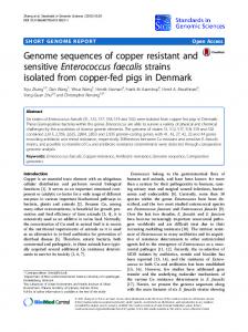
Fig. 1. The alkaline lysis single cell gel electrophoresis (comet) assay for measuring single-strand breaks, alkali-labile sites and crosslinking of DNA in tissue culture cells co-incubated with E. faecalis. Randomly selected cells were scored by fluorescent microscopy using a five point scale: 0, no visible DNA migration from the nucleus; 1, minimal DNA migration with an intact nucleus; 2, moderate DNA migration with reduction in the size of the nucleus; 3, extensive DNA migration with only a pinpoint nucleus remaining; 4, complete migration of DNA into a comet tail with no visible nucleus remaining.
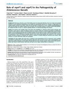
Fig. 2. ESR–spin trapping spectra for OG1RF in McCoy’s with varying percentages of fetal calf serum. Bacteria grown in brain–heart infusion were washed and resuspended in McCoy’s to 109 c.f.u./ml for 30 min. DEPMPO was added to 20 mM. Arrow points up field and length indicates 10 Gauss.
TM1 (aroC::Tn917) and PW18 (menB::p3erm) extracellular O2– production was ⬍1.0 nmol/min/109 c.f.u. (19), and no H2O2 was detectable, implying that H2O2 was entirely generated by disproportionation of O2–. H2O2 damages eukaryotic cell DNA The effect of fetal calf serum on extracellular O2– production by OG1RF was investigated prior to tissue culture studies. OG1RF was resuspended in McCoy’s with varying concentrations of fetal calf serum, incubated for 30 min and O2– and hydroxyl radical measured using ESR–spin trapping. Decreasing signal intensities for DEPMPO–hydroxyl radical and DEPMPO–superoxide adducts were found as the percentage of fetal calf serum increased (Figure 2). This effect was probably due to contamination of fetal calf serum with small amounts of MnSOD as reported previously (31), or from nonenzymatic scavenging of O2– by other serum constituents. As CHO and HT-29 cells grew well in McCoy’s containing 10% fetal calf serum, and McCoy’s supplemented with 5 or
10% fetal calf serum gave the strongest ESR–spin trapping signals, 10% fetal calf serum was selected for all subsequent experiments. CHO and HT-29 cells were incubated for 30 min with 0, 25, 50, 100 or 200 µM H2O2. All mean ridits for all H2O2treated cells were significantly ⬎0.50 using unexposed cells as the control distribution (Table I). These results confirmed the comet assay and ridit analyses as sensitive measures of H2O2-mediated damage to genomic DNA. In addition, increasing mean ridits for greater concentrations of H2O2 were consistent with a dose response. Ridits at each H2O2 concentration were greater for CHO than HT-29 cells, possibly due to an enhanced resistance of HT-29 cells to H2O2-mediated DNA damage. The addition of catalase to CHO and HT-29 cells led to a modest, but statistically significant, decrease in mean ridits (Table II). Mean ridits, however, were unchanged for cells exposed to both 200 µM H2O2 and catalase. No treatment led to cell losses ⬎10%. Under neutral conditions xanthine oxidase and xanthine produce O2– and H2O2 (32). To test whether enzymatically generated reactive oxygen species damaged HT-29 cell DNA, combinations of xanthine, xanthine oxidase and MnSOD were incubated with these cells for 30 min (Table III). Compared with untreated controls, xanthine modestly increased the mean ridit to 0.57 (P ⫽ 0.01), whereas xanthine oxidase alone had no significant effect (ridit ⫽ 0.48, P ⫽ 0.35). The combination of xanthine and xanthine oxidase, however, increased the mean ridit to 0.82 (P ⬍ 0.001), indicating substantial genomic DNA damage. As MnSOD rapidly converts O2– to O2 and H2O2, MnSOD alone afforded presumably no protection to cells if DNA damage was due to H2O2 (mean ridit ⫽ 0.83, P ⬍ 0.001). E. faecalis damages eukaryotic cell DNA To determine whether extracellular O2– from E. faecalis damaged eukaryotic cell DNA, OG1RF and TM1 at 108 and 109 c.f.u./ml were co-incubated with CHO and HT-29 cells for 30 min (Table IV). Mean ridits were calculated using OG1RF as the test distribution and TM1 as the control. For all experiments significantly increased mean ridits, or increased genomic DNA damage, were observed for cells exposed to OG1RF compared with TM1. A dosing effect was apparent with increased mean ridits at the higher concentration of bacteria. Similar levels of genomic DNA damage were found when the incubation time was extended to 60 min (data not shown). Finally, the mean ridit for TM1 at 109 c.f.u./ml was not significantly different from control cells not exposed to any bacteria. Catalase protects against E. faecalis-induced damage of eukaryotic cell DNA To determine which reactive oxygen species generated by E. faecalis damaged tissue culture cell DNA, co-incubations were performed using CHO cells and OG1RF at 109 c.f.u./ml. Beforehand, MnSOD, catalase or MnSOD and catalase were added to bacteria in McCoy’s medium. After a 30 min coincubation, comet assays were performed and supernatants studied by ESR–spin trapping (Figure 3). For MnSOD-treated cells no decrease occurred in the mean ridit. This indicated that MnSOD conferred no protection against genomic DNA damage. In contrast, catalase alone, or in combination with MnSOD, significantly reduced the mean ridits suggesting substantial protection against DNA damage. ESR–spin trapping of tissue culture supernatants containing MnSOD showed no DEPMPO adduct signals for O2– and hydroxyl radical (Figure 3), indicating hydroxyl radical arose 531
M.M.Huycke, V.Abrams and D.R.Moore
Table I. Effect of H2O2 on CHO and HT-29 cell DNA using the comet assay Comet scores
Frequency of scores (%)a CHO cells, H2O2 (µM)
0 1 2 3 4 Ridit P value aThirty
HT-29 cells, H2O2 (µM)
0
25
50
100
200
0
25
50
100
200
38 38 12 8 4 0.50 –
12 32 20 23 12 0.70 ⬍0.001
50 17 23 39 12 0.77 ⬍0.001
1 10 15 44 30 0.88 ⬍0.001
0 0 6 27 67 0.95 ⬍0.001
54 36 9 1 0 0.50 –
35 45 16 3 1 0.61 0.004
21 53 24 2 0 0.68 ⬍0.001
30 35 20 15 0 0.67 ⬍0.001
9 26 28 20 17 0.84 ⬍0.001
minute incubations; assays performed twice with data combined.
Table II. Effect of catalase and H2O2 on CHO and HT-29 cell DNA using the comet assay Comet scores
Frequency of scores (%)a CHO cells
0 1 2 3 4 Ridit P value aThirty
HT-29 cells
Control
Catalase
H2O2
Catalase and H2O2
Control
Catalase
H2O2
Catalase and H2O2
65 28 6 1 0 0.50 –
76 18 4 2 0 0.44 0.006
11 19 27 30 13 0.74 ⬍0.001
62 31 7 0 0 0.52 0.41
30 41 25 3 1 0.50 –
44 36 12 5 3 0.43 ⬍0.001
11 21 29 25 14 0.75 ⬍0.001
40 38 18 4 0 0.44 0.003
minute incubations; catalase, 100 U/ml; H2O2, 200 µM; assays performed twice with data combined.
Table III. Effect of xanthine and/or xanthine oxidase on HT-29 cell DNA using the comet assay Comet scores
0 1 2 3 4 Ridit P value aThirty
Frequency of scores (%)a Control
Xanthine
Xanthine oxidase
Xanthine and xanthine oxidase
Xanthine, xanthine oxidase and MnSOD
42 34 16 6 2 0.50 –
19 60 18 2 1 0.57 0.01
45 38 8 9 0 0.48 0.35
8 21 11 34 26 0.82 ⬍0.001
5 20 11 39 25 0.83 ⬍0.001
minute incubations; xanthine, 2 mM; xanthine oxidase, 100 U/ml; MnSOD, 100 U/ml.
entirely from O2– by iron-catalyzed Fenton reactions. In the presence of catalase, O2– and hydroxyl radical–DEPMPO adducts were still detectable. Loss of ESR signals occurred only when MnSOD had been added. The lack of effect of catalase on hydroxyl radical production from O2– is consistent with previous reports and represents either an accelerated decay of the O2––DEPMPO adduct to a hydroxyl adduct by the tissue culture cells (33), or incomplete scavenging of H2O2 by catalase at the concentration used in this experiment. In aggregate, these data show that H2O2 arises from extracellular O2– produced by E. faecalis, and that H2O2 damages CHO cell DNA following short-term exposure. 532
E. faecalis damages colonic epithelial cell DNA in vitro A rat intestinal colonization model was used to evaluate the in vivo production of reactive oxygen species by E. faecalis. Colon contents from rats that had been colonized with OG1RFSS or PW18-SS for 5 days had similar mean concentrations of enterococci (Table V). Four-fold greater mean concentrations of H2O2 were found in colon contents for OG1RF-SS compared with PW18-SS colonized rats (P ⫽ 0.05). Ileal contents, however, had lower concentrations of both enterococci and H2O2, and no significant differences were noted in these measures between the two groups of colonized rats. ESR– spin trapping of ileal and colon contents for the OG1RF-SS
Enterococcus faecalis damages epithelial cell DNA
Table IV. Damage to CHO and H-29 cell DNA by E. faecalis using the comet assay Comet scores
Frequency of scores (%)a CHO cells Control
0 1 2 3 4 Ridit P value aThirty
16 41 12 19 11
HT-29 cells 108 c.f.u./ml
109 c.f.u./ml
TM1
OG1RF
TM1
OG1RF
24 43 14 14 5 0.50 –
11 46 16 20 7 0.58 0.004
13 50 17 14 6 0.50 –
1 27 30 21 21 0.71 ⬍0.001
Control
38 44 14 4 0
108 c.f.u./ml
109 c.f.u./ml
TM1
OG1RF
TM1
OG1RF
54 17 5 15 9 0.50 –
30 33 19 13 5 0.59 0.01
41 35 17 4 3 0.50 –
3 33 35 17 12 0.78 ⬍0.001
minute incubations; assays performed twice with data combined.
DNA, scrapings from ileal and colon segments were assessed using the comet assay. The mean ridit for cells from ileal segments were not different for rats colonized with OG1RFSS compared with PW18-SS (Table VI). For colon cells, however, the mean ridit was significantly higher for OG1RFSS compared with PW18-SS colonized rats (ridit ⫽ 0.65, P ⬍ 0.001). The results suggest epithelial cell DNA in the colon was damaged through the production of extracellular O2– by E. faecalis. Discussion
Fig. 3. Effect of catalase (1000 U/ml) and/or MnSOD (100 U/ml) on CHO cell DNA co-incubated with OG1RF at 109 c.f.u./ml for 30 min by the comet assay. (A) No addition (black bars); MnSOD (white bars, ridit ⫽ 0.54, P ⫽ 0.13); catalase (spotted bars, ridit ⫽ 0.24, P ⬍0.001); MnSOD and catalase (striped bars, ridit ⫽ 0.25, P ⬍0.001). Ridits were calculated using cells co-incubated only with OG1RF as the control distribution. (B) ESR–spin trapping of aliquots of supernatants from (A) following the 30 min incubation with DEPMPO at 20 mM: 1, no additions; 2, MnSOD; 3, catalase; 4, MnSOD and catalase. Arrow points up field and length indicates 10 Gauss.
colonized rats demonstrated DMPO adducts for both hydroxyl and thiyl radical (Figure 4). The characteristic hyperfine splitting constants for thiyl radical–DMPO adducts presumably arose from partial oxidation by E. faecalis of thiol compounds in stool (34,35). Colonic concentrations of hydroxyl and thiyl–DMPO adducts were 3-fold greater for rats colonized with OG1RF-SS compared with PW18-SS (P ⫽ 0.04). To determine whether the increased production of reactive oxygen species by OG1RF-SS in vivo damaged epithelial cell
No single model has yet been accepted that can account for CIN found in sporadic adenomatous polyps and colorectal cancer (1–4,16,17,23). One novel hypothesis, however, proposes reactive oxygen species from colonic bacteria as direct initiators of genetic mutability (12). A corollary to this theory is potential modulation of bacterial free radical production and alteration of cellular antioxidant defenses by diet, linking genomic instability with existing epidemiological evidence on nutritional factors known to affect the prevalence of colorectal cancer (11,12,15,36). Production of reactive oxygen species by colonic bacteria necessarily assumes the presence of O2 in the colon. This would be expected at the epithelial–luminal interface where O2 passively diffuses from cells into luminal contents. Leaching of O2 from the epithelium is detectable in flatus where a mean pO2 of 30 torr (range 0–119 torr) has been measured for healthy persons (37). Any O2– produced by colonic bacteria would disproportionate to H2O2 in the mildly acidic conditions of the colon, and be available to diffuse back into colonic epithelial cells to cause genomic DNA damage (38). The discovery of extracellular O2– production by E. faecalis, a normal intestinal commensal, suggests a bacterial source for colonic reactive oxygen species. Enterococci have robust reducing properties (39,40) and were initially reported to produce extracellular O2– in 1981 (41). We subsequently found this phenotype typical for E. faecalis, but not other enterococci or facultative bacteria that colonize the colon (18,42). Genetic studies of E. faecalis strain OG1RF show demethylmenaquinone is essential for extracellular O2– production, and that O2– is only produced upon the conditional loss of terminal quinol oxidases that redox cycle this membrane-associated charge carrier (19). 533
M.M.Huycke, V.Abrams and D.R.Moore
Table V. Concentrations of enterococci, H2O2 and DMPO adducts of hydroxyl and thiyl radicals in rat intestinal contentsa Concentration
Ileum
Enterococci (log10 c.f.u./g stool ⫾ SD) H2O2 (µM/g stool ⫾ SD) DMPO-radical adducts (µM/g stool ⫾ SD)
Colon
PW18-SS
OG1RF-SS
P value
PW18-SS
OG1RF-SS
P value
5.3 ⫾ 0.8 0.8 ⫾ 1.2 0.8 ⫾ 0.9
8.6 ⫾ 1.7 1.3 ⫾ 1.6 0.7 ⫾ 1.6
0.25b 0.13c 0.84c
9.1 ⫾ 0.4 3.1 ⫾ 2.7 0.6 ⫾ 0.3
9.1 ⫾ 0.6 11.7 ⫾ 14 2.0 ⫾ 2.2
0.91b 0.05c 0.04c
aTwenty-eight bStudent’s cWilcoxon
rats were colonized, 14 per group. t-test. signed rank test.
Fig. 4. Consecutive ESR spectra of intestinal contents for representative rats colonized with E. faecalis strain OG1RF-SS (A) or PW18-SS (B) using DMPO as a spin trap. Hyperfine coupling constants for DMPO adducts showed: 1, hydroxyl radical (aN ⫽ 14.9 G and aH ⫽ 14.9 G) for ileal (6) and colon (3 and 6) contents; 2, thiyl radical (aN ⫽ 14.3 G and aH ⫽ 16 G) for ileal contents (3); 3, composites of hydroxyl and thiyl radical adducts for ileal (2) and colon (1 and 2) contents. Small signals for ascorbyl radical were detected in ileal contents (2 and 3). All spectra were recorded identically except with the ileal contents for 3 were recorded at 10-fold lower sensitivity. Concentrations of enterococci are shown in parentheses as log10 (c.f.u./g stool). Arrows point up field and length indicates 10 Gauss.
Table VI. Damage to rat colonic epithelial cell DNA by intestinal colonization with E. faecalis using the comet assay Comet scores
Frequency of scores (%)a Ileum
0 1 2 3 4 Ridit P value aSixteen
Colon
PW18-SS
OG1RF-SS
PW18-SS
OG1RF-SS
10 26 27 33 6 0.50 –
4 23 36 27 10 0.53 0.08
10 24 43 15 8 0.50 –
1 18 33 31 17 0.65 ⬍0.001
rats were colonized, eight per group; 50 cells scored per intestinal
segment.
O2– is reactive under many in vitro and in vivo conditions and leads to H2O2 and hydroxyl radical (43). Our findings indicate that E. faecalis can readily generate O2– in the rat 534
intestinal tract milieu. This was indirectly confirmed by ESR– spin trapping of ileal and colon contents where derivative hydroxyl and thiyl radicals were found. Sulfur-centered thiyl radicals most likely represent an oxidation of intestinal contents such as mucin which contains numerous cysteine-rich subdomains (44), glutathione from shed intestinal epithelial cells (45) or cysteine-rich compounds (e.g. mycothiol) synthesized by other intestinal microorganisms (46). In this study we developed a modified glutathione peroxidase assay to measure H2O2 in intestinal contents. This technique, along with the ESR–spin trapping to quantify aqueous free radicals, showed significant differences in the concentrations of reactive oxygen species for rats colonized with the wildtype compared with mutant E. faecalis strains. Of note were lower, but still detectable, concentrations of H2O2 and DMPO adducts in intestinal samples from PW18-SS colonized rats. This may have been due to residual O2– production by PW18-SS through partial complementation of the biosynthetic block in the bacterial 1,4-dihydroxy-2-naphthoic acid synthase by endogenous naphthoate compounds in the intestine (19). Alternatively, a low level of reactive oxygen species might have arisen spontaneously from other intestinal constituents such as vitamin K1, a phylloquinone produced by bacteria that readily forms reactive semiquinone radicals when mixed with bile salts and iron (47). Finally, anaerobic intestinal flora not suppressed by streptomycin or spectinomycin could have been another potential source of H2O2. Anaerobic bacteria are ordinarily present in high concentrations in the intestine and can generate H2O2 through poorly defined mechanisms upon exposure to oxygen (48). What effect, if any, basal levels of oxidative stress from these sources might have on epithelial cell DNA, however, was not addressed in this model. We primarily undertook this investigation to determine whether E. faecalis colonizing the colon could damage epithelial cell DNA. The comet assay was selected to measure genomic DNA damage because: (i) it is among the most sensitive assays for detecting single or double-strand breaks and crosslinking in oxidatively damaged eukaryotic DNA; (ii) demonstrates a dose–response relationship for levels of DNA breakage at the lower end of detection for most other methodologies and (iii) has proven reliability in multiple studies (28,49). In vitro results show that H2O2 arising from O2– produced by E. faecalis rapidly damages tissue culture cell DNA. Similar results were observed in vivo for luminal cells from the colon of rats colonized with E. faecalis. Evidence for DNA damage, however, was not observed for cells from the ileum, perhaps because H2O2 and DMPO adduct concentrations in this portion of the intestine were less than in the colon where E. faecalis colonization was 50–10 000-fold more dense. Although con-
Enterococcus faecalis damages epithelial cell DNA
centrations of enterococci in tissue culture assays and colonizing the rat colon were 10–100-fold higher than what is normally recovered from human stool, these values still fall within the upper range for what has been reported previously for healthy persons (50). Any plausible theory of colorectal carcinogenesis should consider why certain dietary factors are positively and negatively associated with these tumors (16,17). Oxidative stress from colonic flora is attractive in this respect. For example, high-risk diets—such as those rich in meat—could promote colon cancer through increased ingestion of iron. Iron is largely unabsorbed by the intestine and higher concentrations in the colon could accelerate catalysis of O2– to hydroxyl radical (12,51,52), increasing oxidation of polyunsaturated fatty acids in epithelial cell membranes. Peroxidation products of polyunsaturated fatty acids include the long-lived and freely diffusable clastogens 4-hydroxy-2-nonenal and 4-oxo-2-nonenal (53,54). In addition, dietary fats could directly promote free radical propagation via lipid peroxidation (55). Conversely, low-risk diets—such as those rich in fruits, vegetables and fiber— contain diverse antioxidants that could scavenge reactive oxygen species and limit free radical chain reactions (16,17). For example, phytic acid is a common component of fiber that can coordinate iron in a manner that inhibits hydroxyl radical formation (56). Finally, intraluminal reactive oxygen species may promote CIN by transforming dietary procarcinogens into tumor promoters (13,14). We previously investigated the relationship between intestinal colonization with O2–-producing enterococci and the risk for colorectal adenomas and cancer (42). In a prospective cohort study we found 40% of human stool samples from adults presenting for colonoscopy contained enterococci that produced extracellular O2–. No association, however, could be established between colonization with these bacteria and the risk for colorectal adenomas or cancer using multivariate modeling. Follow-up stool cultures from the same subjects one year later revealed significant changes in enterococcal flora. Eleven percent of subjects who had superoxide-producing enterococcal strains in their initial stool samples were no longer colonized with these microorganisms, whereas 14% had acquired a superoxide-producing strain when the initial culture had been negative. Heretofore unrecognized variability in enterococcal intestinal flora highlights difficulties inherent to assessing risk factors for slowly developing malignant lesions. Changes in colonic flora over time would obviously render single point-in-time measurements of intestinal contents inadequate for the determination of long-term risk (50). Despite this, our in vitro and in vivo findings support a role for extracellular O2– production by E. faecalis in colon carcinogenesis. Oxidative stress on the colon epithelium from normal luminal bacteria such as E. faecalis is most likely low level, chronic and subject to modulation by diet and changing flora. It affords a plausible mechanism for CIN and may explain, at least in part, the genomic instability observed at the earliest stages of colorectal tumorigenesis (1,6). Further work, however, is needed to establish a direct link between luminal oxidative stress and the formation of adenomatous polyps and colorectal cancer. Acknowledgements We thank Wendy Joyce for technical assistance and Michael S.Gilmore, Raymond Tice, Yashige Kotake, Jan Pitha and Willis Owen for invaluable
advice. This work was supported by a Merit Review Program from the Department of Veterans Affairs (to M.M.H.) and the Frances Duffy Endowment.
References 1. Stoler,D.L., Chen,N., Basik,M., Kahlenberg,M.S., Rodiguez-Bigas,M.A., Petrelli,N.J. and Anderson,G.R. (1999) The onset and extent of genomic instability in sporadic colorectal tumor progression. Proc. Natl Acad. Sci. USA, 96, 15121–15126. 2. Hanahan,D. and Weinberg,R.A. (2000) The hallmarks of cancer. Cell, 100, 57–70. 3. Lindblom,A. (2001) Different mechanisms in the tumorigenesis of proximal and distal colon cancers. Curr. Opin. Oncol., 13, 63–69. 4. Breivik,J. and Gaudernack,G. (1999) Genomic instability, DNA methylation and natural selection in colorectal carcinogenesis. Cancer Biol., 9, 245–254. 5. Boland,C.R., Thibodeau,S.N., Hamilton,S.R. et al. (1998) A National Cancer Institute workshop on microsatellite instability for cancer detection and familial predisposition: development of international criteria for the determination of microsatellite instability in colorectal cancer. Cancer Res., 58, 5248–5257. 6. Shih,I.-M., Zhou,W., Goodman,S.N., Lengauer,C., Kinzler,K.W. and Vogelstein,B. (2001) Evidence that genetic instability occurs at an early stage of colorectal tumorigenesis. Cancer Res., 61, 818–822. 7. Arribas,R., Capella`,G., T⬘rtola,S., Masramon,L., Grizzle,W.E., Perudcho,M. and Peinado,M.A. (1997) Assessment of genomic damage in colorectal cancer by DNA fingerprinting: prognostic applications. J. Clin. Oncol., 15, 3230–3240. 8. Malkhosyan,S., Yasuda,J., Soto,J.L., Sekiya,T., Yokota,J. and Perucho,M. (1998) Molecular karyotype (amplotype) of metastatic colorectal cancer by unbiased arbitrarily primed PCR DNA fingerprinting. Proc. Natl Acad. Sci. USA, 95, 10170–10175. 9. Duesberg,P., Rausch,C., Rasnick,D. and Hehlmann,R. (1998) Genetic instability of cancer cells is proportional to their degree of aneuploidy. Proc. Natl Acad. Sci. USA, 95, 13692–13697. 10. Thiagalingam,S., Laken,S., Willson,J.K.V., Markowitz,S.D., Kinzler,K.W., Vogelstein,B. and Lengauer,C. (2001) Mechanisms underlying losses of heterozygosity in human colorectal cancers. Proc. Natl Acad. Sci. USA, 98, 698–702. 11. Graf,E., Mahoney,J.R., Bryant,R.G. and Eaton,J.W. (1984) Iron-catalyzed hydroxyl radical formation: stringent requirement for free iron coordination site. J. Biol. Chem., 259, 3620–3624. 12. Babbs,C.F. (1990) Hypothesis paper: free radicals and the etiology of colon cancer. Free Radic. Biol. Med., 8, 191–200. 13. Bardelli,A., Cahill,D.P., Lederer,G., Speicher,M.R., Kinzler,K.W., Vogelstein,G. and Lengauer,C. (2001) Carcinogen-specific induction of genetic instability. Proc. Natl. Acad. Sci. USA, 98, 5770–5775. 14. Sweeney,E.A., Chipman,J.K. and Forsythe,S.J. (1994) Evidence for directacting oxidative genotoxicity by reduction products of azo dyes. Environ. Health Perspect., 102 (suppl 6), 119–122. 15. Erhardt,J.G., Lim,S.S., Bode,J.C. and Bode,C. (1997) A diet rich in fat and poor in dietary fiber increases the in vitro formation of reactive oxygen species in human feces. J. Nutr., 127, 106–109. 16. McLarty,J.W. (1997) Antioxidants and cancer: the epidemiologic evidence. In Garewal,H.S. (ed.) Antioxidants and Disease Prevention. CRC Press, Inc., Boca Raton, FL, pp. 45–65. 17. Sandler,R.S. (1996) Epidemiology and risk factors for colorectal cancer. Gastroenterol. Clin. N. Am., 25, 717–736. 18. Huycke,M.M., Joyce,W. and Wack,M.F. (1996) Augmented production of extracellular superoxide production by blood isolates of Enterococcus faecalis. J. Infect. Dis., 173, 743–746. 19. Huycke,M.M., Moore,D., Shepard,L., Joyce,W., Wise,P., Kotake,Y. and Gilmore,M.S. (2001) Extracellular superoxide production by Enterococcus faecalis requires demethylmenaquinone and is attenuated by functional terminal quinol oxidases. Mol. Microbiol., 42, 729–740. 20. Chevion,M., Berenshtein,E. and Zhu,B.-Z. (1999) The role of transition metal ions in free radical-mediated damage. In Gilbert,D.L. and Colton,C.A. (eds) Reactive Oxygen Species in Biological Systems. Plenum Publishers, New York, NY, pp. 103–131. 21. Wang,D., Kreutzer,D.A. and Essigmann,J.M. (1998) Mutagenicity and repair of oxidative DNA damage: insights from studies using defined lesions. Mutat. Res., 400, 99–115. 22. Henle,E.S. and Linn,S. (1997) Formation, prevention and repair of DNA damage by iron/hydrogen peroxide. J. Biol. Chem., 272, 19095–19098. 23. Kinzler,K.W. and Vogelstein,B. (1996) Lessons from hereditary colorectal cancer. Cell, 87, 159–170. 24. Frew,J.E., Jones,P. and Scholes,G. (1983) Spectrophotometric
535
M.M.Huycke, V.Abrams and D.R.Moore determination of hydrogen peroxide and organic hydroperoxides at low concentrations in aqueous solution. Anal. Chim. Acta, 155, 139–150. 25. Frejaville,C., Karoui,H., Tuccio,B., Le Moigne,F., Culcasi,M., Pietri,S.R.L. and Tordo,P. (1995) 5-(Diethoxyphosphoryl)-5-methyl-1-pyrroline Noxide: a new efficient phosphorylated nitrone for the in vitro and in vivo spin trapping of oxygen-centered radicals. J. Med. Chem., 38, 258–265. 26. Armstrong,M.J., Bean,C.L. and Galloway,S.M. (1992) A quantitative assessment of the cytotoxicity associated with chromosomal aberration detection in Chinese hamster ovary cells. Mutat. Res., 265, 45–60. 27. Zweibaum,A., Pinto,M., Chevalier,G., Dussaulx,E., Triadou,N., Lacroix,B., Haffen,K., Brun,J.-L. and Rousset,M. (1985) Enterocytic differentiation of a subpopulation of the human colon tumor cell line HT-29 selected for growth in sugar-free medium and its inhibition by glucose. J. Cell. Physiol., 122, 21–29. 28. Singh,N.P., McCoy,M.T., Tice,R.R. and Schneider,E.L. (1988) A simple technique for quantitation of low levels of DNA damage in individual cells. Exp. Cell. Res., 175, 184–191. 29. Strauss,G.H.S. (1991) Non-random cell killing in cryopreservation: implications for performance of the battery of leukocyte tests (BLT). I. Toxic and immunotoxic effects. Mutat. Res., 252, 1–15. 30. Fleiss,J.L. (1981) The comparison of proportions from several independent samples: ridit analysis. In Statistical Methods for Rates and Proportions, 2nd edn. John Wiley & Sons, New York, NY, pp. 150–157. 31. Baret,A. and Emerit,I. (1983) Variation of superoxide dismutase levels in fetal calf serum. Mutat. Res., 121, 293–297. 32. Fridovich,I. (1970) Quantitative aspects of the production of superoxide anion radical by milk xanthine oxidase. J. Biol. Chem., 245, 4053–4057. 33. Rouband,V., Sankarapandi,S., Kuppusamy,P., Tordo,P. and Zweier,J.L. (1998) Quantitative measurement of superoxide generation and oxygen consumption from leukocytes using electron paramagnetic resonance spectroscopy. Anal. Biochem., 257, 210–217. 34. Harman,L.S., Mottley,C. and Mason,R.P. (1984) Free radical metabolites of L-cysteine oxidation. J. Biol. Chem., 259, 5606–5611. 35. Buettner,G.R. (1987) Spin trapping: ESR parameters of spin adducts. Free Radic. Biol. Med., 3, 259–303. 36. Graf,E. and Eaton,J.W. (1985) Dietary suppression of colonic cancer; fiber or phytate? Cancer, 56, 717–718. 37. Kirk,E. (1949) The quantity and composition of human colonic flatus. Gastroenterology, 12, 782–794. 38. Baker,M.S. and Gebicki,J.M. (1984) The effect of pH on the conversion of superoxide to hydroxyl free radicals. Arch. Biochem. Biophys., 234, 258–264. 39. Dolin,M.I. (1955) The DPNH-oxidizing enzymes of Streptococcus faecalis. II. The enzymes utilizing oxygen, cytochrome c, peroxide and 2,6dichlorophenol-indophenol or ferricyanide as oxidants. Arch. Biochem. Biophys., 55, 415–435. 40. Barnes,E.M. (1956) Tetrazolium reduction as a means of differentiating Streptococcus faecalis from Streptococcus faecium. J. Gen. Microbiol., 14, 57–68.
536
41. Falcioni,G.C., Coderoni,S., Tedeschi,G.G., Brunori,M. and Rotilio,G. (1981) Red cell lysis induced by microorganisms as a case of superoxideand hydrogen peroxide-dependent hemolysis mediated by oxyhemoglobin. Biochim. Biophys. Acta, 678, 437–441. 42. Winters,M.D., Schlinke,T.L., Joyce,W.A., Glore,W.R. and Huycke,M.M. (1998) Prospective case-cohort control study of intestinal colonization with enterococci that produce extracellular superoxide and the risk for colorectal adenomas or cancer. Am. J. Gastroenterol., 93, 2491–2500. 43. Buettner,G.R. and Mason,R.P. (1990) Spin-trapping methods for detecting superoxide and hydroxyl free radicals in vitro and in vivo. Methods Enzymol., 186, 127–133. 44. Ohmori,H., Dohrman,A.F., Gallup,M., Tsuda,T., Kai,H., Gum,J.R. Jr, Kim,Y.S. and Basbaum,C.B. (1994) Molecular cloning of the aminoterminal region of a rat MUC 2 mucin gene homologue. J. Biol. Chem., 269, 17833–17840. 45. Mårtensson,J., Jain,A. and Meister,A. (1990) Glutathione is required for intestinal function. Proc. Natl Acad. Sci. USA, 87, 1715–1719. 46. Newton,G.L., Arnold,K., Price,M.S., Sherrill,C., Delcardayre,S.B., Aharonowitz,Y., Cohen,G., Davies,J., Fahey,R.C. and Davis,C. (1996) Distribution of thiols in microorganisms: mycothiol is a major thiol in most actinomycetes. J. Bacteriol., 178, 1990–1995. 47. Valko,M., Morris,H., Mazu´ r,M., Rapta,P. and Bilton,R.F. (2001) Oxygen free radical generating mechanisms in the colon: do the semiquinones of vitamin K play a role in the aetiology of colon cancer? Biochim. Biophys. Acta, 1527, 161–166. 48. Gordon,J., Holman,R.A. and McLeod,J.W. (1953) Further observations on the production of hydrogen peroxide by anaerobic bacteria. J. Pathol. Bacteriol., 66, 527–537. 49. Fairbairn,D.W., Olive,P.L. and O’Neill,K.L. (1995) The comet assay: a comprehensive review. Mutat. Res., 339, 37–59. 50. Finegold,S.M., Flora,D.J., Attebery,H.R. and Sutter,V.L. (1975) Fecal bacteriology of colonic polyp patients and control patients. Cancer Res., 35, 3407–3417. 51. Nelson,R.L., Yoo,S.J., Tanure,J.C., Andrianopoulos,G. and Misumi,A. (1989) The effect of iron on experimental colorectal carcinogenesis. Anticancer Res., 9, 1477–1482. 52. Lund,E.K., Wharf,S.G., Fairweather-Tait,S. and Johnson,I.T. (1999) Oral ferrous sulfate supplements increase the free radical-generating capacity of feces from healthy volunteers. Am. J. Clin. Nutr., 69, 250–255. 53. Esterbauer,H., Schaur,R.J. and Zollner,H. (1991) Chemistry and biochemistry of 4-hydroxynonenal, malonaldehyde and related aldehydes. Free Radic. Biol. Med., 11, 81–128. 54. Lee,S.H., Oe,T. and Blair,I.A. (2001) Vitamin C-induced decomposition of lipid hydroperoxides to endogenous genotoxins. Science, 292, 2083–2086. 55. Frei,B. (1994) Natural Antioxidants in Human Health and Disease. Academic Press, Inc., San Diego, CA. 56. Graf,E. and Eaton,J.W. (1993) Suppression of colonic cancer by dietary phytic acid. Nutr. Cancer, 19, 11–19. Received December 11, 2001; accepted December 13, 2001