Zilong Wenn, Katherine Chen$, James E. Darnell,. Jr.1, and Stanley CohenSlI ... neal injection of epidermal growth factor (EGF) into mice resulted in the ...
THE JOURNAL OF BIOLOGICAL CHEMISTRY Vol. 269, No. 35,Issue of September 2, pp. 21933-21935, 1994 0 1994 by The American Society for Biochemistry and Molecular Biology, Inc. Printed in U.S.A.
Communication
peritoneal administration of EGF t o mice resulted in t h e phosphorylation on tyrosine of many proteins in all organs examined (14). In the liver two of these proteins, Statla ( ~ 9 1and ) StatlP (p84), are transcription factors that appear to be phosin the cytoplasmand translocated to the phorylated on tyrosine nucleus following the administration of either EGF or IFN-y to mice (15). Liver nuclear extracts prepared from either EGF- or (Received for publication, June 14, 1994) IFN-y-treated mice specifically bound the SIE element (SISconditioned media inducible element) of the c-fos promoter. Susan Ruff-Jamison$§, Zhong Zhongll, Extracts from EGF-treated animals formed three protein-SIE Zilong Wenn, Katherine Chen$, JamesE. Darnell, complexes, SIF A, B, and C, whereas nuclear extracts from Jr.1, and Stanley CohenSlI IFN-y-treated animals formed only two SIE complexes, SIF B From the @Department of Biochemistry, Vanderbilt and C. Similar complexes were detected in nuclear extracts University School of Medicine, Nashville, Tennessee from A431 cellstreated with EGF or IFN-y (16)and from cells 37232-0146 a n d the ILaboratory of Molecular Cell treated with platelet-derived growth factor or colony stimulaBiology, The Rockefeller University, ting factor-1(17). Veryrecently a number of other extracellular New York, New York 10021 signaling proteins (growth factors, hormones, cytokines) have Previous studies demonstrated that the intraperito- been reported to inducethe tyrosine phosphorylation of Statl neal injection of epidermal growth factor (EGF) into and other related or associated proteins (reviewedin Refs. 18, mice resulted in the appearance, within minutes, of several tyrosine-phosphorylated proteins in liver nuclei. 19, and 22). In the present report we have identified,Western by blotting Two of these proteins have been identified as the tranand immunoprecipitation experiments, another transcription scription factors p91/p84 (Statlcullp) (Ruff-Jamison, factor, Stat3 (18), that is tyrosine-phosphorylated and transloChen, K., and Cohen,S. (1993)Science 261,1733-1736). We have now identified, by Western blotting and im- cated to the nucleus in the livers of mice treated with either munoprecipitation, an additional EGF-modulated tran- EGF or lipopolysaccharide (LPS). Furthermore,Stat3 is capascription factor, Stat3. We find that Stat3 is tyrosine- ble of specifically bindingthe SIE and appears to be responsible phosphorylatedandpresent in mouse liver nuclei for SIF A detected in mobility shift assays.
Epidermal Growth Factor and Lipopolysaccharide Activate Stat3 Transcription Factor in Mouse Liver"
s.,
following either EGF or lipopolysaccharide administraEXPERIMENTALPROCEDURES tion. Gel shift analyses show that Stat3 is capable of Materials-ND4 Swiss Webster micewere obtained from Harlanspecifically binding the SIE (a DNA sequence presentin the c-fos promoter). Three active SIE binding complexes Sprague-Dawley.EGFwas prepared as described (20). Immobilon-P membranes were from Millipore. RC20H (horseradish peroxidase-con(SIF A, B, and C) exist in the nucleus after the adminis- jugated recombinant antibody fragment specific for phosphotyrosine) tration ofEGF: one complex that contains Stat3, one and anti-Statla$ (anti-GAF,polyclonal; anti-ISGF3, monoclonal) were that contains Statl, and athird complex that appears to from Transduction Laboratories. Polyclonal anti-Stat3 was prepared as contain both proteins. Only one active SIE binding com- described (18).ECL (enhanced chemiluminescence reagent) was from plex, containing Stat3, was detected after the adminis- Amersham Corp. Prestained molecularweight standards were from Life Technologies, Inc. The SIE oligonucleotide wasobtained from Olitration of lipopolysaccharide. go's Etc. Poly(d1-dC).poly(dI-dC) was from Pharmacia Biotech Inc. Protein A-Sepharose, LPS (Escherichia coli, serotype 026:B6),and all other reagents were from Sigma. Epidermal growth factor (EGF)' elicits a wide range of physPreparation of Liver Extracts for WesternBZotting and Immunoiological responses from a variety of cell types (reviewed in precipitation-Solutions of EGF 11 mg/ml), LPS (2 mgiml), or phosRefs. 1-3). All of these responses are mediated by the EGF phate-buffered saline were injected intraperitoneally into mice at a dose receptor, whose intrinsic tyrosine kinase activity is enhanced of 10 pug of body weight. Mice were sacrificedby cervical dislocationat by ligand binding (reviewed in Refs. 4-7). Understanding how the indicated times, and the livers wereremoved and immediately frozen in liquid nitrogen. Mouse liver nuclei were isolated, purified by signals generated at the plasma membrane are transduced into centrifugation through 2.2 M sucrose, and extracted with 0.2 M sodium signals that alter cellular physiology and activate transcription chloride as described previously (15). 25-pl aliquots of the nuclear exis an area of intense investigation in many laboratories. tracts were resolved on a SDS-7.5%polyacrylamide gel, transferred to Over the past decadea number of proteins that are phosphomembrane, blocked, and probed with RC2OH as described previously rylated on tyrosine in response to EGF have been identified (15). in Blotswere stripped and reprobed with anti-Stat3 followedby various cell lines (reviewed in Refs. 8-13). I n uiuo, t h e intra- horseradish peroxidase-conjugatedgoat anti-rabbit antibody. Antibody binding was detected by ECL. Aliquots of nuclear extracts (50 p1) from control, EGF-treated, or * This work was supported by United States Public Health Service LPS-treated animals were added to 50 pl of buffera( 10 mM Tris, pH 7.5, Grant HD-00700. The costs of publication of this article were defrayed 3 mM EDTA) and immunoprecipitated with 1 pl of anti-Stat3 for 4 h at in part by the payment of page charges. This article must therefore be 4 "Cfollowedby the addition of 50 pl of protein A-Sepharose (50% hereby marked "aduertisement" in accordance with 18 U.S.C. Section slurry) for 1h. The resulting pellets were washed three times in buffer 1734 solely to indicate this fact. A supplemented with 0.1 M NaCl. "he bound proteins were eluted by 5 Supported by National Institutes of Health Grant CA09582. I( To whom correspondence should be addressed: Dept. of Biochemis- boiling in 100 pl of 2 x Laemmli buffer (21) for 5 min. Equal aliquots try, Vanderbilt University School of Medicine, 607Light Hall, Nashville, were resolved on a SDS-7% polyacrylamide gel, transferred to membrane, and immunoblotted with RC2OH. Blots were stripped and reTN 37232-0146. Tel.: 615-322-3318;Fax: 615-322-4349. 'The abbreviations used are: EGF, epidermal growth factor; IFN, probed sequentially with monoclonal anti-Statla,p followed by horseinterferon; SIE, SIS-conditionedmedia inducible element; LPS, lipopo- radish peroxidase-conjugated goat anti-mouse antibody and anti-Stat3 lysaccharide; ECL, enhanced chemiluminescence;IL, interleukin; PBS, followed by horseradish peroxidase-conjugated goat anti-rabbit phosphate-buffered saline; PAGE, polyacrylamide gel electrophoresis. antibody.
21933
EGF and LPS Stat3 Activate
21934
-
-
m
E
0
L
-
E L
-
0 I) m
-
anti-PY
200
-Free
Probe
B
A
Probe Time (min) Treatment
m I 0
’ c
x
97
-
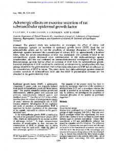
r FIG.1. Mobility shift assay of EGF- and LPS-induced SIE bind69ing proteins. Solutions of EGF (1 mg/ml), LPS (2 mg/ml),or PBS were injected intraperitoneally intomice a t a dose of 10 pl/g of body weight. Mice were sacrificed at the indicated times, and liver nuclei were isolated and extracted with0.2 M NaCl a s described under “Experimental FIG.2. EGF and LPSinduce the appearanceof Stat3 in mouse Procedures.” Aliquots(5 pl) of each extract were assayedfor the ability liver nuclei. Nuclear extracts were prepared from PBS-, EGF-, or to bind a ”P-labeled SIE oligonucleotide as described under “Experi- LPS-treated mice a s described in the legend to Fig. 1.Aliquots (25 pl) mental Procedures” The positions of SIF A, B, and C and free probe arewere resolved by SDS-PAGE and assayed by Western blot analysis with indicated. E , EGF; L,LPS; -, PBS control. either anti-phosphotyrosine (panel A ) or anti-Stat3 (panelB ). E, EGF; L, LPS; -, PBS control. Gel Shift and Supershift Analyses-The sequence of the SIE element used in thegel shift analysis is:5’-GTG CAT ‘R’C CCG TAAATC TTG TCT ACAATT C-3’. All incubations were carried outat room temperature. 5 pl of nuclear extract from control, LPS-treated,or EGF-treated animals were incubated with 2 pg of poly(dI-dC).poly(dI-dC) in 3 mM EDTA, 1mM dithiothreitol, and4% Ficoll in a final volume of 20 pl for 10 min. 32P-Labeled SIEoligonucleotide (1x lo5 cpm, approximately 1 ng) was added to the incubation for 15 min. The reaction wasresolved -A -6 on a 4% native acrylamide gel containing 0.5 x TBE buffer (25mM Tris, ‘C 25 mM boric acid, 0.5 mM EDTA) for 2-3 h at 150 V. The gel was dried and exposed to film. For supershift experiments,gel shift analysis was carried out as described above except that 1 p1of either polyclonal anti-Statla$, or anti-Stat3 was added to the indicated samples in the first 10-min incubation period. RESULTS AND DISCUSSION
We have previously reported the detection of several tyrosine-phosphorylated proteins inmouse livernuclei within minutes following the administrationof EGF (15).Western blotting and immunoprecipitation experiments indicated that the proteins migrating with nominal molecular masses of 91 and 84 kDa that were detected in nuclei after EGF treatment corresponded to the IFN-inducible proteins Statla and Statlp, respectively. Gel shift analysis using the SIE,SIS-conditioned the medium element of the c-fos promoter, revealed that threespecific EGF-induced protein-DNA complexes could be formed. By antibody reactivity Stat1 was detected as a component of two of these complexes (SIF B and C ) ; the protein responsible for the third complex (SIF A) was not identified. A number of laboratories have reported that treatment of responsive cells with a wide variety of extracellular signaling proteins results in the appearance, cytoplasm in and nuclei, of tyrosine-phosphorylated proteins (22). Some of these proteins appear tobe activated transcriptionfactors identical or related to Statla. In an attempt toidentify the protein responsible for the EGF-induced SIF A and to assess the specificity of the response to EGF, we initiated a search for other cytokines that would induce SIE binding activity. Since obtaining sufficient quantities of cytokines for these in situ experiments was not economically feasible, we chose to inject bacterial lipopolysaccharide intraperitoneally. This treatment promotes a strong acute phase response resulting from the release of cytokines including IL-6, tumor necrosis factor, and IL-1 from different cell types (23). To determine if LPS injection is capable of inducing SIE binding activity we performed gel shift analyses using a 32Plabeled SIE element with nuclear extracts from animals that had been injected with PBS, LPS, or EGFfor 15 or 75 min. As shown previously, nuclear extractsfrom mice treated with EGF for 15 minformed three specific binding complexes designated SIF A, B, and C (Fig. 1)(15). These three bands were dimin-
FIG.3. Supershift analysis of SIE binding proteins.Nuclear extracts were prepared from PBS-, EGF-, or LPS-treated mice and assayed for the ability to bind SIE as described in Fig. 1. Antibodies specific for either Statla$ or Stat3 were included in thegel shift reactions as indicated. The positions of SIF A, B, and C and freeprobe are indicated. E, 15 min of EGF; L,75 min of LPS; -, PBS control.
ished by 75 min after EGF injection. Control nuclear extracts and extractsfrom mice treated with LPS for 15 min possessed no SIE binding activity. Interestingly, SIF A could be observed in nuclear extracts from mice treated with LPSfor 75 min (Fig. 1).This apparent delay in induction of SIF A binding activity following LPS treatment is consistentwith the timenecessary to promote the releaseof IL-6 and IL-1. We,therefore, conclude that the induction of SIF A activity probably is not a direct effect of LPS but rather an indirect response to LPS induction of cytokine release, since IL-6 has been shown to induce SIF A in hepatoma cell cultures (16). Since SIF A was seen inresponse to injection of either EGF or LPS, we examined the control and EGF and LPS nuclear extracts for the presence of tyrosine-phosphorylated proteins (Fig. 2, panel A). As previously shown, nuclear extracts from mice treated with EGF for 15 mincontainseveral induced tyrosine-phosphorylated proteins in themolecular mass range of 84-92 kDa, whose extent of phosphorylation is diminished by 75 min. No induced tyrosine-phosphorylated proteinswere evident in nuclear extractsfrom mice treated with eitherLPS or PBS for 15 min. However, one tyrosine-phosphorylated protein that migrated slightly faster than the phosphorylated Statla protein identified earlier was detected in the nuclear extracts prepared 75 min after LPS treatment. In view of the recentdiscovery of a new member of the STAT (signal transduction activator of transcription family) of proteins, Stat3, and theobservation that Stat3 becomes phosphorylated on tyrosine in response to EGF or IL-6 in cultured
EGF and LPS Activate Stat3
21935
ies. The precipitated proteins, after SDS-PAGE and transfer to filters, were immunoblotted with anti-Stat3, anti-phosphotyStat1a.p Stat3 rosine, and anti-Statla$. E L E L E L Treatment Blotting with anti-Stat3 again demonstrated that Stat3 is present in nuclei of EGF- and LPS-treated animals but is not 200 m I detectable or present at only trace levels in nuclei from control 2 97 animals (Fig. 4). The anti-phosphotyrosine antibody demon69f strated the presence of a tyrosine-phosphorylated protein in the anti-Stat3 immunoprecipitates from both EGF- and LPS45 treated mice that migrated indistinguishably from the Stat3 FIG.4. Immunoprecipitation of proteins from nuclear extracts detected by Western blot (labeled p89 in Fig. 4).Reblotting the with anti-Stat3. Nuclear extracts were prepared from PBS-, EGF-, Stat3 immunoprecipitates with anti-Statla,p revealed that and LPS-treated mice as described in Fig. 1. Aliquots (50 pl) of the nuclear extracts were immunoprecipitated with anti-Stat3 for 4 h a t Statl proteins also are immunoprecipitated by anti-Stat3 from nuclear extracts after the administration of EGF but not after 4 "C followed by the addition of protein A-Sepharose for 1 h. The resulting precipitates were recovered, analyzed by SDS-PAGE, and as- LPS (Fig. 4). From the immunoprecipitation experiments we sayed by Western blot with antibodies to phosphotyrosine (PY), conclude that Stat3 is tyrosine-phosphorylated and transloStatla#, or Stat3. The positions of S t a t l a (p91), Stat3 (p89), and S t a t l p ( ~ 8are 4 ) indicated. E, 15 min of EGF; L,75 min of LPS; -, PBS cated to the nucleus in response to both EGF and LPS. It appears that Statla$ isalso immunoprecipitated by the anticontrol. Stat3 antibody but only after activation by EGF, implying that human tumor cells (181, wetested whether anyof the tyrosine- Statl and Stat3proteins may coexist in the same native comphosphorylated nuclear proteins induced by the administration plex after the administration of EGF. of EGF or LPS to intact mice was the newly described Stat3 From the result presented, we draw the following concluprotein. (Anote aboutthe migration of the proteins distinguish- sions: 1)EGF and LPS induce the tyrosine phosphorylation and able by Statl and Stat3 antiserum may be helpful. The fastest nuclear translocation of Stat3; 2) Stat3 ispresent inthe nuclear migrating band is Statlp (thenominal 84-kDa protein), and the complexes that bind SIE in response to EGF and LPS; and 3) slowest is thephosphorylated form of Statla (thenominal 91- Stat3 is a component of the protein complexes that generate kDa protein). Stat3 exists as a major protein that migrates both SIF A and SIFB. faster than thephosphorylated Statla and at about the same position as non-phosphorylated Statla.) Re-examination of the REFERENCES nuclear extracts from EGF- and LPS-treated animals (illus1. Carpenter, G. & Wahl, M. I. (1990)in Peptide Growth Factors and Their Receptors (Sporn, M.D., and Roberts, A. B., eds) pp. 69-171, Springer trated in Fig. 2, panel A ) with specific antisera to Stat3 rePublishing Co., New York vealed the presence of Stat3 innuclear extracts after either15 2. Fisher, D. A. & Lakshamanan, J. (1990)Endocr: Rev. 11,418-442 min of EGF or 75 min of LPS administration (Fig. 2, panel B ) , 3. Laurence, D. J. R. & Gusterson, B. A. (1990)%mor Biol. 11,229-261 Carpenter, G. (1987)Annu. Reu. Biochem. 56,881-914 4 . coincident with the appearance of both tyrosine phosphorylaYarden, Y. & Ullrich, A. (1988)Annu. Reu. Biochem. 57,443478 tion signals and SIFA binding activity (compare Figs. 2 and 1). 5. 6. Ullrich, A. & Schlessinger, J. (1990)Cell 61, 203-212 7. Hunter, T.(1989)Curr. Opin. Cell Biol. 1,1168-1181 We, therefore, performed supershift analysisof the SIEbind8. Carpenter, G. & Cohen, S.(1990)J. Biol. Chem. 265,7709-7712 ing complexes with the 15-min EGF and 75-min LPS nuclear 9. Carpenter, G. (1992)FASEB J. 6,32834289 extracts using both anti-Statla$, and anti-Stat3 antibodies. 10. Glenney, J. R., Jr. (1992)Biochim. Biophys. Acta 1134,113-127 Antibodies specific for StatlaJ shifted SIF B and C from the 11. Pawson, T.& Gish, G . D. (1992)Cell 71,359-362 Pawson, T.(1992)Cum Opin. Genet. Deu. 2,612 EGF extractsbut hadno effect onSIF A in these extractsor the 12. 13. Koch, C. A., Anderson, D., Moran, M. F., Ellis, C. & Pawson, T.(1991)Science 252 668-674 LPS extracts (Fig. 3). However, an anti-Stat3 antibody superR. W. & Cohen, S.(1992)Proc. Natl. Acad. Sci. U.S.A. 89,8477shifted SIF A from the LPS extracts and SIFA and B from the 14. Donaldson, 8481 EGF extracts. Control antibodies had no effect on band shift 15. RuffJamison, S.,Chen, K. & Cohen, S.(1993)Science 261,1733-1736 mobility (data not shown). From this experiment we conclude 16. Sadowski, H. B., Shuai, K., Darnell, J. E., Jr. & Gilman, M. Z. (1993)Science 261, 1739-1744 that Stat3 isa component of SIF A and of SIF B but is not a 17. Silvernnornen, O.,Schindler, C., Schlessinger, J. & Levy, D.E. (1993)Science 261,1736-1739 component of SIF C. These results are very similar to those 18. Zhong, Z., Wen, Z. & Darnell, J. E.,Jr. (1994)Science 264,95-98 obtained in the cultured human tumor cells (18). 19. Stahl, N. & Yancopoulos, G. D.(1993)Cell 74,587-590 To determine if Stat3 is tyrosine-phosphorylated in response 20. Savage, C. R., Jr. & Cohen, S.(1972)J. Biol. Chem. 247,7609-7611 to the injection of EGF or LPS, proteins in nuclear extracts 21. Laemmli, U. K. (1970)Nature 227,680-685 Darnell, J. E., Jr., Kerr, I. M. & Stark, G. R. (1994)Science 264,1415-1421 from control mice or mice injected with EGFfor 15 min or LPS 22. 23. Wegenka, V. M., Buschmann, J., Liitticken, C., Heinrich, P. C. & Horn, F. (1993)Mol. Cell. Biol. 13, 276288 for 75 min were immunoprecipitated with anti-Stat3 antibod-
E a anti-Stat3
-
-
-
-
Adsorbant Blot
E!