Anti-Rh0(D) IgG binds to band 3 glycoprotein of the human erythrocyte membrane. [integral proteins/alkaline extraction/Rho(D) antigen/band 3 ensembles].
Proc. Nati. Acad. Sci. USA Vol. 78, No. 5, pp. 2898-2902, May 1981
Biochemistry
Anti-Rh0(D) IgG binds to band 3 glycoprotein of the human erythrocyte membrane [integral proteins/alkaline extraction/Rho(D) antigen/band 3 ensembles]
EDWARD J. VICTORIA, LAWRENCE C. MAHAN, AND S. P. MASOUREDIS Pathology Department, School of Medicine, University of California, San Diego, La Jolla, California 92093
Communicated by Harvey A. Itano, February 12, 1981
ABSTRACT Alkali-extracted erythrocyte ghost membranes from Rh0(D)-positive and Rho(D)-negative donors were incubated with human immune anti-Rho(D) IgG and nonimmune IgG. After sensitization with IgG, the integral membrane proteins were solubilized in Brij 36T nonionic detergent and chromatographed by gel filtration. There was a distinct resolution of IgG into free and membrane-complexed forms. The IgG-complexed membrane proteins were isolated by the use of a staphylococcal protein A affinity support. The protein A-bound complexes were examined for polypeptide composition by gel electrophoresis after elution. Only Rh0(D)-positive membrane proteins incubated with immune antiRho(D) IgG revealed intact band 3. Control Rh-negative membrane proteins that had reacted with immune anti-Rho(D) IgG and the Rh-positive membranes that had reacted with nonimmune IgG showed only low molecular weight fragments of band 3 that bound nonspecifically to IgG. Arguments are presented supporting a band 3 localization for the Rh antigen.
The Rh antigens are restricted to the erythrocyte plasma membranes of the highest primates, including man. Since the report by Levine in 1939 (1), a wealth of serologic and genetic data have accumulated that suggest, among other things, the importance of Rh to normal erythrocyte survival and its possible involvement in several idiopathic and drug-induced autoimmune hemolytic anemias. An existing paradox is that despite the vast amount ofinformation available about the structure of the erythrocyte plasma membrane (2, 3), it has not been possible to relate this knowledge to the molecular structure of Rh. In part, this probably reflects the lipid-dependent, integral membrane protein character ofthe Rh complex as elucidated mainly by Green (4, 5). Data from radiation inactivation studies (6) and the gel filtration behavior of solubilized membrane fractions (7) have indicated a molecular size range from 174,000 to below 300,000 daltons, respectively, for the Rh0(D) antigen. Membrane proteins can be classified as integral or peripheral (2). In the erythrocyte, the peripheral constituents face the cytoplasm and thus would be unlikely candidates for antigenic activity in the native erythrocyte. They also are capable of associating with integral components after nonionic detergent solubilization under nondenaturing conditions (8). Evidence obtained with a variety of techniques suggests that in the human erythrocyte only the integral polypeptides band 3 [nomenclature of Steck (9)] and the sialoglycoproteins have significant exposure on the cell surface. Our approach to the Rh antigen problem has been to start with an integral protein-enriched erythrocyte membrane fraction obtained by mild alkaline extraction and, after anti-Rho(D) IgG binding, to solubilize and isolate those membrane com-
ponents bound to IgG and by implication associated with the Rh antigen. MATERIALS AND METHODS Preparation of Integral Membrane Protein Fraction. Fresh blood was obtained from normal type 0, Rh0(D)-positive and Rho(D)-negative donors. Ghosts were prepared from washed erythrocytes containing no detectable leukocytes, essentially by the method of Fairbanks et al. (10). The procedure for obtaining integral proteins was a modification of the method of Steck and Yu (11). The important modifications consisted of including EDTA during the alkaline extraction step and subsequent S-alkylation with iodoacetamide after the extraction. These measures were designed to control the tendency of the alkali-treated proteins to aggregate as reflected by the presence of aggregated material or the inability to penetrate gels in the absence of reductant during sodium dodecyl sulfate/polyacrylamide electrophoresis (12). Freshly prepared ghosts were suspended at 3 mg of protein per ml in 5 mM sodium phosphate, pH 8.0 (5P8), containing 1 mM EDTA and preincubated for 10 min at 0°C. After this preincubation, designed to inhibit trace metal-catalyzed sulfhydryl oxidation, the sample was mixed with pH 12 water containing 1 mM EDTA and through which N2 had been bubbled, to a final protein concentration of 0.3 mg/ ml. After maintenance of the pH at 12 for 20 min at 0WC, the suspension was centrifuged (27,000 X g, 30 min), washed by resuspending in 5P8 containing 1 mM EDTA and 5 mM recrystallized iodoacetamide, and centrifuged again. The sample was then resuspended in 5P8/1 mM EDTA/5 mM iodoacetamide and incubated at 0°C in the dark with stirring for 20 min at a final protein concentration of 3-6 mg per ml. After this step, the sample was dialyzed against 0.015 M sodium phosphate/ 0.15 M NaCl, pH 6.5 (BNS 6.5), containing 5 mM iodocetamide in the dark and finally against BNS 6.5 alone before incubation with IgG in BNS 6.5. Immunoglobulin G Isolations. The IgG fractions from hightitered anti-Rho(D) serum and from normal nonimmune serum were obtained by diethylaminoethyl-cellulose anion-exchange chromatography by the method of Levy and Sober (13) and were labeled with [l2I]iodine monochloride as described by Helmkamp et al. (14). The anti-Rho(D) serum was monospecific when tested against a commercial erythrocyte panel and had an antiglobulin titer of 1:16,000 when tested against an R1Rj cell. Incubation of Membrane Proteins with IgG. Immune antiRho(D) IgG was incubated with integral proteins derived from Rho(D)-positive and Rho(D)-negative erythrocytes. In addition, nonimmune IgG was incubated with Rho(D)-positive membrane proteins. For all experiments, the "MI-labeled IgG used Abbreviations: 5P8, 5 mM sodium phosphate, pH 8.0; BNS 6.5, 0.015 M sodium phosphate/0. 15 M NaCl, pH 6.5; IMP, intramembranous
The publication costs of this article were defrayed in part by page charge payment. This article must therefore be hereby marked "advertisement" in accordance with 18 U. S. C. §1734 solely to indicate this fact.
particle. 2898
Biochemistry:
Victoria et al.
Proc. Natl. Acad. Sci. USA 78 (1981)
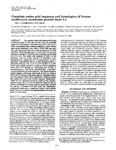
was pretreated with 200 kallikrein inhibitor units of Kunitz pancreatic trypsin inhibitor (Trasylol, obtained from Mobay Chemical, New York) per mg. In a typical experiment, 30 mg of membrane proteins was incubated with 10 mg of "2I-labeled IgG in 10 ml of BNS 6.5 for 1.5 hr at 370C. After the incubation, the suspension was washed twice in cold 5P8 containing 1 mM EDTA. The 27,000 X g (for 30 min) pellet was then solubilized at 370C for 15 min in 30 ml of 0.5% Brij 36T (a lauryl polyoxyethylene ether) nonionic detergent made up in 20 mM Tris HCV1 mM EDTA, pH 7.4. After centrifugation at. 100,000 x g for 60 min, the supernatant was fractionated by gel filtration. Sepharose CL-4B Gel Filtration. The post 100,000 x g supernatant obtained above was fractionated at 15'C on a 420-ml Sepharose CL-4B column equilibrated in the same buffer used to solubilize the sensitized membrane proteins. Five-milliliter fractions were collected at a 0.25-ml/min flow rate. After radioactivity measurements, the free IgG and membrane proteinbound IgG fractions were identified. The latter were pooled and incubated with Staphylococcus aureus protein A-Sepharose CL-4B beads. Protein A Batch Adsorption of Membrane Protein-IgG Complexes (15). Column fractions representing membranecomplexed radiolabeled IgG were pooled (typically about 90 ml) and incubated with 150 mg of protein A on Sepharose CL-4B beads. After room temperature mixing by rotation for 3-4 hr, the beads were washed at least five times each with 15 vol of solubilizing buffer. The centrifuged beads containing only IgGcomplexed membrane proteins were then eluted with 0.5 ml of fresh 2% sodium dodecyl sulfate/6 M (ultra-pure) urea for 20 min at 37°C in a bath-type sonifier. The small volume was designed to achieve desirable protein concentrations for electrophoresis. The conditions did not selectively release adsorbed components. Analytical Procedures. Electrophoretic procedures were performed essentially according to Fairbanks et al. (10). Protein estimations were based on A" of 13A4 for IgG and 12.0 for sodium dodecyl sulfate-solubilized erythrocyte membranes or on the method of Lowry et al. with bovine serum albumin as standard (16). All Sepharose products were from Pharmacia and electrophoresis chemicals were from Bio-Rad. All other chemicals were of the highest commercially available grade. RESULTS In contrast to native ghosts (Fig. 1A), the membrane fraction is highly enriched in band 3 and totally devoid of peripheral proteins as in the original method of Steck and Yu (11). All of the gel data presented in this communication were obtained in the absence of reducing agent, which attests to the nonaggregability of the alkylated membrane proteins. As shown in Fig. 1C, one consequence of alkylation is that upon dialysis against BNS 6.5 there was fragmentation of band 3. This was observed to comparable extents in the experimental sample and both controls. Isotonic buffer conditions were important for the subsequent binding of IgG to membrane proteins. Band 3 fragmentation: (i) did not appear to be due to protease contamination (it could not be blocked by proteolytic inhibitors); (ii) was restricted to band 3 and did not involve the major sialoglycoproteins; (iii) gave rise mainly to two bands of apparent molecular weights 68,000 and 29,000; and (iv) reflected an ionic strengthinduced phenomenon involving band 3 sulfhydryls (unpublished data). No further degradation of band 3 occurred consequent to IgG binding or Brij 36T solubilization; some dimerization sometimes was observed (Fig. 1D). IgG binding was about 10% of the IgG presented and solubilization efficiency was
2899
A
B
D
0.5 Relative mobility
1.0
FIG. 1. Absorbance profiles of Coomassie blue-stained proteins after sodium dodecyl sulfate/polyacrylamide gel electrophoresis. Amount loaded: 22 ,g of protein. No reductant was used. (A) Control ghosts. (B) Alkali-extracted, alkylated membranes in 5P8; the major peak is band 3. (C) After dialysis against BNS 6.5; asterisks denote (from left to right) band 3-derived fragments of apparent molecular weights 68,000 and 29,000. (D) After Brij 36T solubilization, post100,000 x g supernatant; asterisk denotes dimerized band 3.
about 66% for both IgG and membrane proteins. As shown in the gel filtration results of Fig. 2, there was a clear separation of free IgG from membrane protein-bound IgG. Band 3 has been reported to associate noncovalently in nonionic detergent
Troc. Natl. Acad. Sci. USA 78 (1981)
Biochemistry: Victoria et al.
2900
0.4r
15
n -,q
IL2
.t
F
11Ir
0
c0
Q
x
v
0
0
5 Ei v
b
0.11
A
10 6
:
0
00
-
10
A-
gr-
20 30 40 50 60 70 Fraction
Z=
-- -
m
80 90 100
FIG. 2. Detergent gel filtration of membrane proteins and 1251-labeled IgG in Sepharose CL-4B in 0.5% Brij 36T/20 mM Tris HCl/1 mM EDTA, pH 7.4. Void volume occurred at about fraction 25.
to a variety of peripheral proteins, including band 4.2, glyceraldehyde-3-phosphate dehydrogenase, aldolase, ankyrin, and possibly spectrin (17), none ofwhich was present in our starting membrane fraction. It has recently been shown (12) that band 3 eluting at the location offraction 46 exists in a homooligomeric state. No evidence was obtained from detergent gel electrophoresis for a comigrating or strongly noncovalently held polypeptide eluting at this location. These considerations make less likely the possibility that the Rho(D) antigen integral polypeptide represents a membrane constituent that associated noncovalently with band 3 during gel filtration and was subsequently missed after the immunoadsorption procedure. In data not shown, we have found that sialoglycoproteins chromatograph sharply at about fraction 58 (from 55 to 64) and do not bind IgG. The data shown in Figs. 1 and 2 were obtained with proteins derived from Rh0(D)-positive membranes sensitized with immune anti-Rho(D) IgG. The Rh0(D)-negative and nonimmune control experiments yielded very similar results until the protein A step (see below). All three systems had the slight 280 nm-absorbing peak at about fraction 46 of Fig. 2. In the absence of IgG (data not shown), binding of membrane proteins to protein A was insignificant. It ranged between 2.5% and 3.9% of that seen when the membrane proteins were complexed to IgG. In most experiments, over two-thirds of the IgG was bound by protein A. As shown in Fig. 3, all three systems-the experimental, the negative control, and the nonimmune control-show lower molecular weight bands of apparent molecular weight 68,000 and 29,000 in the protein A eluate. These polypeptides probably represent the sulfhydryl-dependent band 3 degradation products (shown by asterisks in Fig. 1C), which show a high nonspecific affinity for IgG. Only in the experimental incubation was there a substantial contribution to the profile by a polypeptide with a mobility similar to that of intact band 3 (indicated by the stippled area in Fig. 3A). There was a total absence of intact band 3 polypeptide in the nonimmune controls and only a small amount in the Rh-negative control. It is important to reiterate that all three systems had comparable amounts of band 3 before the protein A step and that band 3 degradation beyond the level seen upon dialysis against BNS 6.5 was never observed. Thus, the absence or markedly reduced content ofband 3 in the control gel profiles is significant. As discussed below, there are other arguments that make band 3 localization for the Rho(D) antigen a plausible finding. In experiments not shown, we were able to show that none of the polypeptide bands appearing below IgG were "I labeled, thereby excluding IgG breakdown as their origin.
B
C
0.5 Relative mobility
1.0
FIG. 3. Gel electrophoresis of material eluted from protein A. (A) Rh-positive membrane proteins with immune anti-Rho(D) IgG; stippled area corresponds in mobility to band 3; asterisks denote band 3derived fragments as in Fig. 2C; major peak off-scale represents IgG. (B) Rh-negative membrane proteins with immune Rho(D) IgG. (C) Rhpositive membrane proteins with nonimmune IgG.
DISCUSSION Of all the major blood groups, the Rh system has proven the most intractable to a biochemical approach. The results of this investigation obtained with an alkylated integral membrane protein fraction suggest that band 3 may be involved in Rh activity. The experimental model used is hampered by the selfdegradation of band 3, which could not be blocked, resulting in band 3 fragments that bind IgG nonspecifically. Unfortunately, the original procedure of Steck and Yu (11), in which there is no band 3 breakdown, led to disulfide-based aggregate
Biochemistry:
Victoria et al.
formation. The use of a reductant was precluded by the necessity of working in the presence of antibody. Moreover, as previously found in other systems (18, 19), sulfhydryl sensitivity also precluded radioiodination of the alkylated membrane protein fraction [both aggregation and degradation were observed upon iodination (unpublished)]. Despite these limitations, the available evidence is consistent with a band 3 localization for the Rho(D) antigen. A multiplicity of cell functions have now been ascribed to band 3 [see Rothstein (20) and Steck (17) for recent reviews]. The greatest obstacle to reconciling band 3 with Rh antigen activity lies in the discrepancy in numbers, nearly a million copies per cell (17), against less than 40,000 (21), respectively. Whereas studies suggest that band 3 may be heterogeneously glycosylated (22), the preponderance of evidence (16) indicates that the bulk of the polypeptide is homogeneous. Despite this apparent homogeneity, reports have appeared suggesting different protein chemical behavior among band 3 monomers, possibly due to heterogeneous interactions with other membrane components. Thus, for example, Lutz et al. (23) have shown that, in their experiments, only one-fifth of band 3 could undergo disulfide crosslinking. In addition, Nigg and Cherry (24) have recently shown differences among subpopulations of band 3 in connection with linkage to the submembrane cytoskeleton. The discrepancy in numbers could result from the existence of subpopulations of band 3 that have Rho(D) determinants inaccessible to anti-RhU(D). This interpretation is plausible because the rare, but normal, D--/D-- phenotype erythrocyte can have in excess of 200,000 D antigenic determinants (25). D-erythrocyte membranes appear to be identical to those of an R1R1 phenotype on dodecyl sulfate/polyacrylamide gel electrophoresis (data not shown). If the Rh determinants were borne by a minor polypeptide constituent of the membrane, then D-- electrophoretic patterns should contain a prominent new band. They do not. Another possibility for reconciling band 3 with Rh involves invoking an oligomeric state for band 3 which would bring the numbers into agreement. There is general agreement that band 3 exists in ghosts as a dimer (8, 26). Less well known but persuasive is the evidence that band 3 may exist as a tetramer in the membrane and possibly even higher oligomeric forms (27). The arguments supporting a native tetramer state for band 3 have recently been carefully considered (28). Should the Rh antigenic determinant require an oligomeric state of band 3, then band 3 would satisfy the site number requirements for the Rh0(D) antigen. Postulating oligomeric band 3 has served to reconcile the number of band 3 monomers with the number of intramembranous particles (IMPs) revealed by freeze-fracture electron microscopy (27, 28). There is little question but that IMPs contain band 3. This is based on various experiments involving reconstitution, labeling, and sulfhydryl electron cytochemistry (29-31). If one accepts that band 3 is the major constituent of IMPs, then it is possible to bring another argument to bear on the band 3:Rh number discrepancy. It is well known that the density of IMPs differs greatly [as much as by a factor of 5 in unfixed ghosts (32)] between the external and protoplasmic faces. The external face has the much lesser density. Work from this laboratory involving immunoelectron microscopy has previously detected similarities in topological patterns of ferritin-anti-Rh antibody and the reported behavior of IMPs such as clustering after proteolysis (33). A location for Rh on IMPs is therefore plausible on these grounds and experimentally verifiable by freeze-etching studies. Recently, a powerful technique (34) based on classical target
Proc. Natl. Acad. Sci. USA 78 (1981)
2901
theory has reappeared and is being applied to difficult receptor systems such as insulin and glucagon. Radiation inactivation of the Rho(D) antigen in ghost membranes was recently reported to yield a molecular weight of 174,000 ± 10,000 (6), which, within experimental error, is the size of a band 3 dimer (24). It is conceivable that a similar analysis of intact erythrocytes may yield a tetramer size. Using highly specific, radiolabeled anti-Rho(D) eluates (33), we have found in integral proteins obtained by the method of Steck and Yu (11) rigorous immune specificity (data not shown). On the other hand, we have been unable to confirm reports of readily released low molecular weight Rh antigen materials with the labeled antibody technique (35, 36). Because, on the basis of the work of Lorusso et al. (37) and this communication, sialoglycoproteins are not involved in Rh.function, then, if one accepts the radiation inactivation estimate for size of Rh, it can be demonstrated that, other than band 3, there is no single polypeptide component in the alkali-treated ghosts in sufficient quantity to account for its Rh antigen content. As mentioned in the Introduction, the Rh antigen occurs only in primates. It is interesting to note that in a recent study on the immunological reactivity of human band 3 no crossreactivity of anti-band 3 was detectable between primates and nonprimates (38). This is in marked contrast to anti-spectrin, which crossreacted with the most primitive erythrocytes tested (39). Finally, we have not mentioned the other major Rh antigens: C, c, E, and e. We have previously proposed (21), mainly on the basis of immunoelectron microscopy, "that a single membrane component serves as the locus for the Rh antigens." We would like to propose that all Rh antigens are represented as band 3 ensembles of homologous subunits in which the interactions may be governed by sulfhydryl oxidation-reduction (40, 41) and that these interactions may determine Rh specificity. We would expect all the Rh antigens to be the same size, an expectation that could be tested by target analysis. Recently, a similar model was proposed for the acetylcholine receptor consisting of a complex-of homologous subunits (42). We are indebted to Dr. J. M. Bowman of the Canadian Red Cross at Winnipeg for high-titered anti-Rho(D) sera and to Joan DeCherrie for confirmation of the specificity of the antisera used. The San Diego Blood Bank has generously provided us with normal donor blood samples. This work was supported by National Institutes of Health Grants HL-23108 (to E.J.V.) and HL-12994 (to S.P.M.).
1. Levine, P. & Stetson, R. E. (1939) J. Am. Med. Assoc. 113, 126-127. 2. Singer, S. J. (1974) Annu. Rev. Biochem. 43, 805-833. 3. Marchesi, V. T. (1978) Annu. Rev. Med. 29, 593-603. 4. Green, F. A. (1965) Vox. Sang. 10, 32-53. 5. Green, F. A. (1972)J. Biol. Chem. 247, 881-887. 6. Folkerd, E. J., Ellory, J. C. & Hughes-Jones, N. C. (1977) Immunochemistry 14, 529-531. 7. Lorusso, D. J. & Green, F. A. (1975) Science 188, 66-67. 8. Yu, J. & Steck, T. L. (1975)J. Biol. Chem. 250, 9176-9184. 9. Steck, T. L. (1974) J. Cell Biol. 62, 1-19. 10. Fairbanks, G., Steck, T. L. & Wallach, D.F.H. (1971) Biochemistry 10, 2606-2617. 11. Steck, T. L. & Yu, J. (1973)J. Supramol. Struct. 1, 220-232. 12. Victoria, E. J. & Mahan, L. C. (1981) Biochim. Biophys. Acta 644, in press. 13. Levy, H. B. & Sober, H. A. (1960) Proc. Soc. Exp. Biol. Med. 103, 250-252. 14. Helmkamp, R. W., Goodland, R. L., Bale, W. F., Spar, I. L. & Mutschler, L. E. (1960) Cancer Res. 20, 1495-1500. 15. Kessler, S. W. (1975)J. Immunol. 115, 1617-1624. 16. Lowry, 0. H., Rosebrough, N. J., Farr, A. L. & Randall, R. J. (1951) J. Biol. Chem. 193, 265-275. 17. Steck, T. L. (1978)J. Supramol. Struct. 8, 311-324.
2902
Biochemistry: Victoria et al.
18. Zweig, M., Heilman, C. J., Jr. & Hampar, B. (1979) Virology 94, 442-450. 19. Jackson, D. C. (1980)J. Immunol. Methods 34, 253-260. 20. Rothstein, A. (1978) in Molecular Specialization and Symmetry in Membrane Function, eds. Solomon, A. K. & Karnovsky, M. (Harvard Univ, Cambridge MA), pp. 129-159. 21. Masouredis, S. P., Sudora, E. J., Mahan, L. & Victoria, E. J. (1976) Transfusion 16, 94-106. 22. Golovtchenko-Matsumoto, A. & Osawa, T. (1980) J. Biochem. (Tokyo) 87, 847-854. 23. Lutz, H. U., von Daniken, A., Semenza, G. & Bachi, T. (1979) Biochim. Biophys. Acta 552, 262-280. 24. Nigg, E. & Cherry, R. J. (1980) Proc. Natl. Acad. Sci. USA 77, 4702-4706. 25. Hughes-Jones, N. C., Gardner, B. & Lincoln, P. J. (1971) Vox. Sang. 21, 210-216. 26. Nigg, E. & Cherry, R. J. (1979) Nature (London) 277, 493-494. 27. Dorst, H. -J. & Schubert, D. (1979) Hoppe-Seyler's Z. Physiol. Chem. 360, 1605-1618. 28. Weinstein, R. S., Khodadad, J. K. & Steck, T. L. (1980). in Membrane Transport in Erythrocytes, eds. Lassen, U. V., Ussing, H. H. & Wieth, J. 0. (Munksgaard, Copenhagen), pp. 35-50. 29. Yu, J. & Branton, D. (1976) Proc. Natl. Acad. Sci. USA 73, 3891-3895.
Proc. Natl. Acad. Sci. USA 78 (1981) 30. Shotton, D., Thompson, K., Wofsy, L. & Branton, D. (1978)J. Cell Biol. 76, 512-531. 31. Fugimoto, T. & Ogawa, K. (1980) Acta Histochem. Cytochem. 13, 72-89. 32. Pricam, C., Fisher, K. A. & Friend, D. S. (1977) Anat. Rec. 189, 595-608. 33. Victoria, E. J., Muchmore, E. A., Sudora, E. J. & Masouredis, S. P. (1975)J. Clin. Invest. 56, 292-301. 34. Kepner, G. R. & Macey, R. I. (1968) Biochim. Biophys. Acta 163, 188-203. 35. Plapp, F. V., Kowalski, M. M., Izer, L. T., Brown, P. J., Evans, J. & Chiga, M. (1979) Proc. Natl. Acad. Sci. USA 76, 2964-2968. 36. Litten, J., Culpepper, R. & Bakerman, S. (1978) Biochim. Biophys. Acta 543, 226-234. 37. Lorusso, D. J., Binette, J. P. & Green, F. A. (1977) Immunochemistry 14, 503-508. 38. England, B. J., Gunn, R. B. & Steck, T. L. (1980) Biochim. Biophys. Acta 623, 171-182. 39. Pinder, J. C., Phethean, J. & Gratzer, W. B. (1978) FEBS Lett. 92, 278-282. 40. Marzullo, G. & Hine, B. (1980) Science 208, 1171-1173. 41. Massague, J. & Czech, M. P. (1980) Diabetes 29, 945-947. 42. Raftery, M. A., Hunkapiller, M. W., Strader, C. D. & Hood, L. E. (1980) Science 208, 1454-1457.