NEW MICROBIOLOGICA, 33, 393-397, 2010
Establishment of two chicken embryonic cell lines in a newly developed nutrient medium Katsuyuki Kadoi Laboratory of Veterinary Microbiology, College of Bioresource Sciences, Nihon University, 1866, Kameino, Fujisawa, Kanagawa, Japan
SUMMARY Two cell lines, named KCEK and KCEL, were established from chicken embryonic kidney and lung. The basal culture medium was newly developed and the cell growth medium consisted of K1999 supplemented with 10% heat inactivated chicken serum. Both cells were well adapted to grow in vitro and more than 50 passages have been made so far. Once the cell lines were established the cells were easily adapted to grow in other growth media supplemented with fetal calf serum. Neither tumor formation in chicks nor P52 avian leucosis common antigen was detected in these cells. However, the oncogene analysis on these cells has not been performed yet. Both cells were permissive hosts for the Aujeszky’s disease virus, Newcastle disease virus, and vesicular stomatitis virus. KEY WORDS: Chicken embryonic cell line, Novel cell culture medium Received March 19, 2010
Chicken cell lines of non-tumor origin or viral antigen-free are still exceptional. Majority of avian cell lines reported so far is originated from tumor organs (Akiyama and Kato, 1974; Baba et al., 1978; Calnek et al., 1978; Dienglewicz and Parcells, 1999; Morgan and Parsons, 1986; Kawaguchi et al., 1987) and/or transformed cells (Chang and Delany, 2004; Danforth et al., 1994; Desjardins et al, 1986; Kaaden et al., 1982; Kitamoto et al., 1980; Beug et al., 1979) by oncogenic virus transfection (Abujoub and Coussens, 1995; Ikuta et al., 1987; Zinkewich-Peotti et al., 1988; Hill et al., 1985). At present, the exceptional cases reported are one called virus-free cell line (Ogura and Fujiwara, 1987) and another named DF-1 (Himly et al., 1998). The present author developed a basal nutrient medium (BM1999) for chicken cell culture in 1999 (PCT/JP2006/310852) after many trials and errors. During the study on the BM1999 two cell lines originated from chicken embryo (kidney and lung) were established. Corresponding author Katsuyuki Kadoi Istituto G. Caporale Via Campo Boario - 64100 Teramo, Italy. E-mail:
[email protected]
Accepted May 25, 2010
The cell growth medium (GM) consisted of BM1999 supplemented with 10% chicken serum (CS), substituted to fetal calf serum (FCS) after the establishment of cell lines. The most of primary cultures were successfully passaged more than 20 times when cultures were incubated at 38-39°C in stationary condition. The composition of BM1999 is shown in Table 1. All ingredients are dissolved in double distilled water and sterilized by filtration with 220 mm pore membrane (Millipore Co.,). It is stored at 48°C in dark and used within 6 months after the preparation. GM employed for the establishment of cell lines was consisted of 9 parts of BM1999 and 1 part of home-prepared CS. CS was separated from the blood harvested from young broilers by cardiac puncture. CS was clarified, filtered, heated at 55°C for 30 minutes, and stored at 4°C. Antibiotics, ampicillin Na, streptomycin sulfate, and kanamycin sulfate, were added at 100 mg per ml of GM and or cell maintenance medium (MM), BM1999 without supplement of serum. The kidney and lung were collected from a chicken embryo one day before hatching of conventional egg, white leghorn breed (Saitama experimental animal supply center). The tissue was cut into small fragments and treated with 0.1% col-
K. Kadoi
394
TABLE 1 - Basal nutrient medium 1999 for chicken cell culture. Ingredients
(mg per 1 liter)
Sodium chloride
6,200
Potassium chloride
400
Calcium chloride dihydrate
200
Magnesium sulfate
90
Sodium dihydrogenphosphate anhydrous 140 Sodium bicarbonate
2,000
Iron (III) nitrate enneahydrate
0.1
HEPES
250
Glucose
1,000
Galactose
1,000
Fractose
1,000
N-acehyl-D(+)-glucosamine
300
Sodium pyruvate
200
Succinic acid
100
L-Alanine
300
L-Arginine hydrochloride
80
L-Cysteine hydrochloride
70
L-Glutamine
600
Glycine
230
L-Histidine hydrochloride
40
L-Isoleucine
100
L-Leucine
100
L-Lysine hydrochloride
240
L-Methionine
40
L-Ornithine hydrochloride
200
L-Phenylalanine
60
L-Proline
100
L-Serine
64
L-Threonine
490
L-Tryptophan
16
L-Tyrosine, disodium
90
L-Valine
10
Choline bitartrate
10
Folic acid
4
Nicotinamide
4
Calcium pantothenate
4
Pyridoxal hydrochloride
4
Riboflavin
0.4
Thiamine hydrochloride
4
i-inositol
10
Phenol red
5
lagenase solution (cell culture grade, WAKO, Japan) for cell dispersion. The cells were suspended in GM containing c. a half million viable cells per ml, dispensed to flasks (Nunclon delta surface, Sweden), and stationary incubated at 38-39°C. Within seven days incubation, more than 60% confluent monolayer were formed. These cultures were successively passaged in flasks for three times. In this stage only EDTA solution (0.05% EDTA in PBS), without trypsin, was used for cell dispersion. Since cells were stable to grow in vitro, cloning was made by a limited-dilution precedure, seeding a small number of viable cell suspensions into 24-well type microplates (Nunclon). Clones actively grown in the plates were further passaged in flasks. The cells established from kidney were named KCEK and the cells from lung were named KCEL respectively. After 15th passage level, both KCEK and KCEL were adapted to grow in the RPMI-1640, slightly modified to contain extra sugars (2 g of glucose, 0.5 g of galactose, and 0.5 g of fructose per 1,000 ml) supplemented with 10% FCS. These cells have been well preserved for more than 2 years by a manner generally employed. Cellular morphology was studied on slip cultures. Giemsa stainings were made after methanol fixation. KCEK cells show a typical epithelial shape and KCEL cells show a pleomorphic epithelioid morphology. Both cells are distinguished by microscopical observation according to their morphology in the early incubation. Chromosome analysis was done in the usual manner. Cells at log phase growth were incubated for 8 hours in the presence of colchicine (0.02 mg/ml), allowed to swell in 70 mM KCl for 15minutes, fixed in cold methanol-acetic acid (3:1), centrifuged, and resuspended in 40% acetic acid solution. The cells were spread on warm slides and air-dried. After Giemsa staining, the preparations were analyzed and photographed under a light microscope. Fifty metaphase spreads were counted per cell line. Karyotyping estimated for both cells was quasidiploid and major chromosome number was of 2N=76. Sex chromosomes were hardly identified since the size of chromosome was so small. KCEL was estimated to be alveolar macrophages (or secondary alveoli in other name) since the cells were positive
Chicken embryonic cell lines
A
395
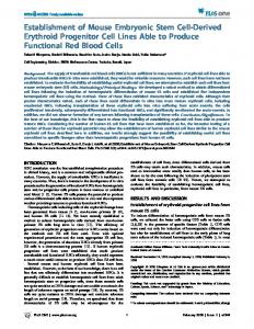
B
FIGURE 1 - Morphology of KCEK and KCEL cells. (A) KCEK cells, passage 44th level, showing an epithelial morphology. Methanol fixed and Giemsa stained. Bar = 100 mm. (B) KCEL cells, passage 49st level, showing epithelioid morphology. Methanol fixed and Giemsa stained. Bar = 100 mm.
for acid phosphatase and weakly positive for nonspecific esterase. Both enzymatic reactions were examined by a commercially available kit (Muto Chem. Japan). None of these enzymes were demonstrated in KCEK cells. Tumorgenicity in vivo and p52 antigen tests were examined as follows. The cell suspension in PBS, containing at ca. two million viable cells per ml, were subcutaneously injected to 10 of one-day old chicks at 1 ml per chick respectively. Clinical observation was made for 3 months, and then autopsy and pathological examinations were performed as usual. The cellular extract prepared from c. one million cells was tested for p52 antigen, know as a common avian leucosis, by the kit (IDEXX Lab. USA) according to the manufacturer’s instruction. Neither tumorgenicity in 1-day-old chicks nor p52 common avian leucosis antigen was demonstrated for both cells. A possibility to certify viral contaminations was tested as follows. The cellular lysate was respectively prepared from both KCEK and KCEL cells, c. 2 million cells each. After clarification they were inoculated to monolayer cell cultures of KSEK6 (Kadoi, 1992), KMP (Kadoi et al., 1997), and KDK-1 cells (Kadoi et al., 1992). These cells were previously confirmed in our hands to be highly sensitive for a variety of viruses. The cultures were maintained at 34°C for 10 days. None of these cultures, KSEK6, KMP, and KDK-1, proved any CPE agents in the first inoculation. However, three serial blind passages were per-
formed for a confirmation. None of viral contaminations was demonstrated after all. Virus susceptibility was tested as follows. Three strains of viruses, Aujeszky’s disease virus (ADV) (Fukusho et al., 1981), Newcastle disease virus (NDV) (Nobuto, 1968), and vesicular stomatitis virus (VSV) (Karstad and Hanson, 1958), were tested for viral susceptibility. All these viruses were known to grow in variety of cells. They were adapted to grow in KSEK6 in advance. Confluent monolayer of both KCEK and KCEL cells were respectively infected with these viruses at MOI =0.05 in the first infection. When infected cells showed clear CPE, second and third virus passages were serially made to inoculate at 1/100 dilution of earlier passages to confirm the replication of infective progenies. The infectivity of third passages was measured by inoculating 10-folddilution of virus in KSEK6 cells grown microplates and expressed in TCID50 per ml according to the CPE occurrence. Both cells were permissive for all viruses tested. Infective progeny virus produced in both cells was at least 107.50 TCID50 per ml for ADV and VSV, and at 105.50 TCID50 per ml for NDV. Adaptation to other media was tested. Trials were also made on the two cell lines on incubation temperatures at 37-39°C, the application of other cell culture media commercially available as Eagle MEM (containing 0.1% glucose) (MEM) and RPMI-1640 (containing 0.1% glucose) (RPMI). KCEK cells were easily adapted to grow in both MEM and or RPMI supplemented with 10%
396
K. Kadoi
FCS. However, KCEL tended to grow better in GM mentioned above. Their growth at 38-39°C was superior than at 37°C. The cell lines of either tumor origin or transformed by oncovirus transfection are not suitable host cells for biological products although some of such cells has been used for virus assay system when their susceptibility is high enough for practical application. A large number of SPF chicken hatching eggs have been utilized worldwide, not only for vaccine production and also diagnostic procedures. The egg supply demands major financial support. In these circumstances established chicken cell lines free from viral contamination or not tumor origins are highly expected. Both KCEK and KCEL have not been analyzed for oncogenes. Therefore at present it is limitedly expressed that two cell lines were established from chicken embryo and the newly developed BM1999 was beneficial for in vitro culture of chicken cells. These cells are novel candidate tools in biosciences including virology. In the author’s limited experience (unpublished data), both cells were good feeder cells for the hybridomas during cloning procedures in monoclonal antibody preparation.
REFERENCES ABUJOUB A., COUSSENS P.M. (1995). Development of a suitable chick cell line infected with Marek’s disease virus. Virology. 214, 541-549. AKIYAMA Y., KATO S. (1974). Two cell lines from lymphomas of Marek’s disease. Biken Journal. 17, 105116. BABA T.W., AND HUMPHRIES E.H. (1984). Differential response to avian leucosis virus infection exhibited by two chicken lines. Virology. 135, 181-188. BABA T.W., GIROIR B.P., HUMPHRIES E.H. (1985). Cell lines derived from avian lymphoma exhibit two distinct phenotypes. Virology. 144, 139-151. BEUG H., VON KIRCHBACH A., DODERLEIN G., CONSCIENCE J.F., GRAT T. (1979). Chicken hematopoietic cells transformed by seven strains of defective avian leukemia viruses display three distinct phenotypes of differentiation. Cell. 18, 375-390. CALNEK B.W., MURTHY K.K., SCHAT K.A. (1978). Establishment of Marek’s disease lymphoblastoid cell lines from transplantable versus primary lymphomas. Inter. J. Cancer. 21, 100-107. CALNEK B.W., SCHAT K.A. (1991). Proliferation of chicken lymphoblastoid cells after in vitro infection with Marek’s disease virus. Avian. Dis. 35: 728-737.
CHANG H., AND DELANY M.E. (2004). Karyotype stability of the DT40 chicken B cell line: Macrochromosome variation and cytogenetic mosaicism. Chromosome Res. 12, 299-307. FUKUSHO A., SHIMIZU M., KUBO M., NANBA K., SHIMIZU Y., KONNO S., SUZUKI K., OTAKI T. (1981). The first outbreak of Aujeszky’s disease in swine in Japan. II. Virus isolation. Bull. Nat. Inst. Anim. Heal. Japan. 82, 5-11. GEERLIGS H., QUANZ S., SUURLAND B., SPIJKERS T.E., RODENBERG J., DAVELAAR F.G., JONGSMA B., KUMAR M. (2008). Efficacy and safety of cell associated vaccines against Marek’s disease virus grown in continuous cell line from chickens. Vaccine. 26, 55955600. HILL M., HILLOVA J., MARIAGE-SAMSON R., MARX M. (1985). Isolation of a line of immortal chicken embryo fibroblasts after transfection with the nuclei of Rous sarcoma virus-transformed Chinese hamster cells. Exp. Cell Res. 156, 127-139. HLINAK A., JAHN S., GRUNOW R., HEIDER G., VON BAEHR R. (1988). Feeder cells from different sources and conditioned media for recloning of human-mouse and mouse-mouse hybridomas. Folia. Biol. (Praha). 34, 105-117. INOUE M., YAMAMOTO H., MATUO K., HIHARA H. (1992). Susceptibility of chicken monocytic cell lines to infectious bursal disease virus. J. Vet. Med. Sci. 54, 575-577. KAADEN O.R., LANGE S., STIBUREK B. (1982). Establishment and characterization of chicken embryo fibroblast clone LSCC-H32. In Vitro. 18, 827832. KADOI K. (1992). Viral susceptibility of an established cell line of swine embryo kidney. Microbiologica. 15, 313-318. KADOI K., MOCHIZUKI A., IKEDA T., KAMATA H., YUKAWA M., INOUE Y. (1992). Susceptibility of a line of dolphin kidney cell culture to several herpesviruses. J. Basic Microbiol. 32, 227-232. KADOI K., MORITA M., KAMATA H. (1997). Viral susceptibility of a newly established cell line derived from peritoneal cavity of Balb/C mouse. Microbiologica. 20, 149-154. KARSTAD L., HANSON R.P. (1958). Primary isolation and comparative titrations of five field strains of vesicular stomatitis virus in chicken embryos, hogs, and mice. Am. J. Vet. Res. 19: 233-236. KAWAGUCHI T., NOMURA K., HIRAYAMA Y., KITAGAWA T. (1987). Establishment and characterization of a chicken hepatocellular carcinoma cell line. LMH Cancer Res. 47, 4460-4464. KELLER L.H., RUFNER R., SEVOIAN M. (1979). Isolation and development of a reticuloendotheliosis virustransformed lymphoblastoid cell line from chicken spleen cells. Infect. Immun. 25, 694-701. KITAMOTO N., IKUTA K., KATO S., HIRAI K. (1980). Persistence of genomes of both herpesvirus of
Chicken embryonic cell lines
turkeys and Marek’s disease virus in a chickenTlymphoblastoid cell line. Biken J. 23, 1-8. LANGLOIS A.J., LAPIS K., ISHIZAKI R., BEARD J.W., BOLOGNESI D.P. (1974). Cancer Res. 34, 1457-1464. LIE X., LEE L.F., SHARMA J.M., NARERIAN K. (1983). Establishment of lymphoblstoid cell lines from Marek’s disease primary tumors. Poult. Sci. 62, 1902-1905. LYON J.A., HINSHAW V.S. (1991). Replication of influenza A viruses in an avian macrophage cell line. J. Gen. Virol. 72, 2011-2013. MENGELING W.L., BOTHE A.D., RITCHIE A.E. (1972). Characteristics of a coronavirus (strain 67N) of pigs. Am. J. Vet. Res. 33, 297-308. MORGAN J.H., PARSONS J.T. (1986). Characterization of c-myc proteins from avian bursal lymphoma cell lines. Virol. 150, 178-186. MUNCH D., HOHLSTEIN L., SEVOIAN M. (1978). In vitro establishment of Marek’s disease herpesvirus-transformed productive and nonproductive lymphoblastoid cell lines. Infect. Immun. 20, 315-318. NOBUTO K. (1968). Newcastle disease in Japan. Bull. L’ Off. Int. Epiz. 70, 435-438. OGURA H., FUJIWARA T., NAMBA M. (1984). Establishment of two chick embryo fibroblastic cell lines. Gann. 75, 410-414. OGURA H., FUJIWARA T. (1987). Establishment and characterization of a virus-free chick cell line. Acta. Med. Okayama. 41. 141-143. OKAZAKI W., WITTER R.L., ROMERO C., NAZERIAN K., SHARMA J.M., FADLY A., EWERT D. (1980). Induction of lymphoid leucosis transplantable tumours and the establishment of lymphoblastoid cell lines. Avian Pathol. 9, 311-329.
397
PARCELLS M.S., DIENGLEWICZ R.L., ANDERSON A.S., MORGAN R.W. (1999). Recombinant Marek’s disease virus (MDV)-derived lymphoblastoid cell lines: Regulation of a marker gene within the context of the MDV genome. J. Virol. 73, 1362-1373. PFEIFER S., KALLIO A., VAHERI A., PETTERSSON R., OKERBLOM N. (1980). Stable bone-marrow-derived cell line producing transforming avian acute leukemia virus OK 10. Int. J. Cancer. 25, 235-242. QURESHI M.A., MILLER L., LILLEHOJ H.S., FICKEN M.D. (1990). Establishment and characterization of a chicken mononuclear cell line. Vet. Immun. Immunopathol. 26, 237-250. RATH N.C., PARCELLS M.S., XIE H., SANTIN E. (2003). Characterization of a spontaneously transformed chicken mononuclear cell line. Vet. Immun. Immunopathol. 96, 93-104. RIDPATH J.F., HUIATT T.W., TRENKLE A.H., ROBSON R.M., BECHTEL P.J. (1984). Growth and differentiation of chicken embryo muscle cell cultures derived from fast- and slow-growing lines. Intrinsic differences in growth characteristics and insulin response. Differentiation, 26: 121-126. ROTH S., KAADEN R.R. (1985). Use of chicken cell line LSCC-H32 for titration of animal viruses and exogenous chicken interferon. Appl. Environ. Microbiol. 49, 634-636. TEMIN H.M., KASSNER V.K. (1975). Replication of reticuloendotheliosis viruses in cell culture: Chronic infection. J. Gen. Virol. 27, 267-274. VAN CAMPEN N., EASTERDAY B.C., HINSHAW V.S. (1989). Virulent avian influenza viruses: their effect on avian lymphocytes and macrophages in vivo and in vitro. J. Gen. Virol. 70. 2887-2895.