Filament Assembly Properties of the Sarcomeric Myosin Heavy Chain1 MACDONALD WICK2 The Ohio State University, Department of Animal Sciences, Columbus, Ohio 43210 ABSTRACT Meat is the edible muscle tissue of animals. The sarcomere is the fundamental functional unit of muscle. Growth and development of muscle is accomplished by the highly ordered accretion and assembly of the constituent proteins in the sarcomere. Primary amino acid sequence elements of the constitutive proteins carry the information necessary for determining the final architecture of the sarcomere. The mechanisms by which the constitutive proteins are assembled and function together to form the sarcomere and produce muscle contraction is just now beginning to be understood.
The predominant protein in the sarcomere, found in the thick filament system, is myosin. In physiological buffers purified myosin spontaneously assembles into a synthetic thick filament with a dramatic resemblance to the native thick filament. Some of the amino acid sequence elements contributing to myosin’s assembly properties may also be critical to myosin’s solubility function, which is so crucial to the manufacture of high quality prepared meat products. This review summarizes recent experimental results contributing to our understanding of the mechanism of sarcomeric muscle myosin assembly.
(Key words: myosin, synthetic filament, parallel dimer, assembly, sarcomere) 1999 Poultry Science 78:735–742
thin. Thin filaments are composed of filamentous actin each anchored at one end in perpendicularly aligned Zdisks. Associated with the thin filaments are the proteins of the contractile regulation system, tropomyosin and troponin, and the giant proteins, titin and nebulin, reviewed elsewhere (Moos et al., 1995). Interdigitating between the thin filaments are bipolar thick filaments, composed almost entirely of the fibrous contractile protein, myosin. The fine structure of the A-band and of the various myosin-binding and other filament-binding proteins of native thick filaments is reviewed elsewhere (Sjostrom and Squire, 1977; Davis, 1988b; Seiler et al., 1996). Early investigations classified muscle fibers into two arbitrary classes, red and white. This classification is incomplete due to the presence in each of the classes of multiple myosin isoforms. Red fibers generally are smaller in diameter, contain more mitochondria, and contain the pigmented protein, myoglobin. In addition, red fibers have a greater blood supply and contain a higher concentration of lipid than white fibers. Red and white muscles have been shown to have different functions in the living animal (Rosser and George, 1986a,b). Other investigators demonstrated that gel binding and water holding capacity (WHC) of myofibrillar proteins are different, depending on whether they were derived from red or white chicken muscle (Xiong,
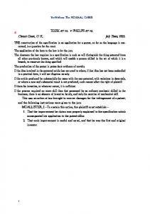
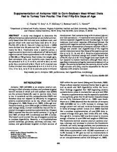
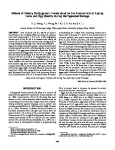
INTRODUCTION Muscle comprises nearly 40% of the body mass of most animals. Figure 1 diagrams the salient features of skeletal muscle cellular organization. Skeletal muscle is made of elongated cells or myofibers specialized for contraction. Each myofiber is approximately 100 mm thick and contains many myofibrils, each about 1 to 2 mm thick. In striated or skeletal muscle tissue myofibrils display a pattern of alternating light (I) and dark (A) bands. The striations arise as a result of the packing arrangement of the filament systems in the sarcomere, the fundamental contractile unit of striated muscle. Striated muscle is found in all animal groups from coelenterates through vertebrates and comprises 80% or more of all muscular tissue (Hickmand, 1970). The predominant cells in muscle are long, multinucleated myofibers. The characteristic banding or striated appearance of skeletal muscle fibers arises from the presence of repeating sets of sarcomeres within specialized contractile organelles, called the myofibrils. The sarcomere is the basic contractile unit of skeletal muscle and is defined as that portion of the myofibril between two Z-disks. Viewed in two dimensions, the sarcomere consists of two sets of filaments, thick and
Received for publication August 1, 1998. Accepted for publication December 22, 1998. 1Salaries and research support provided by state and federal funds appropriated to the Ohio Agricultural Research and Development Center, The Ohio State University. 2To whom correspondence should be addressed:
[email protected].
Abbreviation Key: HC = heavy chain; HMM = heavy meromyosin; LC = light chain; LMM = light meromyosin; MyHC = myosin; TEM = transmission electron microscopy; WHC = water holding capacity.
735
736
WICK
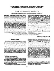
FIGURE 1. Schematic relating the biochemical components to the microscopic structure of muscle tissue. A) Cross section of muscle in the two left micrographs and a longitudinal view of a muscle fiber showing the striated pattern of muscle cells (B). C) The ultrastructure of the sarcomere showing the myofibrillar composition of muscle segment between two Z-bands. D) Magnified view of thick and thin filaments and their associated filament system.
SYMPOSIUM: MUSCLE GROWTH AND DEVELOPMENT
1992, 1994). The precise mechanisms contributing to these differences are not understood; the myosin isoform populations are different in the two types of muscle. White meat, composed of predominantly fast myosin, has greater gel binding and WHC properties than red meat, which is composed of predominantly slow myosin.
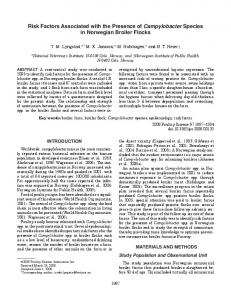
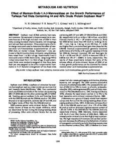
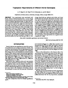
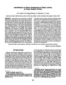
Myosin Sarcomeric myosins belong to the Type II class of myosin proteins that contain an a-helical coiled-coil rod domain that is involved in the assembly of bipolar thick native and synthetic filaments (Cheney et al., 1993; Goodson and Spudich, 1993). Myosin is a relatively large protein with a molecular mass of about 520 kDa (Figure 2). Each myosin molecule is composed of two 220 kDa heavy chains (MyHC) and four light chains (LC), ranging from 17 to 22 kDa (Lowey and Risby, 1971). The entire molecule is approximately 160 nm in length and 2 nm in diameter. The heavy chains interact to form two distinct domains: a pair of globular heads (S1) 15 nm long and 9 nm wide and a a-helical coiled coil rod domain. The rod domain is approximately 150 nm long and 2 nm in diameter and is composed of the C-terminal 1100 amino acids of the MyHC. The myosin rod is a two-stranded coiled-coil motif characterized by two parallel amphipathic a-helical chains that intertwine around each other into a lefthanded superhelix (Crick, 1953; McLachlan and Karn, 1982). Structure/function studies on myosin were greatly aided by the discovery that proteolytic enzymes, such as chymotrypsin, trypsin, and subtilisin, cleave myosin into well-defined, high molecular weight fragments (Mihalyi and Szent-Gyorgyi, 1953). Hydrodynamic and electron microscope studies demonstrated that trypsin, chymotrypsin, and subtilisin cleavage occurs predominantly within the rod domain approximately 80 nm from the C-terminus, generating two fragments. The larger fragment, termed heavy meromyosin (HMM) has a molecular weight of 140 kDa, is soluble in low ionic strength buffers, contains the S1 motor domain, which is associated with the light chains, has adenosine triphosphatase activity, and binds actin. The threedimensional structure of the S1 has recently been determined by x-ray crystallography. Based on this structure, a major update in the mechanism of force generation has been proposed. The smaller fragment, termed light meromyosin (LMM), has a molecular weight of 80 kDa, is composed of the C-terminal two-thirds of the rod domain, and confers the solubility and aggregation properties to the MyHC. The LMM has been shown to form paracrystalline structures at low ionic strength buffers (Harrison et al., 1971; Chowrashi and Pepe, 1977; Strzelecka-Golaszewska et al., 1985; Atkinson and Stewart, 1991a). Each mole of MyHC associates with 2 M of LC (Collins, 1976; Margossian et al., 1983; Baba et al., 1984).
737
MyHC Rod Domain The information necessary for myosin’s assembly functions is carried in the unique repetitive primary sequence elements of the long a-helical coiled coil rod domain. The rod consists of two right hand a-helical coils that intertwine around one another forming a lefthanded coiled coil (Monera et al., 1994). The primary sequence of the rod domain is highly conserved (Parry, 1981; McLachlan and Karn, 1982; Nguyen et al., 1982; Bandman et al., 1994). All a-helical coiled coil proteins have a characteristic seven-residue pattern a, b, c, d, e, f, g, termed a “heptad”. The parallel coiled coil dimeric structure is postulated to be stabilized by the packing of “knobs” formed by the hydrophobic side chains of one helix into “holes” formed by the spaces between side chains of the neighboring helix (Crick, 1953). In this model, hydrophobic residues are spaced every fourth and then third residues apart in the primary sequence of coiled coils (McLachlan and Stewart, 1975). In long chains of ahelices, such as are found in the rod of the MyHC, strong favorable hydrophobic interactions between the a residue of one helix and the a′ of the adjacent coil, in conjunction with the hydrophobic and electrostatic
FIGURE 2. Schematic representation of a sarcomeric myosin molecule. A) Myosin, a 520-kDa hexameric molecule, is shown along with the common proteolytic fragments. Myosin is composed of the two heavy chains (MyHC) and the four associated light chains (LC). The MyHC can be enzymatically cleaved into the low salt insoluble 260-kDa a-helical coiled coil rod domain and the low salt soluble S-1 domain under prolonged enzyme digestion, the MyHC can be enzymatically cleaved into a low salt insoluble 130-kDa light meromyosin (LMM) (B) and a low salt soluble heavy meromyosin (HMM) (C).
738
WICK
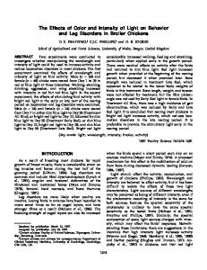
interactions contributed by the d and d′ residues, lead to the formation of a coiled coil (Crick, 1953; O’Shea et al., 1991). Figure 3 is a helical wheel diagram representing the positions of the residues in the helix and positions of the residues in relation to each other in the coiled coil heptad repeat. The a and d positions are involved in hydrophobic interactions to stabilize the coiled coil motif leaving the remainder of the residues exposed to the solvent and available for intramolecular interactions with other myosin rod domains or myofibrillar proteins. In parallel coiled coils, such as are found in the rod of the MyHC, oppositely charged residues commonly occur at positions e and g of adjacent heptads, consistent with the formation of inter-helical ion pairs (McLachlan and Stewart, 1975; Cohen and Parry, 1986; Cohen and Parry, 1990). It has been proposed that conserved e and g residues form intra-helical ion pairing further stabilizing the a-helix (Letai and Fuchs, 1995). The rod exhibits additional repetitive sequences. Previous investigators suggested that the myosin rod shows strong evidence for a 28 amino acid residue unit with a repetitive (Parry, 1981). The rod of sarcomeric myosin is composed of 40 of these 28 amino acid repeats beginning with an invariant proline that is located approximately 838 residues from the N-terminus (McLachlan and Karn, 1982; Strehler et al., 1986). The repeat pattern is interrupted by the insertion of a residue (termed a skip residue) at four d positions along the rod, separated by either seven or eight repeats (McLachlan and Karn, 1982; Strehler et al., 1986; McNally et al., 1989). These skip residues are conserved, and although they do appear to be correlated with characteristic bending in the tail of myosin monomers observed in electron microscopes, they apparently have no role in assembly of myosin. Fourier transform analysis of the charge characteristics of the primary sequence in each a-helix in the LMM has determined that each 28-residue repeat displays a characteristic sinusoidal pattern of alternating positive then negative charged residues in the b and c positions of every other heptad. The b and c residues in the first heptad in every repeat are predominantly occupied by lysine (K) or arginine (R) residues, whereas those residues in the third heptad are occupied by aspartic acid (D) or glutamic acid (E) residues. Thus, the typical repeat is arranged into alternating bands of positive and negative charges (Parry, 1981; McLachlan and Karn, 1982; McLachlan and Karn, 1983), leading to alternating positively and negatively charged zones arrayed along the outer surface of the entire rod (Figure 4). The observation of this pattern has prompted some investigators to suggest that the reduction in the free energy by the neutralization of opposite charges between myosin rods could provide the energy necessary for myosin assembly if the molecules were staggered by odd multiples of 14 residues (Parry, 1981; Matsuda et al., 1982; McLachlan and Karn, 1982; McLachlan, 1983). There is a strong reduction in electrostatic interaction energy when two adjacent rods are aligned with a 98 amino acid residue stagger, or 14.3 nm (McLachlan and
Karn, 1982). This finding has given rise to the proposal that, if neighboring coiled coils are aligned axially by an odd multiple of a charged zone, the reduction in the free energy through electrostatic interactions between oppositely charged zones could provide the driving force for the assembly of higher order oligomers or filaments.
A MECHANISM OF ASSEMBLY
Synthetic Filaments Myosin is extracted from myofibrils in salt solutions greater than 0.3 M. Purified myosin will precipitate by reducing the ionic strength of the salt solution to < 0.2 M and can be recovered by centrifugation at 5,000 × g. If a solution of purified monomeric myosin in 0.6 M NaCl is negatively stained and examined by electron microscopy, no structures or particles are observed. The myosin monomer can be visualized in the electron microscope by the use of shadow-casting (Rice, 1961). As the ionic strength of a purified myosin solution is lowered from 0.6 M to less than 0.2 M NaCl, negatively stained rod-shaped particles, termed synthetic filaments to distinguish them from native thick filaments, are easily
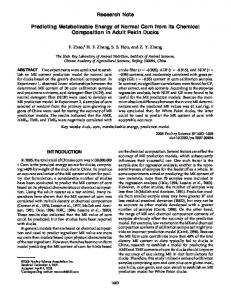
FIGURE 3. A) Helical wheel representation of the a-helical coiled coil motif of the myosin rod. The view is from the N-terminus of the rod domain demonstrating that the a-helical coiled coil is constructed from two right handed a-helices. The heptad positions are labeled a through g. B) Vertical representation of a-helical coiled coil, 2 heptads. The vertical coils indicate the relative positions of the residues from two heptads in each a-helix.
SYMPOSIUM: MUSCLE GROWTH AND DEVELOPMENT
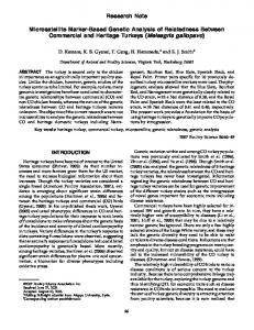
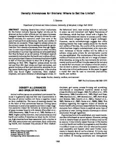
FIGURE 4. Evolutionary conserved charge character in the LMM. The positions of evolutionarily conserved positively charged amino acid side chain residues are represented by black rectangles, and negatively charged amino acid side chain residues by white rectangles. These clusters are arrayed on the solvent or outer surface of the LMM. The diagram is made by unwinding the a-helix in one of the coiled coils. Each repeat (28 amino acids) is stacked individually like rungs on a ladder with the next C-terminal repeat aligned underneath. The numbers to the left indicate the repeat number in the a-helix. The LMM is composed of the C-terminal 22 1/2 repeats of the rod from repeat 17 to repeat 40. Each repeat is composed of 4 heptads, labeled at the top of the figure. Gray boxes indicate neutral, uncharged or hydrophobic amino acid side chain residues. Hatched boxes indicate the position of skip residues in the LMM. Connected ovals at the bottom of the diagram represent the fact that these residues are in a random coil motif.
viewed by transmission electron microscopy (TEM). Synthetic filaments display most of the morphological characteristics of native thick filaments and are a good model to study the mechanism of myofibrillogenesis, myosin assembly, and the architecture of native thick filaments (Huxley, 1963; Josephs and Harrington, 1966; Katsura and Noda, 1971; Pollard, 1975; Pinset-Harstrom and Truffy, 1979; Reisler et al., 1982; Davis, 1985; Davis, 1988a). Synthetic thick filaments of myosin spontaneously assemble when the ionic strength is lowered < 0.2 M KCl. A putative mechanism of synthetic filament assembly (Figure 5) has been proposed based on the studies of Reisler and Davis (Davis et al., 1982; Davis, 1986; Reisler et al., 1986; Davis, 1988a). Monomers initially assemble into parallel dimers. Dimers assemble into antiparallel tetramers, tetramers into octamers. The octamers finally
739
assemble into a minifilament of 16 molecules, which is the nucleation core for additional assembly and corresponds to the central bare zone of the thick filament. Parallel dimers add on to the tips of the nascent filament in a bipolar fashion until the rate of addition of dimers equals the rate of dissociation of dimers, accounting for the narrow length distribution of synthetic filaments (Davis, 1981, 1985, 1986, 1988a; Davis et al., 1982). Employing analytical ultra-centrifugation analysis of salt dissociated synthetic filaments, it has been demonstrated that monomers, dimers, and a structure termed a minifilament, consisting of 16 to 18 myosin molecules arrayed in a tail-to-tail fashion, were intermediates in filament assembly (Reisler et al., 1986a). Furthermore, it has been demonstrated that parallel dimers are dissociated away from the tips of synthetic filaments under the influence of increasing hydrostatic pressure (Davis, 1985). Based on these studies, the following assembly mechanism was proposed: as the ionic strength is lowered, myosin monomers interact to form parallel myosin dimers, which then associate to form a minifilament consisting of myosin associated in an antiparallel arrangement (Reisler et al., 1982, 1986b). Studies on the effects of anti-rod monoclonal antibody Fab fragments on sarcomeric muscle myosin interactions in low salt conditions confirmed that regions in the C-
FIGURE 5. A putative mechanism of myosin assembly. Myosin monomers in high salt (A) spontaneously assemble into parallel dimers (B), minifilaments (C), and finally, synthetic filaments (D) as the ionic strength of the solution is reduced.
740
WICK
terminus of the LMM were responsible for the solubility properties of chicken Pectoralis major muscle myosin (Wick et al., 1997). Unique to these studies were the results that suggest the presence of domains in the N-terminus of the rod controlling the morphology of thick filaments and the architecture of the sarcomere. In contrast to sarcomeric myosin, nonmuscle and smooth muscle type II myosins produce stable minifilament-like structures only (Trybus, 1989; Sinard et al., 1990). Also, unlike sarcomeric myosins, nonmuscle and smooth muscle myosin assembly is regulated by phosphorylation of their heavy or light chains, reviewed elsewhere (Tan et al., 1992). However, sarcomeric myosin minifilaments are not stable under low salt conditions and continue to elongate into bipolar synthetic filaments. The filaments are normally 1.6 mm long and 15 nm in diameter by the addition of parallel dimers on to the ends of the emerging synthetic filament on either side of the central bare zone (Davis et al., 1982; Davis, 1988a). It has been proposed that the mechanism by which parallel dimers are incorporated on to the ends of the nascent filaments is responsible for the narrow size distribution of synthetic filaments observed in vitro. Kinetically, length is regulated by a dissociation rate constant that increases exponentially as the filament grows bidirectionally. Growth ceases at the point of equilibrium between the invariant “on” and the length-dependent “off” rates (Davis, 1986). The mechanism by which the length distribution of native and synthetic thick filaments is achieved is still a
major question in muscle biology. The mechanism has to be consistent with the results of kinetic experiments in which a cumulative property progressively destabilizes the structure of the dimer binding site of the nascent filament. Thermodynamic insight into the mechanism arises from the observation in the electron microscope that native filaments split into three subfilaments in low salt buffers (Huxley, 1963). The splitting is limited to the tips of the filament, where myosin is parallel packed. The central bare zone appears to remain intact. The stable subfilaments in the central bare zone appear to be formed by strong attractive electrostatic interactions, whereas repulsive ionic interactions appear to exist in the regions of the filament in which splits occur. Because filaments formed at pH 8.0 are shorter than filaments formed at pH 7.0, it has been postulated that the repulsive interactions responsible for the splitting of native filaments in low salt are negative in charge (Davis, 1988b).
THE PARACRYSTAL AS A MODEL OF ASSEMBLY In order to determine the location and presence of sequence elements in the rod domain of the MyHC and their roles in the mechanism of fibrillogenesis, sitedirected mutagenesis of recombinant proteins is becoming the tool of choice. However, a full-length functional hexameric recombinant sarcomeric muscle myosin
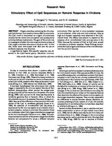
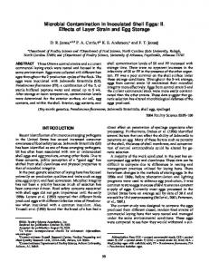
FIGURE 6. Myosin and rod fragments assemble into aggregates with different morphologies. A) A group of purified myosin molecules. B) Transmission electron micrograph shows a typical negatively stained synthetic filament. Synthetic filaments are relatively narrow in length and width distribution. The central bare zone is visible in the center of a tapered bipolar aggregate approximately 1.6 mm in length and 15 nm in diameter. C) Light meromyosin (LMM) fragments are shown to assemble into paracrystals. D) Transmission electron micrograph shows a typical negatively stained paracrystal. Paracrystals exhibit little or no length or width constraints. Bar = 0.5 mm.
SYMPOSIUM: MUSCLE GROWTH AND DEVELOPMENT
molecule has yet to be produced in vitro. Therefore, investigations into the mechanisms of fibrillogenesis currently employ enzymatically generated and genetically engineered recombinant MyHC rod fragments. Muscle and nonmuscle myosin rod fragments have been employed to study the intermolecular interactions responsible for filament assembly and solubility (Chowrashi and Pepe, 1977; Chowrashi et al., 1989; Ward and Bennett, 1989; O’Halloran et al., 1990; Atkinson and Stewart, 1992; Lee et al., 1994). Isolated LMM and rod fragments form paracrystals rather than filaments (Szent-Gyorgyi et al., 1960; Chowrashi and Pepe, 1977; Parry, 1981; Ishii and Lehrer, 1989; Stewart et al., 1989; Ward and Bennett, 1989; Atkinson and Stewart, 1991). In buffers of low ionic strength, the LMM fragment of the rod assemble into highly ordered aggregates termed paracrystals, with axial banding patterns based on odd multiples of 14 nm believed to reflect the architecture of the arrangement of myosin in the thick filament (Figure 6). The LMM fragments exhibit solubility characteristics which are indistinguishable from full length myosin. Hence, LMM fragments have proven to be good tools to investigate the assembly properties of sarcomeric myosins. Deletions of the C-terminal portion of a bacterially expressed recombinant LMM generated a fragment that remained soluble in low salt (Sinard et al., 1989, 1990; Atkinson and Stewart, 1991a). The contribution of the Cterminal 100 amino acids to the solubility and Nterminal sequence elements to the assembly of sarcomeric muscle myosin has been demonstrated with bacterially expressed LMM fragments (Atkinson and Stewart, 1991a). The results of these studies indicated the possibility of subdomains in the LMM that contribute to various low energy states of low salt aggregates of myosin and effect the morphology of native and synthetic filaments.
CONCLUSION MyHC rod fragments assemble into paracrystals, rather than the precisely regulated synthetic filaments, with a narrow distribution of length and width. These observations led to the hypothesis that the bulkiness of the S1 domain of myosin may influence the assembly process. In support of this hypothesis, it was shown that removal of the LC2 from native myosin affected the morphology of synthetic filaments, implicating the myosin head in the mechanism of filament assembly (Chowrashi and Pepe, 1989). We are performing experiments employing bacterially expressed recombinant rod proteins in order to study the sequence elements in the N-terminus of the MyHC involved in myosin’s assembly properties. These studies will lead to the discovery of previously undescribed domains in the MyHC. Analysis of the sequence elements in these domains will contribute our understanding of the intermolecular interactions occurring not only between individual myosin molecules that contribute to thick filament architecture,
741
but also between myosin molecules and with other constitutive myofibrillar proteins in determining the architecture of the sarcomere.
REFERENCES Atkinson, S. J., and M. Stewart, 1991a. Expression in Escherichia coli of fragments of the coiled-coil rod domain of rabbit myosin: influence of different regions of the molecule on aggregation and paracrystal formation. J. Cell Sci. 99: 823–836. Atkinson, S. J., and M. Stewart, 1991b. Molecular basis of myosin assembly: coiled-coil interactions and the role of charge periodicities. J. Cell Sci. 14:7–10. Atkinson, S. J., and M. Stewart, 1992. Molecular interactions in myosin assembly role of the 28-residue repeat in the rod. J. Mol. Biol. 226:7–13. Baba, M. L., M. Goodman, J. Berger-Cohn, J. G. Demaille, and G. Matsuda, 1984. The early adaptive evolution of calmodulin. Mol. Biol. Evol. 1:442–455. Bandman, E., L. A. Moore, M. J. Arrizubieta, W. E. Tidyman, L. Herman and M. Wick, 1994. The evolution of the chicken sarcomeric myosin heavy chain multigene family. Pages 129–139 in: Proceedings of Society of General Physiologists 47th Annual Symposium. The Rockefeller University Press, New York, NY. Cheney, R. E., M. A. Riley, and M. S. Mooseker, 1993. Phylogenetic analysis of the myosin superfamily. Cell Motil. Cytoskel. 24:215–223. Chowrashi, P. K., S. M. Pemrick, and F. A. Pepe, 1989. LC2 involvement in the assembly of skeletal myosin filaments. Biochim. Biophys. Acta 990:216–223. Chowrashi, P. K., and F. A. Pepe, 1977. Light meromyosin paracrystal formation. J. Cell Biol. 74:136–152. Cohen, C., and D.A.D. Parry, 1986. Alpha helical coiled coils— a wide spread motif in proteins. Trends Biochem. Sci. 11: 245–248. Cohen, C., and D.A.D. Parry, 1990. Alpha-helical coiled coils and bundles: how to design an alpha-helical protein. Proteins 7:1–15. Collins, J., 1976. Homology of myosin DTNB light chain with alkali light chains, troponin C and parvalbumin. Nature 259:699–700. Crick, F.H.C., 1953. The packing of a-helices: simple coiledcoils. Acta Cryst. 6:689–697. Davis, J. S., 1985. Kinetics and thermodynamics of the assembly of the parallel- and antiparallel-packed sections of synthetic thick filaments of skeletal myosin: a pressurejump study. Biochemistry 24:5263–5269. Davis, J. S., 1986. A model for length-regulation in thick filaments of vertebrate skeletal myosin. Biophys J. 50: 417–422. Davis, J. S., 1988a. Assembly process in vertebrate skeletal thick filament formation. Ann. Rev. Biophys. Biophys. Chem. 17:217–239. Davis, J. S., 1988b. Interaction of C-protein with pH 8.0 synthetic thick filaments prepared from the myosin of vertebrate skeletal muscle. J. Mus. Res. Cell Motil. 9: 174–183. Davis, J. S., J. Buck, and E. P. Greene, 1982. The myosin dimer: an intermediate in the self-assembly of the thick filament of vertebrate skeletal muscle. FEBS Lett. 140:293–297. Goodson, H. V., and J. A. Spudich, 1993. Molecular evolution of the myosin family: Relationships derived from compari-
742
WICK
sons of amino acid sequences. Proc. Nat. Acad. Sci. USA 90:659–663. Harrison, R. G., S. Lowey, and C. Cohen, 1971. Assembly of myosin. J. Mol. Biol. 59:531–535. Hickmand, C. L., 1970. Integrated Principles of Zoology. 4th rev. ed. The C. V. Mosby Co., Saint Louis, MO. Huxley, H. E., 1963. Electron microscope studies on the structure of natural and synthetic filaments from striated muscle. J. Mol. Biol. 7:281–308. Josephs, R., and W. F. Harrington, 1966. Studies on the formation and physical chemical properties of synthetic myosin filaments. Biochemistry 11:3474–3487. Katsura, I., and H. Noda, 1971. Studies on the formation and physical chemical properties of synthetic myosin filaments. J. Biochem. 69:219–229. Lee, R. J., T. T. Egelhoff, and J. A. Spudich, 1994. Molecular genetic truncation analysis of filament assembly and phosphorylation domains of Dictyostelium myosin heavy chain. J. Cell Sci. 102:2875–2886. Letai, A., and E. Fuchs, 1995. The importance of intramolecular ion pairing in intermediate filaments. Biochemistry 92: 92–96. Lowey, S., and D. Risby, 1971. Light chains from fast and slow muscle myosins. Nature 234:81–85. Margossian, S. S., A. K. Bhan, and H. S. Slayter, 1983. Role of the regulatory light chains in skeletal muscle actomyosin ATPase and in minifilament formation. J. Biol. Chem. 258: 13359–13369. McLachlan, A. D., and J. Karn, 1982. Periodic charge distributions in the myosin rod amino acid sequence match cross-bridge spacings in muscle. Nature 299: 226–231. McLachlan, A. D., and M. Stewart, 1975. Tropomyosin coiledcoil interactions: Evidence for an unstaggered structure. J. Mol. Biol. 98:293–304. McNally, E. M., R. Kraft, M. Bravo-Zehnder, D. A. Taylor, and L. A. Leinwand, 1989. Full-length rat alpha and beta cardiac myosin heavy chain sequences. Comparisons suggest a molecular basis for functional differences. J. Mol. Biol. 210:665–671. Mihalyi, E., and A. G. Szent-Gyorgyi, 1953. Trypsin digestion of muscle proteins. I. Ultracentrifugal analysis of the process. J. Biol. Chem. 201:189–196. Moos, R. L., G. M. Diffee, and M. L. Greaser, 1995. Contractile properties of skeletal muscle fibers in relation to myofibrillar protein isoforms. Rev. Physiol. Biochem. Pharmacol. 126:1–63. Nguyen, H. T., R. M. Gubits, R. M. Wydro, and B. NadalGinard, 1982. Sarcomeric myosin heavy chain is coded by a highly conserved multigene family. Proc. Nat. Acad. Sci. USA 79:5230–5234. O’Halloran, T. J., S. Ravid, and J. A. Spudich, 1990. Expression of Dictyostelium myosin tail segments in Escherichia coli: Domains required for assembly and phosphorylation. J. Cell Biol. 110:63–70. Parry, D. A., 1981. Structure of rabbit skeletal myosin. Analysis of the amino acid sequences of two fragments from the rod region. J. Mol. Biol. 153:459–464. Pinset-Harstrom, I., and J. Truffy, 1979. Effect of adenosine triphosphate, inorganic phosphate and divalent cations on the size and structure of synthetic myosin filaments. J. Mol. Biol. 134:173–188.
Pollard, T. D., 1975. Electron microscopy of synthetic myosin filaments. Evidence for cross-bridge flexibility and copolymer formation. J. Cell Biol. 67:93–104. Reisler, E., P. Cheung, and N. Borochov, 1986a. Macromolecular assemblies of myosin. Biophys J. 49:335–342. Reisler, E., P. Cheung, N. Borochov, and J. A. Lake, 1986b. Monomers, dimers, and minifilaments of vertebrate skeletal myosin in the presence of sodium pyrophosphate. J. Biochem. 25:326–332. Reisler, E., P. Cheung, C. Oriol-Audit, and J. A. Lake, 1982. Growth of synthetic myosin filaments from myosin minifilaments. Biochemistry 21:701–707. Rice, R. V., 1961. Conformation of individual macromolecular particles from myosin solutions. Biochim. Biophys. Acta 52:602–604. Rosser, B.W.C., and J. C. George, 1986a. The avian pectoralis: histochemical characterization and distribution of muscle fiber types. Can. J. Zool. 64:1174–1185. Rosser, B.W.C., and J. C. George, 1986b. Slow muscle fibers in the pectoralis of the turkey vulture (Cathartes aura): an adaptation for soaring flight. Zool. Anz. 217:252–258. Seiler, S. H., D. A. Fischman, and L. A. Leinwand, 1996. Modulation of myosin filament organization by C-protein family members. Mol. Biol. Cell 7:113–127. Sinard, J. H., D. L. Rimm, and T. D. Pollard, 1990. Identification of functional regions on the tail of Acanthamoeba myosin-II using recombinant fusion proteins. II. Assembly properties of tails with NH2- and COOHterminal deletions. J. Cell Biol. 111:2417–2426. Sinard, J. H., W. F. Stafford, and T. D. Pollard , 1989. The mechanism of assembly by three successive dimerization steps. J. Cell Biol. 109:1537–1547. Sjostrom, M., and J. M. Squire, 1977. Fine structure of the Aband in cryo-sections: The structure of the A-band of human skeletal muscle fibres from ultra-thin cryo-sections negatively stained. J. Mol. Biol. 109:49–68. Squire, J. M., and P. J. Vilbert, 1987. Pages 247–281 in: Fibrous Protein Structure. Academic Press, London, U.K. Strehler, E. E., M.-A. Strehler-Page, J.-C. Perriard, M. Periasamy, and B. Nadal-Ginard, 1986. Complete nucleotide and encoded amino acid sequence of a mammalian myosin heavy chain gene: Evidence against introndependent evolution of the rod. J. Mol. Biol. 190:291–317. Strzelecka-Golaszewska, H., L. Nyitray, and M. Balint, 1985. Paracrystalline assemblies of light meromyosins with various chain weights. J. Mus. Res. Cell Motil. 6:641–658. Tan, J. L., S. Ravid, and J. A. Spudich, 1992. Control of nonmuscle myosins by phosphorylation. Ann. Rev. Biochem:721–759. Trybus, K. M., 1989. Filamentous smooth muscle myosin is regulated by phosphorylation. J. Cell Sci. 109:2887–2894. Ward, R., and P. Bennett, 1989. Paracrystals of myosin rod. J. Muscle Res. Cell Motil. 10:34–52. Wick, M., F. Tablin, and E. Bandman, 1997. Effects of antiLMM antibodies on the solubility of chicken skeletal muscle myosin. J. Food Biochem. 20:379–395. Xiong, Y. L., 1992. Thermally induced interactions and gelation of combined myofibrillar protein from white and red broiler muscles. J. Food Sci. 577:582–585. Xiong, Y. L., 1994. Myofibrillar protein from different muscle fiber types: Implications of biochemical and functional properties in meat processing. Crit. Rev. Food Sci. Nutr. 34:293–320.