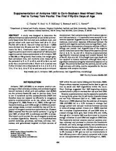
The Effect of Avian Uterine Fluid on the Growth Behavior of Calcite Crystals J. M. Dominguez-Vera,* J. Gautron, J. M. Garcia-Ruiz,† and Y. Nys1 *Department de Quimica Inorganica, †CSIC, University of Granada 18000 Spain, Station de Recherches Avicoles, centre de Tours, 37380 Nouzilly, France ABSTRACT Eggshell formation takes place on the eggshell membrane in an acellular medium, the uterine fluid that contains the inorganic minerals and precursors of the organic matrix. The high degree of eggshell structure could be due to an interaction between calcium carbonate and the organic matrix. The aim of this study was to demonstrate such an interaction by measuring the effect in vitro of uterine fluid collected at various phases of shell formation on precipitation kinetics, size, and morphology of calcite crystals. The SDS-PAGE profiles of the organic constituents differed between the different phases of eggshell formation. The predominant constituents were ovalbumin and ovotransferrin at the initial phase and lysozyme, ovocleidin-17, ovocalyxin-32, 36- and 21kDa bands, and ovocleidin-116 at the growth phase. These proteins were numerous in the terminal phase and
showed an increased staining of the 32- and 66-kDa bands and appearance of very low molecular weight bands. The precipitation lag time was shortened in proportion to the protein concentration at the initial stage. The effect was observed with a lower magnitude in the presence of constituents of growth and terminal phases. Crystal size was smaller in the presence of constituents from the three stages compared with the control. Components from the initial phase induced the formation of twinned crystals and of rounded corners in the rhombohedric crystals. The presence of components from the growth and terminal phases strongly modified the morphology of the calcite crystals. The majority of the corners became rough and developed curved faces. These observations confirm the interaction of the uterine fluid with calcite and its contribution to eggshell structure.
(Key words: hen, eggshell, uterus, calcite, mineralization) 2000 Poultry Science 79:901–907
the result of a precipitation phenomenon on the eggshell membrane. It is hypothesized that the nucleation and growth phases of calcite crystals are both influenced by molecules of the organic matrix (Nys et al., 1999). Therefore, identification of the different macromolecules existing in the crystallization medium (Carrino et al., 1996; Gautron et al., 1999) and knowledge of the type of interaction between them and precritical calcium carbonate nuclei and calcite crystal faces are obligatory steps toward understanding eggshell formation. The eggshell forms into an acellular fluid environment secreted by the uterus. This uterine fluid contains the inorganic components of the eggshell at a supersaturated level relative to calcite (Nys et al., 1991) and the precursors of the eggshell matrix molecules (Gautron et al., 1997a). The uterine protein profile changes during the three different stages of the eggshell formation (Gautron et al., 1997a), namely, a) the initial phase when nucleation of calcite crystals occurs and the mammillary cores form, b) the growth phase when the eggshell is deposited at a constant rate, and c) the final phase when mineral deposition ends.
INTRODUCTION The mineral composition of the eggshell is basically calcite, the most stable polymorph of calcium carbonate. The existence of a remarkable control of calcite crystal morphology throughout the layered eggshell has been emphasized many times from the pioneering work of von Nathasius until the most recent studies (Dennis et al., 1996). This structural organization is accompanied by a textural gradient (Sharp and Silyn-Roberts, 1984; Ka¨lin, Rodriguez-Navarro, Nys and Garcia-Ruiz, unpublished data). The formation of this fabric gradient occurs in a confined acellular space, the uterine lumen, where the eggshell forms. However, there is no doubt that the morphological and kinetic features of calcite precipitation during eggshell formation are in some way tailored. Such a high degree of control including shape, size, location, and specific orientation of the calcite crystals is due to the interaction of calcium carbonate with the organic matrix (Addadi and Weiner, 1992; Arias et al., 1993; Nys et al., 1999). The formation of the eggshell is
Received for publication August 3, 1999. Accepted for publication August 3, 1999. 1 To whom correspondence should be addressed:
[email protected].
Abbreviation Key: PGF2α = prostaglandin
901
902
DOMINGUEZ-VERA ET AL.
The aim of this study was to demonstrate that uterine fluid collected at different phases of eggshell formation exerted a differential influence upon calcite growth. Therefore, a simple and highly reproducible method was developed for studying in vitro the precipitation kinetics and morphology of the resulting calcite crystals in the presence of uterine fluid collected at the three distinct stages of eggshell formation.
MATERIALS AND METHODS Sampling and Analysis of the Uterine Fluids Sixty Brown-egg laying hens (Isa-Brown) were housed individually in cages located in a windowless, air-conditioned poultry house. They were subjected to a cycle of 14 h light:10 h darkness and were fed ad libitum on a layer diet as recommended by the Institut National de la Recherche Agronomique (1989). Cages were equipped with a computerized system to record the daily oviposition times. Ovulation was considered to occur 0.5 h after oviposition. Eggs were expelled in response to an intravenous injection of 50 µg of prostaglandin/hen (PGF2α) at 6 to 9 h (initial phase), 18 to 19 h (growth phase), and 22 to 23 h (terminal phase of shell calcification) after the preceding oviposition (10 to 16 hens per stage). Uterine fluid was collected immediately after egg expulsion, by gravity, into a plastic test tube placed at the entrance of the everted vagina. Aliquots of uterine fluid were diluted 1:1 in SDS-PAGE buffer (0.0625 M TrisHCl, pH 6.8; 2% SDS; 5% β-mercaptoethanol; 25% glycerol; and 0.01% bromophenol blue) just after collection to limit any spontaneous precipitation of calcium carbonate and proteins. Aliquots were also taken to determine the protein concentration according to the Bradford procedure with ovalbumin as a standard. The remainder was frozen in liquid nitrogen for the in vitro crystal growth assay. SDS-PAGE was performed on 5 to 20% gradient gels as described by Gautron et al. (1997a). Proteins were stained with Coomassie blue.
Crystallization Method The crystallization method was based on the sitting drop method. A polystyrene microbridge was filled with 35 µl of the calcium chloride solution (0.2 M). Seven microbridges were fixed to a Petri dish having a hole. The dish was centered on a cylindrical vessel (50 mm in diameter and 30 mm in height) that contained 3 mL of an ammonium bicarbonate solution (0.025 M). The system (shown in Figure 1) was closed, and varying the pH of the solution of calcium chloride controlled supersaturation. The diffusion of ammonia and the asso-
FIGURE 1. Experimental setup used for growing in vitro calcite crystals. The concentration of CaCl2 (microbridge) and of ammonium bicarbonate was 0.2 M and 0.025 M, respectively.
ciated increase in pH resulted in transformation of CO2 into carbonate. If the critical supersaturation for the nucleation of calcite (ks = 2.8 × 10⁻⁹ at 20°C in pure water) is reached, the crucial point is how fast this process occurs. Therefore, the conditions of the kinetics of calcium carbonate equilibration in the drop were initially considered. The main experimental variables were a) the source of ammonia being either ammonium carbonate or bicarbonate and b) the concentration of calcium chloride and ammonia source. The variables were optimized to develop a simple and highly reproducible method yielding a low number of crystals (less than 50), of relatively large size (about 100 µm), and isolated from each other, to allow the effect of additives to be easily discerned. The best results were obtained with the previously described experimental procedure. Three types of samples were studied, corresponding to the uterine fluid collected at the initial, growth, and terminal phases of eggshell formation. Each experimental test comprised seven simultaneous precipitation experiments with three microbridges used as internal controls (only containing the calcium solution) and four microbridges containing the calcium solution plus the effector at different concentrations (10 to 100 µg protein/ mL). The process was followed by optical microscopy2, to determine the precipitation lag time (appearance of the first crystals) and to measure the size of crystals either in the presence or in the absence of the effector. We measured the precipitation lag time as a function of the concentration of effector. The calcite crystals were collected 17 h after the initiation of the experiments, twice rinsed with double-distilled water, dried at room temperature, and then coated with gold and studied by scanning electron microscopy. Two-way analysis of variance (stage and protein concentration) was carried out using the Systat software package3.
RESULTS AND DISCUSSION Characterization of the Uterine Fluid
2
SZH10 Olympus. Systat Inc., Evanston, IL 60204.
3
The SDS-PAGE profiles of the organic constituents of the uterine fluid collected at initial, growth, and terminal
UTERINE FLUID AND CALCITE CRYSTAL GROWTH
FIGURE 2. Electrophoretic profile (SDS-PAGE) of uterine fluid collected at the initial phase (6 to 9 h), growth phase (18 to 19 h), and terminal phase (22 to 23 h) of eggshell formation (Coomassie blue staining).
phases of eggshell mineralization are shown in Figure 2. The uterine fluid composition changed with the stage of calcification, as previously demonstrated (Gautron et al., 1997a). At the initial stage, intense bands corresponded to proteins of the eggwhite, ovalbumin (45 kDa), and ovotransferrin (80 kDa) and at reduced-staining intensity to lysozyme (15 kDa). Eggshell matrix proteins specific to the uterine tissue, such as ovocleidin-17 (Hincke et al., 1995), ovocalyxin-32 (Gautron et al., 2000), and a 21-kDa band (Gautron, Hincke, and Nys, unpublished data), were also observed. Intensely stained bands corresponding to 36-kDa and 21-kDa proteins characterized the uterine fluid collected during the rapid phase of eggshell deposition. The band corresponding to lysozyme was also strongly stained. Another peculiarity at this stage was the appearance of a 116-kDa band that corresponded to ovocleidin-116, the core protein (Hincke et al., 1999) of a proteoglycan rich in dermatan sulfate (Carrino et al., 1997). Moreover, a 66-kDa band was also observed. The electrophoretic profile of the uterine fluid collected at the terminal phase of eggshell calcification showed numerous bands that were also observed during the previous period [lysozyme and ovocalyxin-32, as well as the 21-kDa and 45-kDa (low intensity) bands]. At this stage, we observed increased staining intensity for the 32- and 66-kDa bands and the appearance of very low molecular weight bands (