JOURNAL OF BACTERIOLOGY, Aug. 2001, p. 4477–4483 0021-9193/01/$04.00⫹0 DOI: 10.1128/JB.183.15.4477–4483.2001 Copyright © 2001, American Society for Microbiology. All Rights Reserved.
Vol. 183, No. 15
Eukaryotic Translation Initiation Factor 4E-Dependent Translation Is Not Essential for Survival of Starved Yeast Cells IRIT PAZ
AND
MORDECHAI CHODER*
Department of Molecular Microbiology and Biotechnology, Tel-Aviv University, Ramat Aviv 69978, Israel Received 18 September 2000/Accepted 15 May 2001
The eukaryotic translation initiation factor 4E (eIF4E) interacts with the mRNA 5ⴕ cap structure (m7GpppX) and is essential for the appropriate translation of the vast majority of eukaryotic mRNAs. Most studies of the yeast Saccharomyces cerevisiae CDC33 gene product, eIF4E, have been carried out with logarithmically growing cells, and little is known about its role in starved, nonproliferating cells that enter the stationary phase (SP). It has previously been found that the rate of translation in SP cells is more than 2 orders of magnitude lower than it is in dividing yeast cells. Here we show that this low rate of translation is essential for maintaining the viability of starved yeast cells that enter SP. Specifically, starved cells whose eIF4A is inactive or treated with cycloheximide rapidly lose viability. Moreover, after heat inactivation of the cdc33 temperature-sensitive product, the synthesis of most proteins is abolished and only a small group of proteins is still produced. Unexpectedly, starved cdc33 mutant cells whose eIF4E is inactive and which therefore fail to synthesize the bulk of their proteins remain viable for long periods of time, indistinguishable from their isogenic wild-type counterparts. Taken together, our results indicate that eIF4E-independent translation is necessary and sufficient for survival of yeast cells during long periods of starvation. expression and its complex regulation in dividing cells (e.g., see reference 19). In the past several years, findings that suggest that, during the stationary phase (SP) of the yeast growth cycle, regulation of gene expression is different from that studied in dividing cells have been gradually and slowly accumulating. Thus, in nondividing cells expression of most genes is repressed both at the transcriptional (9) and the translational (13) level. Nevertheless, expression of a small group of genes is maintained (13). Expression of these genes during SP is probably controlled differently than that prevailing in dividing cells. As SP in yeast is considered to be analogous to the G0 state in higher eukaryotic cells (reference 30 and references therein), understanding gene expression in starved nondividing yeast may serve as a model for studying gene expression during the G0 state. In mammals, starvation can lead to a partial inactivation of eIF4E by either affecting its phosphorylation status or increasing its association with eIF4E-binding proteins (29). Nevertheless, the consequence of eIF4E repression on the regulation of translation is not fully understood. Recently, the TOR-mediated signal transduction pathway has been implicated in signaling the status of nutrient availability in yeast by controlling cap-dependent translation. Inhibition of the two yeast TORs, TOR1 and TOR2, by rapamycin results in a global inhibition of cap-dependent translation (4). Interestingly, rapamycin treatment of logarithmically growing cells leads to the acquisition of many parameters characteristic of SP (4), suggesting that the TOR-mediated repression of cap-dependent translation is naturally associated with, or even signals, the entry into SP. Moreover, we have recently found that starved, but not dividing, yeast cells have the capacity to identify IRESs and to carry out translation independently of the 5⬘ end of the mRNA. This finding raises the possibility that cap-independent translation plays an important role in gene expression during SP. Taken
Regulation of translation initiation is a key process in gene expression in eukaryotes. Much is known about the complex pathway of the initiation process in vivo through the numerous studies that have used growing cells as model systems (reviewed in references 16 and 19). It is well established that in growing cells the cap structure, located at the 5⬘ end of the eukaryotic mRNA, plays a pivotal role in recruiting the ribosome to the mRNA. The cap-dependent recruitment of the translation initiation apparatus near the 5⬘ end of the mRNA is followed by a scanning process until the first initiation codon is met and the translation process begins (16, 19). The initial recognition of the cap structure is carried out by the eukaryotic translation initiation factor 4F (eIF4F) complex, composed of (i) eIF4E, which physically interacts with the cap structure (8, 18) and which in Saccharomyces cerevisiae is encoded by a single gene, CDC33 (1, 7); (ii) eIF4G, which serves as a scaffold protein that binds several initiation factors as well as the mRNA (15); and (iii) eIF4A, which, in conjunction with eIF4B, catalyzes the ATP-dependent melting of the RNA secondary structure (24). Inactivation of either component of the eIF4F complex in yeast leads to inhibition of cap-dependent translation (19). Although cap-dependent translation is the major mechanism of translation initiation, other mechanisms have also been documented. The most studied cap-independent mechanism is internal ribosome entry sequence (IRES)-mediated translation (reviewed in references 6 and 26). The yeast S. cerevisiae is one of the popular model systems for studying gene expression in eukaryotes. The advanced genetics and molecular biology of the yeast system have been used to discover many novel factors that play a role in gene * Corresponding author. Mailing address: Department of Molecular Microbiology and Biotechnology, Tel-Aviv University, Ramat Aviv 69978, Israel. Phone: 972–36409030. Fax: 972–25334140. E-mail:
[email protected]. 4477
4478
J. BACTERIOL.
PAZ AND CHODER
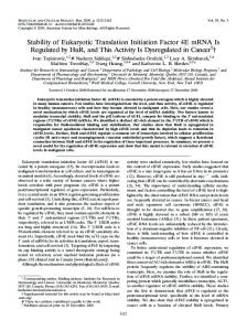
FIG. 1. The eIF4E level decreases following the shift from log phase to SP. Wild-type (WT) cells and their isogenic cdc33-42 counterparts (see Materials and Methods) were grown in YPD at 25°C. Samples of equal amounts of cells were harvested at either mid-logarithmic growth phase (1 ⫻ 107 cells/ml) (lane Log), 24 h later when cells had just entered SP (1.5 ⫻ 108 cells/ml) (lane SP0), or 9 days later (1.5 ⫻ 108 cells/ml) (lane SP9). Proteins were extracted, and 100-g samples were analyzed by Western analysis with anti-eIF4E antibodies that recognize one major band at the expected molecular weight, as detailed in Materials and Methods. Equal loadings were verified by ponsau S staining (not shown) and by the presence of a faint band corresponding to a 40-kDa protein that cross-reacted with the anti-eIF4E antibodies (marked by an arrow). Overexposure was required to detect the cross-reactive protein. (A) Analysis of eIF4E extracted from the wild type. (B) Analysis of mutant eIF4E extracted from cdc33–42 mutant cells. (C) Quantitative analysis. One hundred micrograms of protein (left lanes) or an equal volume of the indicated dilutions (right lanes) was analyzed by the Western blot technique as described for panels A and B except that in this experiment the film was exposed for various lengths of time. Shown is the longest exposure time used to detect the faint band in lane SP9. The values indicated at the bottom of panels A and B are calculations of the fold reduction in the level of eIF4E in SP cells compared with its level in log-phase cells. The values in panel A were determined by scanning the film with an ImageScanner (Amersham Pharmacia Biotech) and using the ImageMaster 1D prime program (Pharmacia). The values in panel B were based on the data shown in panel C. For calculating the fold reduction, the film was scanned and the final results were based on both the fold dilution and results obtained by the scanner (whose linear range is limited).
together, these observations provoked us to examine the role of eIF4E in regulating translation during SP. Here we show that, in SP, eIF4E-dependent translation is responsible for most of the synthetic activity in SP. Yet, the synthesis of a protein(s) that is essential for coping with longterm starvation periods seems to be mediated by eIF4E-independent translation. MATERIALS AND METHODS Yeast strains and media. CWO4 (MAT␣ ura3-1 ade2-1 his3-11,15 trp1-1 leu23,11) (3) was used as the wild type in all experiments. cdc33-42 and tif1-1 mutants are temperature-sensitive derivatives of CWO4 and are described in reference 11. Cells were grown on a synthetic complete medium lacking the appropriate amino acids or in YPD medium (2% Bacto Peptone, 1% yeast extract [Difco Laboratories], 2% dextrose) at 25°C (28). Growth in liquid medium was as described previously (20). The beginning of SP was defined as the point during the growth cycle when the number of cells no longer increased. After entry into SP, cultures were further shaken or rotated in a roller drum for the time periods indicated in the figures. Cell viability assay. Cell viability was determined by plating a known number of cells on rich medium (YPD) plates and counting the number of colonies produced after at least 1 week of incubation at 25°C, as was described previously (27). Incorporation kinetics. Cells were grown on a synthetic medium lacking methionine at the temperatures indicated in the figures. Cells were collected by centrifugation and resuspended in the supernatant to a final concentration of 1 ⫻ 108 to 1.5 ⫻ 108 cells/ml. An aliquot of 50 l (2 ⫻ 105 cells) was taken and equilibrated for 10 min at the temperatures indicated in Fig. 3. 35S Trans-Label (Amersham) was then added to a final concentration of 430 Ci/ml. Samples (2 l) were withdrawn at the time points indicated in the figures and spotted onto a dry 3MM paper which had been soaked in 10 mM EDTA and then air dried. After all samples were collected, the 3MM paper was subjected to trichloroacetic acid precipitation as described previously (23). Protein extraction and Western blot (immunoblot) analysis. Proteins were extracted as described previously (10, 27). Equal volumes of protein samples (100 g) were subjected to electrophoresis in 5-to-15% gradient polyacrylamide gels
(1.5 mm by 16 cm by 20 cm). Western analysis was done as described previously (10, 27). Anti-eIF4E antibodies (9B12 monoclonal antibody) were a generous gift of N. Sonenberg and used at a 1:10 dilution.
RESULTS Previously, Werner-Washburne and her colleagues found that, following the shift from logarithmic growth phase to the SP, the overall rate of translation in yeast cells decreases substantially. It gradually decreases even further as cells progress into later stages of the SP, and it is then more than 2 orders of magnitude lower than it was during the logarithmic growth phase (13). There were two possible explanations for this decrease in translation: the relatively low levels of transcripts or another mechanism of translational repression. Using a polysomal fractionation technique, we found that, in SP, most of the poly(A)containing mRNAs are not associated with ribosomes (results not shown), indicating that the level of the global mRNA is not a limiting factor. This observation suggests that, in SP, the translational initiation of the bulk mRNA is repressed. Since eIF4E is a key factor that regulates translation initiation, we monitored its level throughout growth to SP. Figure 1A shows that the level of eIF4E in SP cells was significantly lower than it was in logarithmically growing cells. Nevertheless, this decrease (two- to fourfold) is smaller than the apparent repression of the overall translation, suggesting that eIF4E is not a limiting factor for translation during SP. However, it is still possible that eIF4E activity was repressed by means other than by controlling its steady-state level, such as by interacting with a repressor or by modifying posttranslational processes
VOL. 183, 2001
eIF4E-INDEPENDENT TRANSLATION IN STARVED YEAST
4479
FIG. 2. The level of the cdc33–42 product declines when growing or starved nongrowing cells are exposed to 34°C. Wild-type (WT) cells and their isogenic cdc33–42 counterparts were grown in YPD at 25°C. At mid-logarithmic growth phase (1 ⫻ 107 cells/ml), cell samples were taken and the rest of the culture continued to grow to SP. The log-phase samples were either incubated at 25°C or shifted to 34°C. Four hours later, equal numbers of cells were harvested and their proteins were extracted. One day after cells had entered SP, the procedure was repeated. Samples (100 g) were analyzed by Western analysis as described in the legend to Fig. 1. Equal loadings were verified as described for Fig. 1.
(reviewed in reference 19). An extreme case of repression would be such that eIF4E activity is completely blocked so that this factor does not play an active role in translation. To determine unambiguously whether or not eIF4E is still required for translation during SP, a temperature-sensitive allele of eIF4E, cdc33-42 (2), was employed. Unlike other known temperature-sensitive alleles of CDC33, cdc33-42 is already inactivated at 32°C (an almost optimal temperature for yeast growth) (5). We chose this allele for our experiments to minimize uncertainties that might have arisen due to the exposure of cells to high temperatures. Note that, in order to make sure that heat inactivation did occur, we used 34°C as our nonpermissive temperature, a still slightly stressful temperature. Monitoring of the steady-state level of the cdc33-42 product at the permissive temperature (25°C) by Western analysis revealed that during the logarithmic phase it was comparable to that of wild-type eIF4E but that during SP it was substantially lower than that of the wild type (Fig. 1B). Quantitation based on data shown in Fig. 1C indicates that, by 9 days in SP, the level of the cdc33-42 product was 280-fold lower than it was during logarithmic growth. The low level of the mutant form of eIF4E was probably due to its being recognized by some SP-active degradation machinery as a defective protein even at the permissive temperature. This likely degradation is not common in SP (see Discussion). To ascertain that the cdc33-42 product became inactive when SP cells were exposed to the nonpermissive temperature (34°C), we took advantage of the observation that heat inactivation led to degradation of the cdc33-42 product (17). Indeed, the protein level decreased following exposure of growing cells to the nonpermissive temperature (Fig. 2, left panel), which is consistent with previous observations (17). When cells in SP were exposed to the nonpermissive temperature, the cdc33-42 product level, which was lower than that of the wild type, decreased even further (Fig. 2, right side), suggesting its inactivation. To examine heat inactivation of the cdc33-42 product by a more direct and functional means, the incorporation kinetics of [35S]methionine was determined. Figure 3A shows that, following exposure of logarithmically growing cdc33-42 cells to
the nonpermissive temperature, the incorporation kinetics markedly decreased. These results are in accord with previously published results (2, 5). As expected (13), the incorporation kinetics in SP cells was markedly lower than it was in logarithmically growing cells and the kinetics were similar in the two starved strains (Fig. 3B). However, unlike the detectable translational capacity of both strains at the permissive temperature, at the nonpermissive temperature, protein synthesis in the mutant cells was undetectable by the means and conditions that we used. These results demonstrate that the cdc33-42 product became inactive both in logarithmically growing cells and in SP cells and indicate that eIF4E is required for the bulk of protein synthesis during SP. Is eIF4E required for the synthesis of all proteins? To determine whether some residual protein synthesis occurs in cdc33-42 cells at the nonpermissive temperature, we used a synthetic medium lacking methionine, instead of the methionine-containing medium (YPD) that was used in the experiments shown in Fig. 3A to D. Under these labeling conditions, a significant, albeit very low, level of incorporation was detected (Fig. 3E). We then analyzed the proteins that were synthesized at 34°C by polyacrylamide gel electrophoresis and found that synthesis of the vast majority of the proteins was completely blocked upon heat inactivation of eIF4E. However, a small group of proteins were synthesized in the mutant cells (Fig. 3E, inset). The results demonstrate that de novo synthesis of proteins was carried out in the starved cdc33-42 cells at the nonpermissive temperature. Relative to the rate of protein synthesis in logarithmically growing cells, the rate of protein synthesis during SP is marginal (reference 13 and our unpublished results) (Fig. 3) and might not be important. We therefore wished to determine whether this residual translation activity is required for maintaining the viability of cells in SP. As most of the translational activity in SP is dependent on eIF4E (Fig. 3), we used a cdc33-42 mutant protein to block the synthesis of the residual proteins. To this end, cdc33-42 cells and the isogenic wild-type counterparts were grown at 25°C to mid-log phase or the beginning of SP and then shifted to 34°C. Cell viability was then monitored by determining plating efficiency as a function of
4480
PAZ AND CHODER
J. BACTERIOL.
FIG. 3. Inactivation of eIF4E in both growing and starved cells results in substantial translational inhibition. (A to D) Wild-type (WT) cells and their isogenic cdc33–42 counterparts were grown in YPD at 25°C to mid-logarithmic growth phase (A and B) or until 12 h after they entered SP
VOL. 183, 2001
eIF4E-INDEPENDENT TRANSLATION IN STARVED YEAST
time at the nonpermissive temperature. As expected, when logarithmically growing cells were shifted to the nonpermissive temperature, cdc33-42 mutant cells died while isogenic wildtype cells remained viable (Fig. 4A). Surprisingly, when a temperature shift up was executed after cells had entered SP, the death curve of the mutant cells was identical to that of the wild-type cells (Fig. 4B). Importantly, the lack of effect of eIF4E inactivation on viability during SP is not dependent on the genetic background and is not allele specific (see the legend to Fig. 4). To determine whether protein synthesis is required for maintaining viability in SP cells, translation elongation was blocked by the addition of cycloheximide. Results clearly indicate that protein synthesis is an essential activity for maintaining viability not only during log phase (Fig. 4A) but also during SP (Fig. 4B). The importance of the translation process for survival in SP was verified using tif1-1 cells, which express a temperature-sensitive mutant form of eIF4A, another subunit of eIF4F that is essential for both cap-dependent and capindependent translation (22). After shifting either growing (Fig. 4C) or stationary (Fig. 4D) tif1-1 cells to the nonpermissive temperature, a relatively rapid death was observed. Rapid death of starved cells was also obtained when cells expressing a temperature-sensitive mutant form of the poly(A)-binding protein (Pab1pF364L) (25) were examined at the nonpermissive temperature (results not shown). Figure 4 shows that protein synthesis is required for surviving long periods of starvation during SP but that the synthesis of proteins that are essential for viability in SP is carried out by an eIF4E-independent and eIF4A-dependent mechanism. DISCUSSION Dividing cells require a continuous synthesis of proteins, mainly for supplying building blocks to daughter cells, for growth in size, and for the control of the complex process of the cell division cycle. Naturally, quiescent cells that enter G0 do not need to synthesize proteins for these purposes. Therefore, the global protein synthesis shutoff that occurs after cells enter SP has probably evolved, at least in part, to preserve precious energy that is required for synthesizing these proteins. Previously, we demonstrated that, in SP, transcription of most genes is actively repressed (9). However, in spite of transcription repression, the mRNA level is not a limiting factor for protein synthesis during SP (see Results and below). Thus, we
4481
conclude that translation is also repressed. The detailed mechanism underlying the robust decline in protein synthesis remains to be determined. Concomitantly with transcriptional and translational repression, yeast cells have evolved a mechanism to maintain constant mRNA and protein levels during SP by repressing mRNA degradation (14) and most likely by repressing protein degradation (13, 21). Note, however, that not all mechanisms of protein degradation are repressed in SP. For example, here we show that the heat-inactive cdc33-42 product is degraded during SP. Nevertheless, it seems that yeast has evolved a simple strategy to cope with the energy limitations elicited by starvation by preserving energy on one hand and maintaining critical levels of stable macromolecules on the other hand. A question that might have arisen, then, is whether or not de novo protein synthesis is essential for coping with starvation. Experiments reported here clearly demonstrate that translation is required to sustain life during SP. Nevertheless, the bulk of this (low-level) translation can be blocked by inactivating eIF4E, with no detectable effect on the survival of the starved cells. Why, then, are starved yeast cells engaged in such a dispensable translation activity? Since protein synthesis in starved cells is extremely low (see the introduction), it is possible that the translation that is observed during SP is due to an incomplete repression of cap-dependent translation (“leaky translation”). Alternatively, it is possible that residual protein synthesis is important to cope with some natural conditions of starvation that cannot be mimicked in the test tube. The long-term survival of starved cells whose cdc33-42 product is heat inactivated strongly suggests that proteins whose synthesis is dependent on eIF4E are not required for maintaining viability in SP. Hence, a protein(s) whose synthesis is essential to survive starvation is synthesized by eIF4E-independent translation. Alternatively, it is possible that heat inactivation was not complete so that the residual activity of eIF4E was sufficient to sustain long-term starvation. We regard this second possibility as unlikely for the following reasons. (i) Heat inactivation of eIF4E blocked translation in starved cells and rendered it almost undetectable. Polyacrylamide gel electrophoresis analysis demonstrated that synthesis of the vast majority of the proteins was completely blocked upon heat inactivation of eIF4E. Thus, heat inactivation of eIF4E was very efficient. Was it efficient enough? The answer to this question can be provided by comparing the translation rates in starved cdc33-42 cells at the nonpermissive temperature and in starved
(C and D), when half of each sample was rapidly shifted to 34°C while the other half was left at 25°C. Cell samples were shaken at the indicated temperatures for an additional 4 h, and then each sample was treated as follows. (A and B) Cells (5 ⫻ 107) were collected by centrifugation, resuspended in 0.5 ml of the supernatant, and immediately incubated at either of the indicated temperatures. For determining incorporation kinetics, 50 l of a cell sample was taken, put in an Eppendorf tube, and incubated at the indicated temperatures for 5 min. Radioactive material was then added, and incorporation kinetics were determined as described in Materials and Methods. (C and D) Samples (50 l) of cells in SP were put in an Eppendorf tube and incubated for 5 min at the indicated temperatures and further treated as described for panels A and B. (E) Wild-type cells and their isogenic cdc33–42 counterparts were grown in synthetic complete medium lacking methionine to SP. One day after entering SP, the culture was shifted to 34°C and shaken for an additional 4 h. Samples (50 l) of cells were put in an Eppendorf tube and incubated for 5 min at 34°C and further treated as described for panels A and B, except that twice as much radioactive material was added. Note that, since the medium lacked methionine, the specific activity of the radioactive methionine was much higher than it was in the experiments whose results are shown in panels A to D. In the inset, wild-type cells and their isogenic cdc33–42 counterparts were grown in synthetic complete medium lacking methionine. Three days after cells entered SP, the culture was shifted to 34°C and shaken for an additional 16 h. Samples (500 l) of cells were labeled for 1 h at 34°C with 300 Ci/ml. Because protein degradation in SP is repressed (13, 21), labeling of proteins for 1 h reflects mainly, if not exclusively, their synthesis rates (21). Proteins were extracted as detailed in Materials and Methods, and 10 g of protein was loaded per lane, electrophoresed, and fluorographed as described in Materials and Methods. Note the different scales of the y axes of the various panels.
4482
PAZ AND CHODER
J. BACTERIOL.
FIG. 4. During SP, eIF4A-mediated and cycloheximide-sensitive translation is essential for cell viability but inactivation of eIF4E has no effect on viability. Wild-type (WT) cells or the indicated isogenic mutant cells were grown in YPD at 25°C. In mid-logarithmic growth phase (1 ⫻ 107 cells/ml) (A and C) or after cells had entered the SP (B and D), the culture was shifted to the nonpermissive temperature. Cultures used in the experiments whose results are shown in panels A and B were then divided, and cycloheximide (CHX) was added to the indicated samples. Viability was determined by plating efficiency and is expressed as the number of CFU per milliliter as a function of time at the nonpermissive temperature. Colonies were allowed to grow on YPD plates at 25°C for 7 days before being counted. Each experiment was done at least twice, with essentially identical results. The experiment whose results are shown in panel D was done four times with identical results. Note that, in addition to the experiments described for panel D, a viability test was performed with yet another cdc33 allele, cdc33–1, using a strain with a genetic background that is different than that of CWO4. When cdc33–1 cells in SP were shifted to the nonpermissive temperature, their death curve was similar to that of the isogenic wild type (results not shown). We also note that viability tests were done also at 25°C as controls for the viability tests described for panels B and D. In these experiments, both wild-type and mutant cells remained viable for at least 1 month (results not shown).
tif1-1 cells at the nonpermissive (and, in this case, also lethal) temperature. We found that, when the two strains were incubated at the nonpermissive temperature, translation in the cdc33-42 mutant was substantially less efficient than that in the tif1-1 mutant (results not shown); yet, the cdc33-42 mutant remained viable at the nonpermissive temperature while the tif1-1 mutant died at the nonpermissive temperature. We therefore conclude that inefficient inhibition of translation was not the reason for the survival of the starved cdc33-42 cells. Rather, proteins whose synthesis is dependent on eIF4E are not required for viability in SP, whereas some of the proteins
whose synthesis is dependent on eIF4A are required for viability in SP. (ii) Shortly after exposure of starved cells to the nonpermissive temperature, the mutant eIF4E became unstable and its level declined to an undetectable level (Fig. 2). Thus, during the many days of incubation at the nonpermissive temperature and in the absence of de novo protein synthesis, eIF4E degradation is likely to proceed (almost) to completion. The proteins whose synthesis is required to sustain viability in SP remain to be determined. Previous work demonstrated that a small group of proteins are being synthesized specifically in SP (13). Some of them are heat shock protein chaperones
VOL. 183, 2001
eIF4E-INDEPENDENT TRANSLATION IN STARVED YEAST
that are essential for survival during SP, and some are not heat shock proteins (13). Ubi4p is a stress-induced protein that was found to be essential for viability in SP (12). We have constructed a fusion of one repeat of the UBI4 gene (including its promoter) and the lacZ gene. Expression of this construct in cdc33-42 cells at the nonpermissive temperature demonstrated that, as previously published (7), UBI4 mRNA is translated independently of the cap structure (results not shown). Moreover, UBI4-lacZ expression was induced ⬃50-fold after the shift from logarithmic growth to SP (results not shown; see also reference 12). Ubi4p is therefore one of the proteins whose eIF4E-independent synthesis is necessary for survival of starved yeast cells. In addition, Ssa1p, a member of the HSP70 family of chaperones, was shown to be translated independently of the cap structure (5). However, the translation of two other members of the HSP70 family, Ssa2p and Ssa3p, whose synthesis is also induced upon entry into SP, was demonstrated to be carried out by a cap-dependent mechanism (5). In consideration of the results reported here, we propose that the cap-dependent synthesis of some SP-induced proteins (e.g., Ssa2p and Ssa3p) is required only during entry into SP but that later their continuous synthesis is dispensable for maintaining long-term viability in SP. Only the synthesis of a subset of SP-induced genes, those whose synthesis is cap independent, is required for long-term viability during SP. The results presented in this paper suggest that there are proteins whose continuous synthesis is required to maintain viability during long periods of starvation in SP. The synthesis of these proteins is carried out by an eIF4E-independent and eIF4A-dependent mechanism. One likely eIF4E-independent and eIF4A-dependent mechanism is IRES-mediated translation. Indeed, we have shown previously that starved S. cerevisiae cells have the capacity to carry out IRES-mediated translation (20). The eIF4E-independent mechanism(s) remains to be elucidated. ACKNOWLEDGMENTS We thank N. Sonenberg for anti-eIF4E antibodies and M. Altmann and A. Sachs for yeast strains. This work was supported by a grant to M.C. from the Israel Science Foundation, founded by the Israel Academy of Sciences and Humanities. REFERENCES 1. Altmann, M., C. Handschin, and H. Trachsel. 1987. mRNA cap-binding protein: cloning of the gene encoding protein synthesis initiation factor eIF-4E from Saccharomyces cerevisiae. Mol. Cell. Biol. 7:998–1003. 2. Altmann, M., N. Sonenberg, and H. Trachsel. 1989. Translation in Saccharomyces cerevisiae: initiation factor 4E-dependent cell-free system. Mol. Cell. Biol. 9:4467–4472. 3. Banroques, J., A. Delahodde, and C. Jacq. 1986. A mitochondrial RNA maturase gene transferred to the yeast nucleus can control mitochondrial mRNA splicing. Cell 46:837–844. 4. Barbet, N. C., U. Schneider, S. B. Helliwell, I. Stansfield, M. F. Tuite, and M. N. Hall. 1996. TOR controls translation initiation and early G1 progression in yeast. Mol. Biol. Cell 7:25–42.
4483
5. Barnes, C. A., M. M. MacKenzie, G. C. Johnston, and R. A. Singer. 1995. Efficient translation of an SSA1-derived heat-shock mRNA in yeast cells limited for cap-binding protein and eIF-4F. Mol. Gen. Genet. 246:619–627. 6. Belsham, G. J., and N. Sonenberg. 1996. RNA-protein interactions in regulation of picornavirus RNA translation. Microbiol. Rev. 60:499–511. 7. Brenner, C., N. Nakayama, M. Goebl, K. Tanaka, A. Toh-e, and K. Matsumoto. 1988. CDC33 encodes mRNA cap-binding protein eIF-4E of Saccharomyces cerevisiae. Mol. Cell. Biol. 8:3556–3559. 8. Carberry, S. E., D. E. Friedland, R. E. Rhoads, and D. J. Goss. 1992. Binding of protein synthesis initiation factor 4E to oligoribonucleotides: effects of cap accessibility and secondary structure. Biochemistry 31:1427–1432. 9. Choder, M. 1991. A general topoisomerase I-dependent transcriptional repression in the stationary phase in yeast. Genes Dev. 5:2315–2326. 10. Choder, M. 1993. A growth rate-limiting process in the last growth phase of the yeast life cycle involves RPB4, a subunit of RNA polymerase II. J. Bacteriol. 175:6358–6363. 11. de la Cruz, J., I. Iost, D. Kressler, and P. Linder. 1997. The p20 and Ded1 proteins have antagonistic roles in eIF4E-dependent translation in Saccharomyces cerevisiae. Proc. Natl. Acad. Sci. USA 94:5201–5206. 12. Finley, D., E. Ozkaynak, and A. Varshavsky. 1987. The yeast polyubiquitin gene is essential for resistance to high temperatures, starvation, and other stresses. Cell 48:1035–1046. 13. Fuge, E. K., E. L. Braun, and M. Werner-Washburne. 1994. Protein synthesis in long-term stationary-phase cultures of Saccharomyces cerevisiae. J. Bacteriol. 176:5802–5813. 14. Jona, G., M. Choder, and O. Gileadi. 2000. Glucose starvation induces a drastic reduction in the rates of both transcription and degradation of mRNA in yeast. Biochim. Biophys. Acta 1491:37–48. 15. Keiper, B. D., W. Gan, and R. E. Rhoads. 1999. Protein synthesis initiation factor 4G. Int. J. Biochem. Cell Biol. 31:37–41. 16. Kozak, M. 1999. Initiation of translation in prokaryotes and eukaryotes. Gene 234:187–208. 17. Lavoie, C., R. Tam, M. Clark, H. Lee, N. Sonenberg, and P. Lasko. 1994. Suppression of a temperature-sensitive cdc33 mutation of yeast by a multicopy plasmid expressing a Drosophila ribosomal protein. J. Biol. Chem. 269:14625–14630. 18. Marcotrigiano, J., A. C. Gingras, N. Sonenberg, and S. K. Burley. 1997. Cocrystal structure of the messenger RNA 5⬘ cap-binding protein (eIF4E) bound to 7-methyl-GDP. Cell 89:951–961. 19. McCarthy, J. E. G. 1998. Posttranscriptional control of gene expression in yeast. Microbiol. Mol. Biol. Rev. 62:1492–1553. 20. Paz, I., L. Abramovitz, and M. Choder. 1999. Starved Saccharomyces cerevisiae cells have the capacity to support internal initiation of translation. J. Biol. Chem. 274:21741–21745. 21. Paz, I., J. R. Meunier, and M. Choder. 1999. Monitoring dynamics of gene expression in yeast during stationary phase. Gene 236:33–42. 22. Pestova, T. V., I. N. Shatsky, and C. U. Hellen. 1996. Functional dissection of eukaryotic initiation factor 4F: the 4A subunit and the central domain of the 4G subunit are sufficient to mediate internal entry of 43S preinitiation complexes. Mol. Cell. Biol. 16:6870–6878. 23. Rosenheck, S., and M. Choder. 1998. Rpb4, a subunit of RNA polymerase II, enables the enzyme to transcribe at temperature extremes in vitro. J. Bacteriol. 180:6187–6192. 24. Rozen, F., I. Edery, K. Meerovitch, T. E. Dever, W. C. Merrick, and N. Sonenberg. 1990. Bidirectional RNA helicase activity of eucaryotic translation initiation factors 4A and 4F. Mol. Cell. Biol. 10:1134–1144. 25. Sachs, A. B., R. W. Davis, and R. D. Kornberg. 1987. A single domain of yeast poly(A)-binding protein is necessary and sufficient for RNA binding and cell viability. Mol. Cell. Biol. 7:3268–3276. 26. Sachs, A. B., P. Sarnow, and M. W. Hentze. 1997. Starting at the beginning, middle, and end: translation initiation in eukaryotes. Cell 89:831–838. 27. Sheffer, A., M. Varon, and M. Choder. 1999. Rpb7 can interact with RNA polymerase II and support transcription during some stresses independently of Rpb4. Mol. Cell. Biol. 19:2672–2680. 28. Sherman, F., G. R. Fink, and J. B. Hicks. 1986. Methods in yeast genetics. Cold Spring Harbor Laboratory, Cold Spring Harbor, N.Y. 29. Sonenberg, N., and A. C. Gingras. 1998. The mRNA 5⬘ cap-binding protein eIF4E and control of cell growth. Curr. Opin. Cell Biol. 10:268–275. 30. Werner-Washburne, M., E. L. Braun, M. E. Crawford, and V. M. Peck. 1996. Stationary phase in Saccharomyces cerevisiae. Mol. Microbiol. 19:1159– 1166.