Steps towards the evaluation of coral reef restoration by using small .... added to each vial to dissolve the calcium salt, followed by 2.5 ml of Instagel scintillation ...
Marine Biology (2000) 136: 807±812
Ó Springer-Verlag 2000
B. Rinkevich
Steps towards the evaluation of coral reef restoration by using small branch fragments
Received: 17 August 1999 / Accepted: 15 February 2000
Abstract ``Gardening'' of denuded coral reef habitats is a novel restoration approach in which sexual and asexual recruits are used. The present study aimed at the evaluation of the potentiality for restoration use of different types of small fragments subcloned from the Red Sea coral species Stylophora pistillata. In situ short-term (24 h, 45Ca method) and long-term (1 year, alizarin Red S vital staining) experiments revealed high variation (up to 70%) in growth rates between up-growing branches of a speci®c genet, and that tip ratios in dichotomous branches (n = 880) dier signi®cantly between newly formed and older branches, further emphasizing the within-colony genetic background for spatial con®guration. Small, isolated branches (87% within 7 months in the northern Red Sea area (van Treeck and Schuhmacher 1997). The above studies and others (Rinkevich 1995) further revealed that local conditions, the type of substrate and coral species chosen may signi®cantly aect the results. We have studied the rehabilitation of Red Sea reefs by using both sexual and asexual recruits. The common branching hermatypic coral Stylophora pistillata (Fig. 1) was the studied species. Colonies of this species exhibit an axially rod-like growth form. New, up-growing branches (UGBs) are primarily added by dichotomous ®ssion at the tip of a branch. In addition to these UGBs, many lateral (inside and outside) branches are formed. The lateral branches facing outside the colony elongate similarly to the up-growing branches, so the resulting symmetry of a typical S. pistillata colony approximates a sphere (Loya 1976). This formation pattern is main-
tained even after symmetry is lost through partial branch breakage. The lateral branches facing the internal volume of the colony (internal-side branches = ISBs; Fig. 1) are developed from the middle and bases of the UGBs, and it could be predicted that after prolonged elongation, ISBs would encounter and fuse with UGBs. However, such fusion was never observed in unharmed colonies growing in the reef (Rinkevich and Loya 1985). While fragmentation may serve as an important tool for coral reef restoration (Rinkevich 1995), the intracolony variation in growth modes and the documented variation in fragment size versus survival (Highsmith 1982; Lewis 1991; Bruno 1998) may obscure the eectiveness of any approach applied. Additionally, there should be a calculated tradeo measurement for either adapting the protocol of pruning many small fragments from a single colony or subcloning a few large ones. Subcloning large fragments from colonies may be maladaptive due to the stress in¯icted on these colonies. Subcloning small fragments may reduce the stress in¯icted on the colonies but may increase ramet mortality. We evaluate here variations in growth rates and the effectiveness of using small branch fragments of Stylophora pistillata for restoration purposes. Although this species is not known to reproduce asexually under natural conditions, previous studies on allorecognition and physiological characterization of S. pistillata were successfully conducted on small, isolated fragments.
Materials and methods Coral sampling The study was carried out in front of the Marine Biological Laboratory at Eilat, Gulf of Eilat, Red Sea. Mature and healthy specimens of Stylophora pistillata were chosen in shallow (5 m depth) and in deep water (25 to 30 m). A hammer and chisel were used to detach the colonies from the substratum. The colonies were carefully transferred underwater and placed in a new site where they were tied with plastic cords to concrete plates, at the same depth as they had been naturally growing. Specimens harmed by this procedure were excluded. All underwater work was carried out by SCUBA diving. Alizarin vital staining
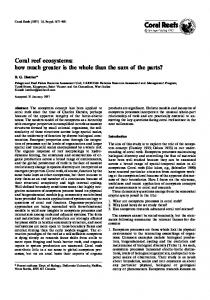
Fig. 1 Typical shape of a Stylophora pistillata colony, schematically drawn from a live specimen growing at 5 m depth (a up-growing branch tips; b dichotomous branch tips; c lateral, inside and outside growing branches)
Stylophora pistillata colonies were incubated in situ with alizarin Red S solution for 24 h (10 ppm; Barnes 1970) by enclosing them within plastic bags. After a postlabeling acclimation period of 24 h, branches were removed by wire cutters and were held vertically on 20 ´ 20 cm concrete plates with preglued plastic clothespins. The tip of the branch, the essential site of calci®cation, was marked by the red dye during incubation. New skeleton was then added as white calcium carbonate above the red area. After 1 year, the plates with the branches were brought to the laboratory. Tissues were removed by H2O2 (Rinkevich and Loya 1984), and the branches were sun-dried for at least 24 h. Each dried skeleton was photographed, weighed (to the nearest 0.1 g) and measured (to the nearest 0.1 mm). Thereafter, the branches were trimmed by wire cutters above the alizarin-marked zones and measured again. The net growth of each branch and the net weight were calculated. We use the term ``tips'' for the �0.5 cm terminal portion of the branch. Branch bases are not (in most cases) the actual bases, since sampled
809 S. pistillata branches in this study were usually 0.05)
Between-colony analyses (Table 5) clearly re¯ect the variability in growth rates characteristic of Stylophora pistillata colonies. Both characters studied (millimeters length added per tip; total weight added per branch, in grams of CaCO3) diered signi®cantly between the ®ve genets studied and also between single-tip and dichotomous-tip branches. For example, while in the case of dichotomous branches, Colonies 3, 4 and 5 were grouped together for length added per tip and for total weight added per branch (in the last case, Colony 1 was
Statistical analysis Length added per tip
Duncan grouping for colonies 1 Single-tip branches
Dichotomous branches
Total weight added per branch
Single-tip branches Dichotomous branches
2
3
4
5
811
also grouped together). In the single tip branches, no such grouping was achieved.
Discussion The present study aimed at evaluating the potential for restoration using small branch fragments (mostly