PATHOLOGY ONCOLOGY RESEARCH
Vol 11, No 2, 2005
Article is available online at http://www.webio.hu/por/2005/11/2/0108
ARTICLE Expression of Osteopontin and CD44 Molecule in Papillary Renal Cell Tumors Koviljka MATUSAN,1 Gordana DORDEVIC,1 Vladimir MOZETIC,2 Ksenija LUCIN1 Department of Pathology, Rijeka University School of Medicine, 2Department of Urology, Clinical Hospital Center Rijeka, Rijeka, Croatia
1
The aim of the study was to analyze the expression of CD44 adhesion molecule and its ligand osteopontin in papillary renal cell tumors, and to assess the possible prognostic significance of CD44 and osteopontin expression in papillary renal cell carcinomas. The expression of the standard and v6 exon containing isoforms of CD44 molecule, as well as of its ligand osteopontin, was immunohistochemically evaluated in 43 papillary renal cell tumors, which included 5 adenomas and 38 carcinomas. In order to assess their prognostic significance, the results obtained in papillary renal cell carcinomas were compared to usual clinicopathological parameters such as tumor size, histological grade, pathological stage, and Ki-67 proliferation index. Normal renal tissue was negative for CD44s and v6 isoforms, while the expression of osteopontin was found in distal tubular epithelial cells in the form of cyto-
plasmic granular positivity. CD44s and v6 isoforms were upregulated in 22 (58%) and 12 (32%) out of 38 carcinomas, respectively. Among all clinicopathological parameters examined, we only found significant association of CD44s-positive carcinomas with lower pathological stage (p=0.026). Papillary renal cell adenomas were generally negative for CD44s, except for focal positivity found in one sample. The osteopontin protein was detected in all adenomas and all papillary renal cell carcinomas, except one. Our results show constitutive expression of osteopontin in papillary renal tumors, including papillary renal cell adenomas. The upregulation of CD44s and v6 isoforms, although found in a considerable number of papillary renal cell carcinomas, does not appear to have any prognostic value in this type of renal cancer. (Pathology Oncology Research Vol 11, No 2, 108–113)
Key words: papillary renal cell carcinoma, cell adhesion, CD44s antigen, CD44v6 antigen, osteopontin, immunohistochemistry
Introduction The present classification of renal tumors is based on cytological appearance and the cell type of origin in combination with growth pattern and genetic alterations.8 According to this classification, papillary renal cell carcinoma is recognized as a distinct subtype of renal carcinoma, which comprises 10-15% of cases in surgical series. It is characterized by predominant papillary or tubulopapillary histolReceived: Aprl 12, 2005; accepted: May 5, 2005 Correspondence: Ksenija LUCIN, Department of Pathology, Rijeka University School of Medicine, Brace Branchetta 20, 51 000 Rijeka, Croatia, Tel.: +38551325805, Fax: +38551325810, E-mail:
[email protected] This work was supported by the Ministry of Science, Education and Sport of the Republic of Croatia (grant 0062066).
© 2005 Arányi Lajos Foundation
ogy with fibrovascular cores, including solid variant with sheets of cells that otherwise resemble papillary renal cell carcinoma.2,8 Genetic features are characterized primarily by trisomies or tetrasomies 7 and 17, and Y chromosome loss in males, as well as additional gains of chromosome 3q, 12, 16, and 20.11,16 Delahunt and Eble have proposed subclassification of papillary renal cell carcinoma into type 1 and type 2, according to the cytoplasmic volume and thickness of the lining cells.7,8 There is debate concerning the relative outcome of papillary renal cell carcinoma compared to clear cell type, which could probably be resolved by adopting this recently proposed subclassification. Cell adhesion molecules are involved in various physiological and pathological processes including cancer. Among them, CD44 molecule has received much interest as a major cell adhesion and signaling molecule involved
Osteopontin and CD44 in Papillary Renal Cell Tumors
109
Table 1. Relationship between CD44s expression and tumor size, stage, grade and proliferation index CD44s expression (No.;%)
Tumor size (cm; mean±SD)
Pathological stage‡ No. (%) 1,2
Negative (16; 42.1) Positive (22; 57.9) p *
6.8±3.3 6.6±3.9 0.860*
Fuhrman’s nuclear grade; No.(%)
3,4
11 (34.4) 5 (83.3) 21 (65.6) 1 (16.7) 0.026†
1, 2
Ki-67 index (mean±SD)
3, 4
12 (46.2) 4 (33.3) 14 (53.8) 8 (66.7) 0.457†
5.5 ± 7.9 5.2 ± 3.6 0.888*
Student t-test; † Pearson’s χ2 test; ‡TNM classification of UICC12
in tumor progression.26 It is a widely distributed cell surface glycoprotein expressed as many isoforms arising from a single gene by alternative splicing. One of them is CD44v6, a variant that has been identified as a marker of cancer progression.13 A principal ligand for the standard form of CD44 is hyaluronan, but ligands for the variant isoforms are not as well characterized. One of the proposed ligands is osteopontin, a secreted adhesive glycoprotein, expressed by various mesenchymal and epithelial cells, and involved in a variety of physiological functions, as well as in tumorigenesis and metastasis.23 Osteopontin contains binding sites for several receptors, including CD44 and integrin αvβ3, cell surface molecules playing a major role in mediating cell migration and adhesion.20 An increase in osteopontin expression levels has been shown to correlate with enhanced malignancy in several in vitro and in vivo studies.1,5,21,24 High expression of both adhesion molecules, CD44 and osteopontin, has been shown to correlate with poor prognostic parameters in clear cell type of renal cell carcinoma, which is the most common type of renal cancer.10,18,19,22 However, to our knowledge, there are no reports on the expression of these two adhesion molecules in papillary renal cell carcinoma. The current study was undertaken to investigate the expression of osteopontin and CD44 molecule in papillary renal cell carcinoma, in order to better characterize this rare type of renal cancer, and to compare the results with prognostic parameters. Materials and methods Tumor samples We examined 50 consecutive nephrectomy specimens in which papillary renal cell tumors had been diagnosed during the routine pathological work from January 1989 till September 2004. Since typing and grading of renal cell carcinoma have markedly changed in the past decade, all hematoxylin-eosin-stained sections from each case were reviewed by two pathologists, followed by exclusion of 3 cases, which did not meet criteria for papillary renal cell carVol 11, No 2, 2005
cinoma diagnosis according to WHO classification.8 The tumors were classified as adenoma or carcinoma, after examination of hematoxylin-eosin-stained tissue sections, according to WHO criteria, and graded using the Fuhrman nuclear grading system.16 Four cases were excluded on the basis of incomplete clinical data or inadequate archival material, so a total of 43 specimens were finally included in the study. Five tumors consisted of tubulopapillary structures lined by small, cuboidal cells with rounded, uniform nuclei lacking cytological atypia. Tumors were circumscribed, but not encapsulated, without stromal reaction, mitoses were absent or rare. These tumors were classified as adenomas, and the other 38 samples, exhibiting at least 75% papillary or tubulopapillary architecture, which did not meet the above mentioned criteria, were classified as papillary renal cell carcinoma. Carcinoma samples were further subdived into the type 1 (single-layered small cells with pale cytoplasm and small oval nuclei with inconspicuous nucleoli) and type 2 (pseudostratified large cells with abundant eosinophilic cytoplasm and large nuclei with prominent nucleoli), according to criteria proposed by Delahunt et al.7 Tumor stage was defined according to the International Union Against Cancer (IUCC) 1997 tumor-node-metastasis (TNM) classification.12
Immunohistochemistry For each case of papillary renal cell carcinoma, a representative slide of the tumor and the corresponding paraffin block was selected. Five-micron sections were cut on glass slides (DakoCytomation, Glostrup, Denmark), and air-dried during the night. Following deparaffinization in xylene and rehydration in alcohol, heat-induced epitope retrieval was achieved by immersing slides in 10 mM citrate buffer (pH 6.0) and boiling for 10 minutes in a pressure cooker. Slides were allowed to cool for 45 minutes, and then pre-incubated with blocking solution containing normal donkey serum (Santa Cruz Biotechnology, Santa Cruz, CA, USA) for osteopontin staining, or normal goat serum (DakoCytomation) for CD44 staining, respectively, for 30 minutes. The staining was performed by indirect immunoperoxidase
110
MATUSAN et al
a
b
c
d
e
f
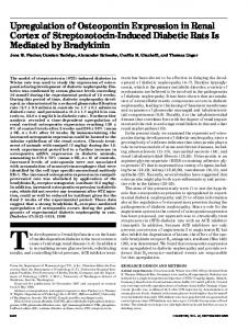
Figure 1. Immunohistochemical expression of CD44s and osteopontin in papillary renal cell carcinoma. Tumor cells are negative for CD44s, while foamy macrophages in tumor stroma are positive (a). Tumor cells are strongly positive for CD44s (b) and CD44v6 (c). Normal renal tissue showing staining of epithelial cells in distal tubules and luminal calcifications (d). Cytoplasmic granular positivity in type 1 (e) and type 2 (f) papillary carcinoma cells.
method in an automated immunostainer (DakoCytomation, TechMateTM Horizon, Glostrup, Denmark) using DakoCytomation LSAB2 HRP system, according to the manufacturer’s protocol. Murine monoclonal antibodies against CD44s (clone SFF304, dilution 1:15000) and CD44v6 (clone VFF18, dilution 1:15000) were purchased from Bender MedSystems (Vienna, Austria), and applied overnight at +4°C. For
negative control, an irrelevant murine monoclonal IgG antibody was used (DakoCytomation). For positive controls, a staining of intratumoral lymphocytes for CD44s, and urothelium for v6 isoform, was used. Osteopontin was detected by goat anti-human monoclonal antibody (clone K-20, Santa Cruz Biotechnology, dilution 1:100), followed by donkey anti-goat IgG as secPATHOLOGY ONCOLOGY RESEARCH
Osteopontin and CD44 in Papillary Renal Cell Tumors
ondary antibody (Santa Cruz Biotechnology, dilution 1:250). For a negative control, an irrelevant goat IgG was used (Santa Cruz Biotechnology). Staining of the luminal portion of distal tubular cells and calcifications within renal parenchyma served as a positive control. In some doubtful cases, staining with murine anti-CD68 antibody (clone KP-1, DakoCytomation, 1:200) was performed to distinguish between tumor cells and histiocytes, which, in activated state, are also positive for osteopontin and CD44s. MIB-1 antibody (DakoCytomation, dilution 1:50) was used to analyze nuclear expression of the Ki-67 cell proliferation antigen. Evaluation of immunohistochemistry Immunohistochemical staining results were examined independently by two pathologists, without knowledge of the nuclear grade or other clinicopathological parameters of each individual case. Cases without any detectable staining were considered negative, as well as those with only focal positivity, found in less than 1% of tumor cells. Positive staining for CD44 molecule and osteopontin was defined as membranous or granular cytoplasmic positivity, respectively, found in more than 1% of tumor cells. Tumor growth kinetics was determined by immunohistochemical detection of Ki-67 antigen. Ki-67 antigen expression was assessed in areas with the highest density of positive cells, and expressed as Ki-67 labeling index (percentage of positive cells) by scoring 500 tumor cells at high power field. The counting was performed on image analyzer using ISSA 3.1 software (Vams, Zagreb, Croatia).
Statistical analysis Statistical analysis was performed using Statistica 6.1 software (StatSoft, Inc., Tulsa, OK, USA). Pearson’s χ2 test was used to assess the significance of associations between categorical data. The mean values of continuous data, such as Ki-67 proliferation index and tumor size, were compared by Student’s t-test. Statistical differences with p value less than 0.05 were considered significant. Results Clinicopathological data The sex distribution was as follows: 4 men and 1 woman for papillary renal cell adenomas, and 30 men and 8 women for papillary renal cell carcinomas. Tumor size for papillary adenomas was 0.6±0.2 cm, while papillary carcinomas measured 6.6±3.6 cm in diameter. The Fuhrman grading distribution was as follows: 3 grade 1, 23 grade 2, 8 grade 3, and 4 grade 4 tumors. Pathological staging according to the TNM classification was pT1 in 19 casespT2 in 13 cases, and pT3 in 6 cases. Vol 11, No 2, 2005
111
Immunohistochemical staining in normal renal tissue and papillary renal cell adenomas In the normal renal parenchyma no staining for CD44s or v6 isoforms was observed, except for a few CD44s- and v6positive tubules, usually in the close vicinity to the tumor tissue. Stromal cells, like lymphocytes and macrophages were also positive for CD44s. Staining was of membranous type, in contrast to staining pattern of osteopontin, which was granular and cytoplasmic. The expression of osteopontin was seen in distal tubular epithelial cells, and was usually most prominent along the luminal side of the cell. Calcifications and stromal cells like macrophages and plasma cells were also positive (Figure 1d). Papillary renal adenomas were negative for CD44s and CD44v6 isoforms, except for one case in which a focal staining of a few tumor cells with anti-CD44s was observed. Staining for osteopontin protein was present in all tumors, although it was generally of lower intensity, and in smaller proportion of tumor cells, comparing to papillary renal cell carcinoma.
Immunohistochemical staining in papillary renal cell carcinomas Sixteen samples were negative for CD44s (42%), while upregulation of CD44s molecule was found in 22 cases (58%) (Table 1). Staining pattern was heterogeneous, ranging from weak positivity in part of the tumor to strong and diffuse staining of almost all tumor cells (Figure 1b). It was always of membranous type, and usually present along the basolateral surface of the cell. CD44v6 was expressed in 12 (32%) tumors, with 8 tumors of type 1 and 4 tumors of type 2 morphology (Figure 1c). All papillary renal cell carcinomas, except one, were positive for osteopontin (Figure 1e, f). Staining was present in the cytoplasm in the form of granules of various sizes and staining intensity. The granules were distributed either evenly, or were showing perinuclear or perimembranous distribution. The pattern of staining varied between samples and ranged from staining of small number of tumor cells with low intensity to strong and diffuse positivity throughout the tumor. The proportion of positive cells varied with predominant expression in more than 50% of tumor cells. Association between CD44 expression and clinicopathological characteristics The expression of CD44 molecule was compared to usual prognostic parameters in renal cell carcinomas such as tumor size, Fuhrman nuclear grade, pathological stage, and Ki-67 proliferation index (Table 1). We could find statistically significant association of CD44s expression only with pathological stage (p=0.026), with higher incidence of CD44s positivity in tumors confined within kidneys (i.e., pT1 or pT2),
112
MATUSAN et al
compared to those invading beyond the kidneys (i.e., pT3 or pT4). Other clinicopathological variables were not associated with the expression of the CD44 molecule, neither CD44s (Table 1), nor CD44v6 (data not shown).
Clinicopathological characteristics of papillary renal cell carcinomas in relation to histological type The results of clinicopathological parameters in relation to 2 morphological types of papillary renal cell carcinoma are shown in Table 2. Mean diameter of type 1 carcinomas was smaller then that of type 2 carcinomas (6.2±3.3 cm versus 7.7±4.4 cm), but the difference was not significant (p=0.254). There was a significant difference in the distribution of nuclear grades and pathological stages between type 1 and type 2 papillary renal cell carcinomas (p