Molecular Human Reproduction Vol.7, No.3 pp. 279–285, 2001
Expression of ZO-1 and occludin in normal human placenta and in hydatidiform moles D.Marzioni1, M.Banita1,6, A.Felici2,7, F.J.Paradinas3, E.Newlands4, M.De Nictolis5, J.Mu¨hlhauser2 and M.Castellucci1,8 1Institute
of Normal Human Morphology, Faculty of Medicine, University of Ancona, Via Tronto, 10/A, I-60020 Ancona, of Vascular Pathology, Istituto Dermopatico dell’Immacolata, IDI, Rome, Italy, 3Department of Histopathology, Imperial College, Charing Cross Hospital Campus, London, 4Department of Medical Oncology, Charing Cross Hospital, London, UK, and 5Institute of Pathological Anatomy and Histology, Faculty of Medicine, University of Ancona, Ancona, Italy 6Permanent address: Institute of Histology, Faculty of Medicine, University of Craiova, Craiova, Roumania 7Present address: Laboratory of Cellular and Molecular Biology, NCI, NIH, Bethesda, USA 2Laboratory
8To
whom correspondence should be addressed. E-mail:
[email protected]
Zonula occludens-1 (ZO-1) and occludin are key molecules in cell–cell contacts. They are tight junction constituents and therefore play a pivotal role in tissue differentiation and organogenesis. In the present report we have investigated the expression of ZO-1 and occludin in normal human placentae and in hydatidiform moles using immunohistochemical and Western blot analyses. In normal placentae, ZO-1 and occludin were mainly localized in the apical part of the syncytium, in cell–cell contacts between syncytium and villous cytotrophoblastic cells as well as between the latter. Extravillous cytotrophoblast of cell islands and cell columns was positive for ZO-1 and occludin in the cell layers proximally located to the villous stroma whereas the cytotrophoblastic cells, distally located from the villous stroma, were totally negative. Furthermore, fetal vessels showed a positive staining pattern for ZO-1 throughout gestation, whereas a positive reaction for occludin was produced mainly at term. A striking result was the altered expression of ZO-1 and occludin in partial and complete moles. In 11 moles, these two molecules were not expressed at all, while in the other nine, their expression was only cytoplasmic in syncytium and villous cytotrophoblastic cells. These findings suggest that ZO-1 and occludin participate in normal placental development, maintaining the organization and functions of different tissue components. The down-regulation and/ or dysregulation of these two molecules may be related to phenotypic changes associated with epithelial cell transformation of the chorionic villi in partial and complete moles. Key words: complete mole/occludin/partial mole/placenta/zonula occludens-1
Introduction Cell–cell adhesion systems are essential for the normal organization and function of the organism and they are regulated in response to various physiological and tissue-specific needs (Wong and Gumbiner, 1997). It has been shown that adhesion molecules of cell–cell junctions influence intracellular functions that control extracellular cell behaviour (Hedrick et al., 1993). Indeed, some of these cell–cell adhesion molecules may function as sensors of the cell’s environment through their interactions at the cell surface, and transmit that information to the cell’s interior (Hedrick et al., 1993). The first protein identified as a tight junction constituent was zonula occludens-1 (ZO-1) with a molecular weight of 220 kDa (Stevenson et al., 1986; Anderson et al., 1988). ZO-1 belongs to a family of multi-domain proteins known as the membraneassociated guanylate kinase homologues (MAGUKs). It is a © European Society of Human Reproduction and Embryology
peripheral membrane protein bound to the cytoplasmic surface of junctional contacts and it is expressed in all tight junctions (Balda and Anderson, 1993; Fanning et al., 1998). ZO-1 has been demonstrated to interact with the transmembrane protein occludin (which is an integral membrane protein localizing at tight junctions with four transmembrane domains and a long COOH-terminal cytoplasmic domain), and also with ZO-2 and F-actin (Furuse et al., 1994; Fanning et al., 1998; Tsukita and Furuse, 1999). Fanning et al. (1998) have indicated that both ZO-2 and occludin interact with specific domains within the N-terminal half of ZO-1, whereas the unique proline-rich C-terminal half of ZO-1 co-sediments with F-actin. Recently, it has been shown that ZO-1, under certain conditions of cell growth, can be detected in the nucleus (Gottardi et al., 1996). Since cell–cell contacts are specialized sites for signalling pathways implicated in growth and differentiation, it has been 279
D.Marzioni et al.
suggested that the nuclear accumulation of ZO-1 may be relevant to its role in MAGUK homologue signal transduction (Gottardi et al., 1996). In the present investigation, we studied the expression of ZO-1 and occludin by immunohistochemistry and Western blot analysis to investigate the molecular state of tight junctions in normal human placentae of first and third trimester of gestation as well as in two gestational trophoblastic lesions with a low degree of aggressiveness, i.e. partial and complete moles. These two placental lesions are characterized by enlarged, vesicular chorionic villi associated with a variable amount of proliferative trophoblast (Mazur and Kurman, 1994). The possible sequelae of these pathologies, particularly of the complete mole, are the invasive mole and/or choriocarcinoma (Mazur and Kurman, 1994). Under normal conditions, most of the invasive processes of the trophoblast are performed by the extravillous trophoblastic cells developing from cell columns, whereas the villous trophoblast which covers the placental villi does not show invasive behaviour (Benirschke and Kaufmann, 2000). The mechanisms of normal trophoblast invasion in vivo are not yet fully understood but important features of this process are cell–cell contacts and the controlled invasion of the extravillous cytotrophoblast into the uterine wall. Since alterations of the cell–cell contacts and of the controlled invasion of trophoblast could be caused by compromised integrity of tight junctions, we propose a potential role for changes to ZO-1 and/or occludin in the tendency of partial and complete moles to persist and invade.
Materials and methods Tissues A total of 19 human placentae aged 7 (n ⫽ 3), 8 (n ⫽ 2), 10 (n ⫽ 1), 12 (n ⫽ 5), 38 (n ⫽ 5) and 40 (n ⫽ 3) weeks postmenstruation were collected from clinically normal pregnancies, interrupted by curettage (aspiration technique) for psycho-social and medical reasons which were unlikely to affect placental structure and function, or terminated by Caesarean sections and normal vaginal deliveries. Specimens of 10 partial moles (15–33 weeks postmenstruation) and 10 complete moles (10–13 weeks postmenstruation) were collected from the pathology files of the Institute of Pathology, University of Ancona (Italy) and of the Centre for Trophoblastic Diseases (Charing Cross Hospital, London, UK). All the cases were documented with clinical and histopathological data. From each placenta, some specimens were frozen in liquid nitrogen for biochemical analysis (see below). Other tissue blocks were fixed for 12 h at 4°C in 4% buffered formalin and routinely processed for paraffin embedding at 56°C, as previously described (Mu¨ hlhauser et al., 1993). Biochemical analysis Protein extracts Human umbilical vein endothelial cells (HUVEC; Clonetics, San Diego, CA, USA) and human epidermoid carcinoma A431 cells (kindly provided by Dr R.Falcioni, Istituto Regina Elena, Rome, Italy) were grown to confluence, for use as positive controls for ZO-1 and occludin Western blot analysis (Van Itallie et al., 1995; Burns et al., 1997). In addition, human primary fibroblasts (kindly provided by Dr G.Zambruno, I.D.I., Rome, Italy) were also cultured for use as negative controls. To prepare protein extracts, confluent
280
HUVEC and A431 cells as well as proliferating fibroblasts were washed with ice-cold phosphate-buffered saline (PBS), harvested and resuspended in lysis buffer [1% (v/v) Triton X-100/0.5% (w/v) sodium deoxycholate/0.2% (w/v) sodium dodecyl sulphate (SDS)/ 150 mmol/l NaCl/10 mmol/l HEPES, pH 7.4/1 mmol/l phenyl methyl sulphonyl fluoride (PMSF)/1 µg/ml pepstatin/25 µg/ml leupeptin/ 10 µg/ml aprotinin/10 mmol/l NaF/2 mmol/l EDTA]. These suspensions were homogenized with a stainless steel (10 strokes) and then centrifuged at 30 000 g for 20 min at 4°C. Supernatants were immunoprecipitated (2 mg) using a polyclonal anti-ZO-1 antibody (Zymed Laboratories Inc, San Francisco, CA, USA) or used as such (100 µg protein/lane) and absorbed on protein-A-Sepharose beads (Pharmacia, Milano, Italy) in Western blot analysis. First and third trimester frozen placental samples were homogenized with a stainless steel (30 strokes) and then centrifuged at 30 000 g for 20 min at 4°C. Supernatants were immunoprecipitated using protein-A-Sepharose (Pharmacia) according to the manufacturer’s instructions. SDS–PAGE and Western Blotting All samples (HUVEC, A431cells, fibroblasts and placental tissues) were fractionated on 8% SDS–polyacrylamide gels (SDS–PAGE) as previously described (Laemmli, 1970), electrophoretically transferred (Trans-Blot-Cell; Bio-Rad Laboratories Inc, Richmond, CA, USA) to nitrocellulose membranes, and subjected to Western blot analysis. Non-specific protein binding was blocked with 0.5% (w/v) non-fat dry milk (Nestle` Food Company, Glendale, CA, USA) in PBS/0.05% Tween (PBS-T). Blots were incubated with affinity purified rabbit anti-ZO-1 (Zymed Laboratories,) diluted 1:2000; or rabbit polyclonal antibody to Occludin (Zymed Laboratories) diluted 1:10 000. After washing, blots were incubated with anti-rabbit secondary antibody conjugated to horseradish peroxidase (Amersham Italia s.r.l., Milano, Italy) diluted 1:10 000. Detection of bound antibodies was performed with the ECL-Western blotting detection kit (Amersham) according to the manufacturer’s instructions. Morphological analysis Immunohistochemistry Paraffin serial sections (5 µm) were cut and stretched at 45°C, allowed to dry, and stored at 4°C until use. Sections were deparaffinized and rehydrated via xylene and a graded series of ethyl alcohol, and treated with 0.1% trypsin (Sigma Chemical Co, St Louis, MO, USA) in distilled water for 5–10 min at 37°C. Sections were incubated for 30 min with 0.3% hydrogen peroxide in methanol to inhibit endogenous peroxidases and then for 30 min at room temperature with non-immune serum. Afterwards the sections were incubated overnight at 4°C with one of the following primary antibodies: affinity purified rabbit anti ZO-1 (Zymed Laboratories) diluted 1:200 v/v; a rabbit polyclonal antibody to occludin (Zymed Laboratories) diluted 1:200 v/v; a monoclonal mouse anti-human endothelial cell, CD31 (Dako, Glostrup, Denmark) diluted 1:40 v/v; a mouse monoclonal antibody anti-human pancytokeratin (Dako) diluted 1:500 v/v. After washing with PBS, the bound antibody was visualized by the peroxidase ABC method (Hsu and Raine, 1981) using the following biotinylated secondary antibodies: for the primary polyclonal antibodies, a goat anti-rabbit IgG (Vector Laboratories, Burlingame, CA, USA) diluted 1:200 v/v; for the primary monoclonal antibodies, a horse anti-mouse IgG, (Vector Laboratories) diluted 1:200 v/v. 3⬘,3⬘diaminobenzidine hydrochloride (Sigma Chemical Co.) was used as chromogen. The sections were rinsed with distilled water, counterstained with Mayer’s haematoxylin and mounted in Eukitt (Kindler GmbH & Co, Freiburg, Germany). For the above immunohistochemical procedures, controls were performed by replacing the primary antibody with PBS. Further controls were performed omitting the secondary antibody. The controls
ZO-1 and occludin in placenta
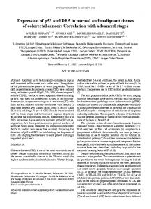
Figure 1. Western blot analysis of (a) zonula occludens-1 (ZO-1) and (b) occludin expression. (a) Protein lysates prepared from first trimester (lane 1) and third trimester (lane 2) placentae were immunoprecipitated with an anti ZO-1 polyclonal antibody. ZO-1 protein expression was detected in first trimester placentae and was markedly increased in third trimester placental tissues. A431 cell line (lane3) and human primary fibroblasts (lane 4) were used as positive and negative controls respectively. (b) Protein lysates prepared from first trimester (lane 1) and third trimester (lane 2) placentae were immunoprecipitated with an anti occludin polyclonal antibody. Occludin protein expression was detected in first trimester placentae and was increased in third trimester placental tissues. Human primary fibroblasts (lane 3) and human umbilical vein endothelial cells (HUVEC, lane 4) were used as negative and positive controls respectively.
were always negative. Non-immune serum was used as a negative control.
Results Western blotting The expression of ZO-1 and of occludin was studied for the first time in human placenta. To evaluate the expression of ZO-1 in normal human placenta, we performed Western blot analysis of first and third trimester placental samples after immunoprecipitation with a specific rabbit anti-ZO-1 antibody (Figure 1). We found that ZO-1 protein was expressed in first trimester placentae and that its expression was strikingly increased in third trimester placentae. As expected, ZO-1 migrated as a doublet with a molecular mass of 223 and 214 kDa (Figure 1, lanes 1, 2 and 3) representing the two isoforms α⫹/α– (Balda and Anderson, 1993). The same analysis with a specific anti-occludin antibody revealed expression of occludin protein (65 kDa) in first trimester placental tissue and a marked increase of this protein in third trimester placental samples. Immunohistochemistry First trimester ZO-1 immunoreaction was weakly visible at the apical part of the cytoplasm and in various nuclei of the syncytiotrophoblast. Also some nuclei of the villous cytotrophoblastic cells showed a positive reaction product for this antigen (Figure 2). Only some tracts of the cellular membrane of villous cytotrophoblastic cells showed an evident reaction product. In the stroma of the chorionic villi, all the fetal vessels (identified in parallel sections by the CD31 antibody immunostaining) showed a very strong ZO-1 immunorectivity at endothelial cell–cell contacts (Figure 2). The plasma membrane of the extravillous
cytotrophoblastic cells in cell islands and cell columns was strongly positive for ZO-1 in the cell layers located proximally to the villous stroma while the extravillous cytotrophoblastic cells distally located from the villous stroma were completely negative (data not shown). Occludin immunoreactivity was observed in the apical plasma membrane of the syncytium. In villous cytotrophoblastic cells an evident and regular reaction product was detected along the apical (in contact with syncytium) and lateral (in contact with neighbour cytotrophoblastic cells) cell membrane at cell–cell contacts (Figure 3). Villous stroma was mainly negative. Only a few fetal vessels showed a weak positive reaction product in the endothelial cells. In the cell islands and cell columns, only two or three layers of extravillous cytotrophoblastic cells proximally located to the villous stroma showed a strong immunoreactivity for occludin. The extravillous cytotrophoblastic cells distally located were negative for this antigen (Figure 4) Thus, immunoreactivity of this antigen in the cytotrophoblastic cell columns was similar to ZO-1 immunostaining. Third trimester The syncytiotrophoblast was weakly and irregularly immunostained for ZO-1. Villous cytotrophoblastic cells were positive at the apical part of the plasma-membrane in contact with the syncytium. ZO-1 protein was localized in the endothelium of fetal vessels at areas of cell–cell contacts (Figure 5). In the basal plate, some extravillous cytotrophoblastic cells showed a weak immunoreactivity in the cytoplasm and in the plasmamembrane for ZO-1. Uterine vessels were positive for this antigen. Occludin was expressed in the syncytiotrophoblast while some villous cytotrophoblastic cells were positive (Figure 6). In the villous stroma the endothelium of fetal vessels was 281
D.Marzioni et al.
immunostained for occludin and the staining pattern was particularly evident in the large fetal vessels (Figure 6). In the basal plate, some extravillous cytotrophoblastic cells (identified by staining with a monoclonal antibody to cytokeratins) showed weak cytoplasmic immunostaining for occludin. Partial and complete moles ZO-1 and occludin showed very variable expression patterns in partial and complete moles. In 11 moles examined in this study (four partial and seven complete) these two molecules
282
were negative (Figure 8) or very weakly positive, whereas in other moles (six partial and three complete) ZO-1 (Figure 9) and occludin (Figure 10) showed a cytoplasmic expression in villous and extravillous cytotrophoblastic cells. The vessels of the pathological villi of the partial moles and all the vessels of the complete moles (identified by positive CD31 staining) were totally negative for these two antigens. However, some vessels of the normal villi of the partial moles showed a positive immunostaining for ZO-1 and occludin.
ZO-1 and occludin in placenta
Discussion In the present study, the immunolocalization of proteins ZO-1 and occludin have been used as a marker of the molecular organization of placental tight junctions in normal human placentae of first and third trimester gestation and in two pathological conditions, i.e. partial and complete moles. There are few data available concerning tight junction ultrastructure in the human placenta (Cavicchia, 1971; Metz et al., 1979; Metz and Weihe, 1980; Reale et al., 1980). Previous freezefracture and thin-section studies have shown the presence of tight junctions of limited extension within apical invaginations of the syncytiotrophoblastic layer. These invaginations were in turn similar to flattened vesicles (also associated with junctions) located in the underlying syncytioplasm. Tight junctions of a limited extension (fasciae occludentes) were also found between the syncytiotrophoblast and villous cytotrophoblastic cells, as well as between villous cytotrophoblastic cells (Metz and Weihe, 1980; Reale et al., 1980). In the placenta, the trophoblastic layer (syncytiotrophoblast and cytotrophoblastic cells) separates maternal and fetal circulations and is involved in the active transport of selected substances (Mitchell et al., 1995). The present study shows ZO-1 and occludin localized in the junctions between villous cytotrophoblastic cells and between syncytium and underlying cytotrophoblastic cells. Thus, these molecules could contribute to the organization and function of the trophoblastic layer. On the other hand, occludin and (small amounts of) ZO-1 localized in the syncytium apical zones could play a minor role in the function of the trophoblastic barrier. They may be remnants of tight junctions displaced from their original location (Reale et al., 1980). Displacement could occur during syncytial growth, which continues until full term and occurs by fusion of villous cytotrophoblastic cells with the overlying syncytium (Reale et al., 1980). Recently, it has been shown that alterations in occludin protein expression may be a mechanism by which vascular permeability is regulated (Antonetti et al., 1998). We have shown that the expression of occludin and ZO-1 increases
during gestation. We can speculate that the increase in these molecules may be related to modifications of placental vascular permeability during gestation, i.e. fetal vessels in third trimester placentae are much more abundant than in the first trimester placentae. Interestingly, it has been pointed out that fetal vessels of the first trimester placenta do not have a welldeveloped basement membrane (Benirschke and Kaufmann, 2000) which, however, is present in the last trimester. Thus, in the human placenta, the tight junctions of the fetal vessels are probably more effective in the last trimester of gestation when both junctions and basement membrane are definitively organized. It has been claimed that the extravillous cytotrophoblastic cells of cell columns located proximally to the villous stroma are proliferating cells, whereas the cytotrophoblastic cells located distally to the villous stroma are no longer proliferative and show invasive behaviour, especially in the first trimester (Kaufmann and Castellucci, 1997). No freeze-fracture studies have been performed on these cells and therefore no consistent data are available on tight junctions between these cells. Extravillous cytotrophoblastic cells of cell islands and cell columns of first trimester placentae showed high expression of ZO-1 and occludin in the proximal cells belonging to the proliferative phenotype, whereas the most distal cells, invading the basal plate were negative for these antigens. These findings support the idea that occludin and ZO-1 show cell–cell adhesion activities in the proliferative extravillous cytotrophoblastic cells, but not in the invasive ones, which are committed to migrate into the maternal side of the placenta. Further important results concern the localization of ZO1 and occludin in partial and complete moles. In these pathologies, ZO-1 and occludin expressions are greatly altered in the trophoblastic layer, i.e. in 11 moles these molecules were not expressed and in another nine moles their expression was mainly cytoplasmic. Thus, our findings suggest that the early loss or alterations of tight junction integrity in these two pathologies with a low degree of aggressiveness could represent the initial stage of more
Figure 2. Immunostaining for zonula occludens-1 (ZO-1) in a first trimester placenta. The syncytiotrophoblast is weakly stained. Various nuclei of the trophoblastic layer are positive for this antigen. Arrows indicate staining for ZO-1 in some tracts of plasma membranes of adjacent cytotrophoblastic cells and between cytotrophoblast and syncytiotrophoblast. Blood vessels are indicated by arrowheads and are positive for ZO-1. Bar ⫽ 22 µm. Figure 3. Immunostaining for occludin in a first trimester placenta. The apical plasma membrane of the syncytiotrophoblast is positive for this antigen. Cytotrophoblastic cells show an evident and regular reaction product along the apical and lateral cell membrane at cell-cell contacts. Nuclei show a strong haematoxylin staining. They are negative for occludin. Bar ⫽ 22 µm. Figure 4. Placental villi and a cell column from a first-trimester placenta. Immunostaining for occludin. (a) In the cell column (*) note the positive reaction product of the extravillous cytotrophoblastic cells located proximally to the villous stroma, whereas the extravillous cytotrophoblastic cells (ET) distally located are negative for this antigen. The framed area delimits part of the cell column. Bar ⫽ 55 µm. (b) Enlargement of the area framed in (a). Occludin is strongly expressed in the extravillous trophoblastic cells proximal to the villous stroma (VS). Bar ⫽ 9 µm Figure 5. Third trimester placental tissue. Immunostaining for zonula occludens-1 (ZO-1). The syncytiotrophoblast shows a weak reaction product. Note the positive staining of the apical plasma membrane (arrow) of a cytotrophoblastic cell in contact with the syncytium. Fetal vessels (V) show ZO-1 expression in the endothelium at areas of cell-cell contacts. Bar ⫽ 9 µm Figure 6. Third trimester placental tissue. Immunostaining for occludin. The trophoblastic layer (arrows) and the endothelium, particularly of the large fetal vessels, are positive for this antigen. Bar ⫽ 55 µm Figure 7. Control section. No immunostaining is observable in the villous and extravillous trophoblastic cells (*) and in the stroma of the chorionic villi. First trimester of gestation. Bar ⫽ 55 µm.
283
D.Marzioni et al.
severe conditions, e.g. invasive mole and choriocarcinoma. Our data are corroborated by a recent study (Li and Mrsny, 2000) that demonstrates the functional role of occludin in controlling the phenotypic changes associated with epithelial cell transformation. It is of interest to note that ZO-1 and
occludin were not expressed in the few small vessels of the pathological villi. Alterations of tight junction proteins of fetal vessels may increase permeability and contribute to the formation of hydropic villi in partial moles. The molecules analysed in this study play an important role in co-ordinating the structural and functional events of tight junctions in maintaining the morphology and the function of different tissue components of the human placenta. Given the central role that ZO-1 and occludin play in controlling normal cellular adhesion processes and tight junction functions, we suggest that the dysregulation of ZO-1 and occludin in partial and complete moles may play a critical role in the pathogenesis and behaviour of these gestational diseases.
Acknowledgements We are indebted to Professors E.Reale (Hannover, Germany) and H.Bartels (Mu¨ nchen, Germany) for useful discussions. B.M. was supported by a fellowship from the Romanian Ministry of Public Education. This study was supported by funds from the Italian Ministry of University and Scientific Research (MURST), the University of Ancona to M.D. and C.M. and funds from I.D.I. and the Italian Ministry of Health to M.J.
References Anderson, J.M., Stevenson, B.R., Jesaitis, L.A. et al. (1988) Characterization of ZO-1, a protein component of the tight junction from mouse liver and Madin–Darby canine kidney cells. J. Cell. Biol., 106, 1141–1149. Antonetti, D.A., Barber, A.J., Khin, S. et al. (1998) Vascular permeability in experimental diabetes is associated with reduced endothelial occludin content. Diabetes, 47, 1953–1959. Balda, M.S. and Anderson, J.M. (1993) Two classes of tight junctions are revealed by ZO-1 isoforms. Am. J. Physiol., 264, C919–C924. Benirschke, K. and Kaufmann, P. (2000) Pathology of the Human Placenta. Springer Verlag, New York, USA. Burns, A.R., Walker, D.C., Brown, E.S. et al. (1997) Neutrophil transendothelial migration is independent of tight junctions and occur preferentially at tricellular corners. J. Immunol., 159, 2893–2903. Cavicchia, J.C. (1971) Morphologic evidence of syncytial formation from the cytotrophoblastic cells. Obstet. Gynecol., 108, 339–346. Fanning, A.S., Jameson, B.J., Jesaitis, L.A. et al. (1998) The tight junction protein ZO-1 establishes a link between the transmembrane protein occludin and the actin cytoskeleton. J. Biol. Chem., 273, 29754–29753. Furuse, M., Itoh, M., Hirase, T. et al. (1994) Direct association of occludin with ZO-1 and its possible involvement in the localization of occludin at tight junctions. J. Cell Biol., 127, 1617–1626. Gottardi, C.J., Arpin, M., Fanning, A.S. et al. (1996) The junction-associated protein, zonula occludens-1, localizes to the nucleus before the maturation and during the remodelling of cell–cell contacts. Proc. Natl Acad. Sci. USA, 93, 10779–10784.
Figure 8. Complete mole. Immunostaining for zonula occludens-1 (ZO-1). No reaction product is observable either in the syncytiotrophoblastic collections (*) or in the villous stroma. A similar result was obtained for occludin. Figure 9. Complete mole. Zonula occludens-1 (ZO-1) is expressed in the villous cytotrophoblastic cells (arrows). ZO-1 shows an irregular cytoplasmatic staining in the syncytiotrophoblast. Note the positive reaction product in the extravillous cytotrophoblastic cells (*). Figure 10. Partial mole. The trophoblastic covering is labelled for occludin. Note the positive reaction product in the cytoplasm of the trophoblast. Bar ⫽ 55 µm applies to Figures 8, 9 and 10.
284
ZO-1 and occludin in placenta Hedrick, L., Kathleen, R. Cho and Vogelstein B. (1993) Cell adhesion molecules as tumor suppressors. Trends Cell Biol., 3, 36–39. Hsu, S.M. and Raine, L. (1981) Protein A, avidin and biotin in immunohistochemistry. J. Histochem. Cytochem., 29, 1349–1353. Kaufmann, P. and Castellucci, M. (1997) Extravillous trophoblast in the human placenta. Trophoblast Res., 10, 21–65. Laemmli, U.K. (1970) Cleavage of structural proteins during the assembly of the head of bacteriophage T4. Nature (London), 227, 680–685. Li, D. and Mrsny, R.J. (2000) Oncogenic Raf-1 disrupts epithelial tight junctions via downregulation of occludin. J. Cell. Biol., 148, 791–800. Mazur, M.T. and Kurman, R.J. (eds) (1994) Gestational trophoblastic disease and related lesions. In Blaustein’s Pathology of the Female Genital Tract. Springer Verlag, New York, USA, pp. 1049–1093. Metz, J., Weihe, E. and Heinrich, D. (1979) Intercellular junctions in the full term human placenta. I. Syncytiotrophoblastic layer. Anat. Embryol., 158, 41–50. Metz, J. and Weihe, E. (1980) Intercellular junctions in the full term human placenta. II. Cytotrophoblast cells, intravillous cells and blood vessels. Anat. Embryol., 158, 167–178. Mitchell, A.M., Yap, A.S., Payne, E.J. et al. (1995) Characterization of cell
polarity and epithelial junctions in the choriocarcinoma cell line, JAR. Placenta, 16, 31–39. Mu¨ hlhauser, J., Crescimanno, C., Kaufmann, P. et al. (1993) Differentiation and proliferation patterns in human trophoblast revealed by c-erbB-2 oncogene product and EGF-R. J. Histochem. Cytochem., 41, 165–173. Reale, E., Wang, T., Zaccheo, D. et al. (1980) Junctions on the maternal blood surface of the human placental syncytium. Placenta, 1, 245–258. Stevenson, B.R., Siliciano, J.D., Mooseker, M.S. et al. (1986) Identification of ZO-1: a high molecolar weight polypeptide associated with the tight junction (zonula occludens) in a variety of epithelia. J. Cell. Biol., 103, 755–766. Tsukita, S. and Furuse, M. (1999) Occludin and claudins in tight-junction strands: leading or supporting players? Trends Cell Biol., 9, 268–273. Van Itallie, C.M., Salda, M.S. and Anderson, J.M. (1995) Epidermal growth factor induces tyrosine phosphorylation and reorganization of the tight junction protein ZO-1 in A431 cells. J. Cell. Sci., 108, 1735–1742. Wong, V. and Gumbiner, B.M. (1997) A synthetic peptide corresponding to the extracellular domain of occludin perturbs the tight junction permeability barrier. J. Cell. Biol., 136, 399–409. Received on October 26, 2000; accepted on December 4, 2000
285