Extracellular Polysaccharides of Penicillium vermiculatum Grigorij Kogana,*, Ma´ria Matulova´a and Eva Michalkova´b a b
Institute of Chemistry, Slovak Academy of Sciences, Du´bravska´ cesta 9, 842 38, Bratislava, Slovakia. Fax: +4 21-2-59 41 02 22. E-mail:
[email protected] Biotika, a.s. 976 13 Slovenska´ L’upcˇa, Slovakia
* Author for correspondence and reprint requests Z. Naturforsch. 57 c, 452Ð458 (2002); received December 26, 2001 Extracellular Polysaccharide, Isolation, Characterization Extracellular polysaccharide mixture that forms a viscous mucilate in the fermentation medium during industrial cultivation of Penicillium vermiculatum was isolated by ethanol precipitation and its structural characteristics were investigated by a combination of physicochemical methods. The mixture contained two structurally different polysaccharides similar to those previously described for some fungal species. This is the first report of the fully structurally characterized extracellular polysaccharides of Penicillium vermiculatum.
Introduction Penicillium vermiculatum (anamorph of Talaromyces flavus) is a filamentous fungus that produces several low-molecular weight metabolites which reveal antibiotic activity. For this reason, the strain initially discovered as a wood-rot fungus growing on the timbering in the uranium mines, is now used in pharmaceutical industry for production of such antibiotic metabolites upon fermentation in aqueous media containing sucrose as carbon source. Although these intracellular metabolites have been extensively structurally characterized (Fuska et al., 1972, 1979a, 1979b, 1991; Proksa et al., 1992a, 1992b, 1996), no attempt has been made to determine the nature of the extracellular compounds that were abundantly excreted into the medium during the fermentation, produced viscous slime-like mass and thus hindered isolation of the desired antibiotic. Upon the laboratory fermentation of P. vermiculatum (then called Talaromyces vermiculatum) in the semisynthetic medium containing glucose, peptone and phosphate, Mizuno et al. (1974) isolated a polysaccharide that revealed antimicrobial activity against several bacteria, yeasts and fungi in mice. This polysaccharide antibiotic named Talaron, however, contained over 9% nitrogen which can imply around 50% protein content, which was also corroborated by a strong absorption at ca. 280 nm in the ultraviolet absorption spectrum (Mizuno et al., 1974). Thus, although authors claimed Talaron to be unique polysaccharide antibiotic, it 0939Ð5075/2002/0500Ð0452 $ 06.00
is rather a proteoglycan and its observed activity could originate as well from its protein part. In the present work, we have for the first time isolated the pure polysaccharide from the industrial cultivation broth of P. vermiculatum and characterized its complex structure. Materials and Methods Microorganisms Penicillium vermiculatum Dang. CCM F-276 was stored on Czapek-Dox medium at 4 ∞C. 500 ml flasks containing 100 ml of inoculation medium were seeded with 3 ml spore suspension and subsequently cultivated at 28 ∞C for 24 h on a rotary shaker. The inoculum was transferred into 500 ml flasks containing 100 ml of production medium and cultivated on a rotary shaker at 28 ∞C for 7 days to produce vermiculine. The composition of inoculation medium was previously described (Adamcova´ et al., 1992), while for production was used the same medium enriched with 0.025 g/l CuSO4 · 5H2O. Preparation of the extracellular polysaccharides After the fermentation, biomass was separated by centrifugation at 104 ¥ g. The polysaccharides were precipitated from the supernatant by a fourfold volume of ethanol, dissolved in water and repeatedly precipitated with ethanol. After the third precipitation, the polysaccharide was dissolved in
” 2002 Verlag der Zeitschrift für Naturforschung, Tübingen · www.znaturforsch.com ·
D
G. Kogan et al. · Extracellular Polysaccharides of Penicillium vermiculatum
453
water and freeze-dried to give slightly yellowish fluffy flakes.
from the peak area of the respective alditol trifluoroacetates.
Size-exclusion chromatography
Nuclear magnetic resonance (NMR) spectroscopy
The polysaccharides were separated on a Sepharose CL-6B glass column (150 ¥ 2 cm) using water as a mobile phase. Analytical characterization of the polysaccharides Molecular weight of the polysaccharides was determined using high performance liquid chromatography (HPLC) instrument that contained a high-pressure pump (LCP 3001; Laboratornı´ prˇı´stroje, Prague), an eight-port switching valve equipped with two 100 µl loops (model PK 1, Vy´vojove´ dı´lny, Czechoslovak Academy of Sciences, Prague) and two in series connected stainless steel HPLC columns (250 ¥ 8 mm) packed with Separon HEMA-BIO 1000 sorbent (mean particle size 10 µm; Tessek Ltd, Prague). The chromatographic process was monitored with a differential refractometric detector (RIDK 102; Laboratornı´ prˇı´stroje, Prague). The mobile phase was 0.1 m NaNO3. The flow rate was 0.4 ml minÐ1. The HPLC system was calibrated with a set of pullulans (Shodex Standard P-82, Macherey-Nagel, Düren, Germany) and for this reason the molecular weight values should be considered relative to the pullulan reference matierial (Sˇolte´s et al., 1993). Elemental analysis of the polysaccharides was performed using the C, H, N-analyzer EA 1108 (FISONS Instruments, UK). Total acidic hydrolysis was carried out by heating the polysaccharide in 2 m trifluoroacetic acid for 4 h. After hydrolysis, the solution was evaporated several times with addition of distilled water. The residue was dissolved in water and sodium borohydride was added to reduce the monosaccharides to the respective alditols, which were subsequently acylated with trifluoroacetic acid anhydride to produce alditol trifluoroacetates. The mixture of the products was analyzed by gas chromatography on a Hewlett Packard Series II 5980 gas chromatograph, and the nature of the monosaccharides was established based on the comparison of the respective retention times with those of the standards. Quantification of the monosaccharide composition was made
1 H and 13C NMR spectra of the polysaccharides were acquired at 25 ∞C in D2O using the 300 MHz Bruker Avance DPX FT-NMR spectrometer, equipped with selective excitation unit and a kit for gradient enhanced spectroscopy, in 5 mm 1H, 13 C, 31P, 15N multinuclear probe with inverse detection with z-gradients. Chemical shifts were referenced to internal acetone standard with δ 2.025 in 1H and δ 31.07 in 13C NMR spectra. Following techniques were used for signal assignments: 2D COSYDQF (Davies et al., 1991), TOCSY (Parella et al., 1997), HSQC (Schleucher et al., 1994), and HMBC (Zhu et al., 1994). Glycosidic linkage types between individual sugar residues were established on the basis of 2D NOESY and 1D gradient-enhanced transient NOESY experiments using Gaussian pulses for selective excitation (Stott et al., 1997).
Infrared (IR) spectroscopy, optical rotation The IR spectra were measured using a NICOLET MAGNA-IR 750 instrument. The polysaccharide samples were pressed into KBr pellets at sample: KBr ratio 1:100. To obtain better resolution and more exact band positions, Fourier selfdeconvolution techniques (FT) was applied using the OMNIC 3.2 software (bandwith 50 cmÐ1; enhancement factor 2.3). The spectra were recorded in both absorbance and transmittance modes. [α]D was measured at 20 ∞C in 1% solutions in distilled water using a Perkin Elmer 241 polarimeter. Results and Discussion P. vermiculatum Dang. CCM F-276 is used for industrial preparation of macrodiolide antibiotic vermiculin when cultivated on saccharose medium (Fuska et al., 1972). Growing in the presence of glucose, the strain produces the phthalido-pyranone vermistatin (Fuska et al., 1979b). These compounds are the major metabolites of P. vermiculatum and because of their antibiotic and immunomodulatory properties major attention was paid to their isolation and characterization. Several other low molecular weight metabolites such as
454
G. Kogan et al. · Extracellular Polysaccharides of Penicillium vermiculatum
vermicillin (Fuska et al., 1979a), dehydroaltenusin (Fuska et al., 1991), vermilutin and (Ð)-mitorubrinol (Proksa et al., 1992a and b), and vermixocins A and B (Proksa et al., 1992c) have been isolated from the cultivation medium of P. vermiculatum using extraction with organic solvents. This procedure is however significantly complicated by a high content of polymeric substances contained in the medium which makes it very viscous and hard to handle. Eliminating or decreasing the production of extracellular polysaccharides might therefore be important for extraction of desired low molecular metabolites at their industrial production by fungal fermentation. In order to isolate the extracellular product and determine its nature, we used the fermentation broth left after the extraction of vermiculin and subjected it to precipitation with a four-fold volume of ethanol. The precipitate was redissolved in water and reprecipitated with ethanol. The procedure was repeated four times. The final product was freeze-dried. The yield of the dry product obtained from 500 ml of fermentation broth was 2.4 g. Elemental analysis of the pale yellowish flaky product revealed only 35.04% C and 5.35% H. Neither nitrogen nor phosphorus were detected. This is in contrast with the data obtained by Mizuno et al. (1974) for an extracellular “polysaccharide” Talaron which contained 9.36% N and 1.97% P. Such high nitrogen content implies presence of a significant protein moiety, the fact that was corroborated by absorption in ultraviolet region of the spectrum.
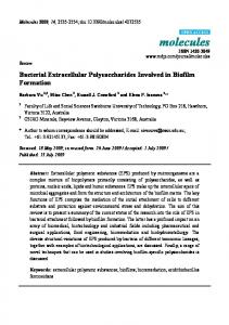
Upon total acidic hydrolysis of the obtained polysaccharide, only two constituent monosaccharides were detected: 61.2% glucose and 38.8% galactose with traces of mannose, which was again in contrast with very heterogenous composition of Talaron, which contained at least six different pentoses and hexoses. Since value of optical rotation [α]20 D was found to be Ð4.5∞, the prevalent configuration of the saccharide units is assumed to be β-anomeric. This assumption is corroborated by the data of 13C NMR and IR spectra (Figs. 1 and 2). 13C NMR spectrum revealed signals in the anomeric region at 108.8 and 103.7 ppm that are characteristic for β-d-galactofuranosidic and β-d-glucopyranosidic units, respectively (Gorin, 1981). Infrared spectrum showed absorption bands at 890 cmÐ1 typical for βlinked polysaccharides and 805 cmÐ1 that could be attributed to galactofuranosyl units (Ruperez and Leal, 1987). Beside the signals of the carbohydrate units, 13C NMR spectrum showed two signals of the carboxylic groups at ca.174 ppm as well as the signals in the region around 61Ð63 ppm that are specific for a methylene group. This data implied that the isolated extracellular polysaccharide contained substitution with malonyl groups, which could be present either in malonogalactofuranan similar to those isolated from the culture broth of Penicillium citrinum Thom 1131 (Kohama et al., 1974) or in luteic acid, β-d-glucan esterified with malonic acid, an acidic polysaccharide produced in the cultivation medium by Penicillium luteum Zukal (Birkinshaw and Raistrick, 1933). The prominent absorption band at 1734 cmÐ1 observed in the IR spectra that was characteristic of the ester bond which corrobo-
Fig. 1. 13C NMR spectrum of the exopolysaccharide of P. vermiculatum. A detail of DEPT spectrum is also shown.
G. Kogan et al. · Extracellular Polysaccharides of Penicillium vermiculatum
455
Fig. 2. Infrared spectrum of the exopolysaccharide of P. vermiculatum.
rated malonyl esterification of the galactofuranan or of the β-glucan component could not discriminate between these two possibilities. The obtained NMR spectral data could be compared with those published by Parra et al. (1994) for the glucogalactofuranan from the mild alkaline extract of the cell walls of Talaromyces flavus, however, presence of the signals typical for β-d-glucopyranosidic units observed in the NMR spectra of the isolated polysaccharide, as well as the presence of high amount of glucose in its hydrolyzate indicated that the isolated extracellular polysaccharide was actually a mixture containing several polysaccharides of different nature. Using size-exclusion chromatography, the reprecipitated extracellular product was purified and its molecular mass was determined to be ca. 77,000. Interestingly, no separation of the mixture was achieved and the coexisting polysaccharides co-eluted in a broad peak, which is similar to the situation observed with two different water-soluble cell-wall polysaccharides described by Parra et al. (1994). Also, observation of the relatively small amount of mannose in the extracellular material was most probably due to the presence of cell-wall material, since it was described that cell walls of Penicillium species contained mannoglucogalactans (Ruperez and Leal, 1987) or galactomannans (Parra et al., 1994). To determine the structural features of the isolated exopolysaccharide, several homo- and heterocorrelated NMR techniques were applied, that provide information on the origin of the proton
chemical shifts and the carbon atoms to which they are linked (Kogan and Uhrı´n, 2000). Fig. 3 shows heterocorrelated 1HÐ13C NMR spectrum of the exopolysaccharide, where the 1H spectrum is shown on the top of the figure (axis x) and the 13C spectrum along axis y, while the crosspeaks appear in the positions corresponding to the mutually bound proton and carbon atoms. As can be seen, there are six prominent signals situated in the anomeric regions of the spectra (designated AÐF), which correspond to the constituent monosaccharide units found in the exopolysaccharide, a weak signal located at ca. 5.17/101 ppm that probably belongs to a contaminant, and a signal located in the anomeric part of the 1H spectrum at 5.03 ppm, which however corresponded to a nonanomeric carbon atom with a shift of ca. 80 ppm (designated E3 for the reasons explained below). Assignment of the proton and carbon signals of the units AÐF is presented in Table I A. The assignment was performed using the standard methods of homo- and heterocorrelated NMR spectroscopy. Glycosidic linkages between the units AÐD were established using 1D transient as well as 2D NOESY spectra (the NOE responses corroborating the linkage position are provided in Table I B). On the basis of these data it was possible to establish the structure of the polysaccharide having the following tetrasaccharide repeating unit structure (letters AÐB correspond to the peaks observed in the 1H NMR spectrum, while f and p designate fu-
456
G. Kogan et al. · Extracellular Polysaccharides of Penicillium vermiculatum
Fig. 3. 1HÐ13C heterocorrelated HSQC spectrum of the exopolysaccharide of P. vermiculatum.
ranosidic and respectively):
pyranosidic
ring
conformation,
B C . . . 5 6)-β-d-Galf-(1 5 5)-β-d-Galf-(1 5 . . . 2 O 1 α-d-Glcp-(1 5 2)-α-Galf D A Interestingly, the similar structure was found by Parra et al. (1994) for the cell wall polysaccharide of Talaromyces flavus and by Jansson and Lindberg, (1980) for an extracellular polysaccharide elaborated by Penicillium varians which they called varianose. Our NMR data are in full agreement with the previously published data which enables to confirm the structure of glucogalactofuranan component of the exopolysaccharide of P. vermiculatum. An interesting and unusual feature of the polysaccharide is the presence of both α- and β-d-galactofuranosyl residues in the structure. Moreover, the observed minor signal in the proton spectrum at 5.17 ppm could be assigned by comparison to the anomeric signal of (156)-linked α-d-mannopyranose, the backbone unit of the galactomannan, the second cell wall polysaccharide component described by Parra et al. (1994).
A group of two signals (E and F) observed in the β-anomeric region of the 1H NMR spectrum showed no relation to the rest of the observed signals and therefore belonged to a different polysaccharide present in the exopolysaccharide mixture. Both signals obviously belong to the (156)-linked β-d-glucopyranose units, however, for E residue 2D TOCSY spectra revealed an unusual strongly downfield shifted signal of H-3 (E3 signal in Fig. 3): to 5.03 ppm in comparison with “normal” 3.49 ppm value found for H-3 in residue F). Such downfield shift was caused by malonyl substitution at position C-3 of residue E. This was confirmed also by 1D transient and 2D NOESY spectra of unit E where after irradiation of H-1, resonances of H-6 and H-6⬘ at 4.25 and 3.88 ppm, H-3 at 5.03 ppm, as well as a broad signal at 3.73 ppm were detected, the latter including H-5 and CH2 protons of the malonyl substituent. Thus, the structure of the second component polysaccharide in the exopolysaccharide mixture of P. vermiculatum can be depicted as follows: E F . . . 5 6)-β-d-Glcp-(1 5 6)-β-d-Glcp-(1 5 6)-β-d-Glcp-(1 5 . . . 3 O 1 malonyl
G. Kogan et al. · Extracellular Polysaccharides of Penicillium vermiculatum
457
Table I.1H and 13C NMR chemical shifts (δ, ppm) and coupling constants (Hz, in brackets) for the monosaccharide units of the exopolysaccharide mixture of P. vermiculatum (A) and NOE responses obtained in 1D transient NOESY spectra after selective irradiation of the anomeric signals (B). A Unit
H-1/C-1
H-2/C-2
H-3/C-3
H-4/C-4
A
5.37/ 99.65 (3.9) 5.33/106.59 (