www.kjurology.org http://dx.doi.org/10.4111/kju.2013.54.12.834
Urological Oncology
Extramammary Paget Disease of External Genitalia: Surgical Excision and Follow-up Experiences With 19 Patients Jae Hyun Jung, Cheol Kwak, Hyeon Hoe Kim, Ja Hyeon Ku Department of Urology, Seoul National University Hospital, Seoul, Korea
Purpose: There are only a few reports of extramammary Paget disease (EMPD) of the external genitalia because it is a rare malignancy. We investigated patients with EMPD of the penis and scrotum and report the outcome of surgical management. Materials and Methods: From 2000 to 2012, a total of 19 patients diagnosed as having penile and scrotal EMPD underwent wide local excision with or without intraoperative frozen biopsy or preoperative mapping biopsy. The medical charts of these patients were reviewed and analyzed retrospectively. Mean follow-up was 22.5 months (range, 1 to 60 months). Results: The mean age of the patients was 68 years (range, 57 to 82 years). In some patients, the lesions were misdiagnosed as either eczema or some other benign skin lesion at outside institutions, with a mean delay in diagnosis of 43.5 months (range, 1 to 198 months). Intraoperative frozen biopsy or preoperative mapping biopsy was performed in 18 patients. The resection margin was negative in 9 patients (47.4%) and positive in 10 patients (57.6%). Intraepithelial EMPD without dermis invasion was observed in 5 patients (26.3%), whereas diseases with dermis invasion were noted in 14 patients (73.7%). During the follow-up period, recurrences occurred in four patients, and two patients with dermis invasion and recurrence died from the disease. Conclusions: Diagnosis of EMPD should not be delayed to allow for prompt management. Our findings suggest that intraoperative frozen biopsy or preoperative mapping biopsy cannot guarantee negative margins on final pathology. However, preoperative mapping biopsy and wide local excision with intraoperative frozen biopsy demonstrates good prognosis of EMPD, especially in those cases without dermal invasion. Keywords: External genitalia; Extramammary Paget disease; Penis; Scrotum; Surgery This is an Open Access article distributed under the terms of the Creative Commons Attribution Non-Commercial License (http://creativecommons.org/licenses/by-nc/3.0) which permits unrestricted non-commercial use, distribution, and reproduction in any medium, provided the original work is properly cited.
Corresponding Author: Ja Hyeon Ku Department of Urology, Seoul National University Hospital, 101 Daehak-ro, Jongno-gu, Seoul 110-744, Korea TEL: +82-2-2072-0361 FAX: +82-2-742-4665 E-mail:
[email protected]
lesion and symptoms like pruritus may precede the appearance of clinically visible lesions. Because of its rare incidence (1/3.7 million males annually) [3], the pathophysiology, staging, prognosis, and treatment of EMPD have not been clarified. The present study reports the clinical characteristics of EMPD of the penis and scrotum and the outcome of wide local excision of EMPD.
INTRODUCTION Since Crocker first described extramammary Paget disease (EMPD) of the scrotum and penis [1], EMPD has been reported as a rare malignancy. Patients generally present with EMPD between the ages of 50 and 80 years. The disease is seen most frequently in Caucasians and more commonly in women than in men [2]. The most common sites of EMPD include the female genitalia and perianal regions; the penoscrotal area has been less commonly reported, with most cases described in Asia. Penoscrotal EMPD usually presents grossly as a well-demarcated erythematous Korean Journal of Urology Ⓒ The Korean Urological Association, 2013
Article History: received 13 September, 2013 accepted 10 October, 2013
MATERIALS AND METHODS A single-institution retrospective review identified 44 male patients who had visited Seoul National University
834
Korean J Urol 2013;54:834-839
835
Extramammary Paget Disease of External Genitalia
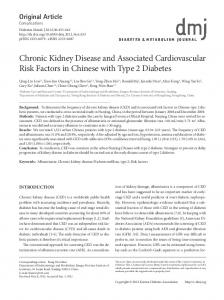
Hospital (Seoul, Korea) for EMPD of the external genitalia between January 2000 and December 2012. Of these 44 patients, the analysis was performed for 19 patients who had undergone wide local excision. Excluded patients had undergone radiotherapy and chemotherapy without surgical excision or had undergone surgical excision in other hospitals. The study was approved by the Institutional Review Board granting approval at the institution. The mean age of the patients at diagnosis was 68 years (range, 57 to 82 years). Most patients underwent computed tomography of the abdomen/pelvis, chest X-ray, and tumor marker evaluation (carbohydrate antigen 19-9, carcinoembryonic antigen [CEA], prostate-specific antigen, and alpha-fetoprotein). Patients in whom gastrointestinal malignancy was suspected underwent esophagoduodenoscopy and colonoscopy. Wide local excision was performed with or without preoperative mapping biopsy (Fig. 1) and intraoperative frozen biopsy, except in one patient who underwent wide local excision with grossly uninvolved lateral margins of 1 cm. In patients who did not undergo preoperative mapping biopsy, the initial excision margin was decided as 2 cm away from the grossly demarcated margin. All excisions were carried deep to the subcutaneous fat. Prophylactic lymph node dissection was not performed, except in one patient who was already known to have locoregional lymph node invasion without distant metastasis. Reconstruction was performed by split-thickness skin graft, full-thickness skin graft, local scrotal flap, or by primary closure. All participants were followed up for a mean duration of 22.5 months (range, 1 to 60 months). Routine surveillance biopsies were not performed in asymptomatic patients. However, if any new suspicious lesions were found on physical examination, further punch biopsies were considered, and most patients underwent computed tomography or ultrasonography to evaluate for recurrence or progression.
The Fisher exact test was used for statistical analyses of factors described by p-value. A difference was considered statistically significant for p-values of less than 0.05. All statistical analyses were performed by using commercially available software (IBM SPSS ver. 18.0, IBM Co., Armonk, NY, USA).
RESULTS The clinical characteristics of the patients are shown in Table 1. All patients had presented with pruritus and erythematous lesions, which were the most common exam findings. In some patients, the EMPD lesions had been misdiagnosed as eczema or some other benign skin lesion at outside clinics and were thus managed with supportive treatment. This misdiagnosis delayed proper management by 43.5 months on average, ranging from 1 month to 198 months. To see whether this delay in treatment predicted worse outcomes, the patients were divided into two groups by a 30-month duration from onset of symptoms to treatment. This comparison did not reveal statistical differences in depth of invasion (p=0.108) or disease progression (p=0.263) between the early (<30 months) and delayed treatment (≥30 months) groups. The lesions were located at the penoscrotal junction in 10 patients (52.6%), at the penoscrotal junction with spread to the pubic area in 4 (21.1%), at the scrotum in 4 (21.1%), and at the penis in 4 (4.8%). Of the 19 patients, 6 patients had presented to our institution with recurrent EMPD. Of these six, three patients had undergone the initial surgical excision at an outside institution. Among the remaining three patients, one had cryotherapy at an outside hospital, and two were treated with applications of imiquimod at the Department of Dermatology in our hospital. Of the whole, one patient had a locoregional invasive disease and another had metastatic disease at diagnosis,
FIG. 1. Location of mapping biopsies marked 2 cm distal to the gross margin. (A) Erythematous lesion of extramammary Paget disease at the junction of the penis and scrotum. The margin of the lesion is identified grossly. The dashed mark represents the location of punch biopsies and the dots represent a 2-cm safety margin. (B) Numbers 22 and 23 represent the lesion of additional punch biopsy because number 19, which was the site of the initial punch biopsy, was extramammary Paget disease (+) on the pathologic report. Korean J Urol 2013;54:834-839
836
Jung et al
TABLE 1. Patient characteristics Patient no.
Age (y)
Previous treatment
Duration of symptom (mo)
Underlying malignancy
Immunochemistry (CEA/CK-7)
1 2 3 4 5 6 7 8 9 10 11 12 13 14 15 16 17 18 19
74 75 71 82 66 71 63 66 57 59 71 72 71 63 61 74 71 62 63
Local excision Cryotherapy No Excisional biopsy No No No No No No No No No No WLE No No Imiquimod Imiquimod
2 24 10 2 38 36 1 36 6 26 85 39 38 8 3 36 184 198 55
No No No Colon cancer No No No No No No No No Cholangiocarcinoma No Parotid gland cancer No No No No
NA/NA NA/+ NA/NA NA/+ NA/+ +/+ NA/NA +/+ +/+ +/+ +/+ +/+ +/+ NA/+ +/+ NA/+ +/+ NA/NA +/+
Stage Localized Localized Unknown Localized Localized Metastasis Localized Localized Unknown Unknown Localized Localized Localized Unknown Unknown Unknown Unknown Regional localized
CEA, carcinoembryonic antigen; CK, cytokeratin; NA, not available; WLE, wide local excision.
TABLE 2. Clinical information on treatment and prognosis Patient no.
Type of marginal biopsy
LN dissection
Type of reconstruction
Resection margin
Invasion depth
R/P
1 2 3 4 5 6 7 8 9 10 11 12 13 14 15 16 17 18 19
Frozen Frozen Frozen Both Frozen None Both Frozen Both Mapping Mapping Mapping Mapping Mapping Mapping Both Both Frozen Mapping
No No No No No No No No No No No No No No No No No Yes No
STSG FTSG Flap STSG Repair Flap Flap Repair STSG STSG STSG Repair Flap Repair STSG STSG STSG Repair STS
Neg Pos Pos Pos Pos Neg Pos Neg Neg Pos Neg Neg Pos Neg Pos Neg Pos Pos Neg
Epidermis Epidermis Epidermis Epidermis Epidermis Dermis Dermis Dermis Dermis Dermis Dermis Dermis Dermis Dermis Dermis Dermis Dermis b Dermis Dermis
No/no Yes/no Yes/no No/no No/no Yes/yes No/no No/no No/no No/no No/no No/no No/no No/no No/no No/no No/no Yes/yes No/no
Biopsy during FU/ Overall further management survival (mo) No/no Yes/WLE No/WLE No/no No/no No/chemotherapy No/no Yes/no No/no Yes/no No/no No/no No/noa No/no No/no No/no a No/no No/radiotherapy No/no
NED (60) Disease (60) Disease (61) NED (25) NED (24) Died (17) NED (16) NED (26) NED (12) NED (15) NED (12) NED (18) NED (17) NED (12) NED (15) NED (12) NED (1) Died (6) NED (18)
Other patients with positive resection margin were just followed up closely. LN, lymph node; R, local recurrence; P, progression; FU, follow-up; STSG, split-thickness skin graft; Neg, negative; NED, no evidence of disease; FTSG, full-thickness skin graft; Pos, positive; WLE, wide local excision; Flap, local flap; Repair, primary repair. a :Re-excision for residual tumor cells before discharge. b:Subcutaneous fat invasion.
respectively. Three patients had underlying malignancies that were diagnosed and treated before the management of EMPD. The details of treatment and outcomes in this cohort are summarized in Table 2. Except for one patient (No. 6), all Korean J Urol 2013;54:834-839
of the patients had undergone preoperative mapping biopsy or intraoperative frozen biopsy (Fig. 1). Seven patients underwent preoperative mapping biopsy without intraoperative frozen biopsy. In seven patients, intraoperative frozen biopsy was positive and thus additional excisions
837
Extramammary Paget Disease of External Genitalia
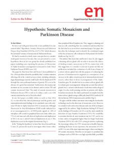
FIG. 2. Kaplan-Meier curve. Kaplan-Meier curve for recurrence-free survival (A) and overall survival (B).
were performed until a negative margin was obtained, except in one patient (No. 2) for whom margin-free resection would have created too large of a soft tissue defect. Tumor cells were detected in resected margins in 10 patients (52.6%) in final pathologic reports. Additional excision was not performed for these patients, who were closely followed up instead. There were four recurrent cases (21.1%), and two of these patients (10.5%) died from the disease. There were no recurrences in patients who underwent preoperative mapping biopsy. The performance of preoperative mapping biopsy was not correlated with the resection margin (p=0.628) or local recurrence (p=0.071). The lesion invaded the dermis in 14 patients (73.7%) and invaded the subcutaneous fat layer in 1 patient (5.3%). The resection margin (p=0.279) and invasion depth (p=0.235) did not correlate with local recurrence. In addition, invasion depth did not correlate with disease progression (p=0.532). Fig. 2 illustrates the cumulative incidence of recurrence and death. Two of nine patients with a positive resection margin underwent additional excision of the positive lesion and there were no recurrences in these patients. Patients 2 and 3, in whom local recurrence was observed at 2 years and 8 months postoperatively, respectively, underwent secondary operations and have been followed up with no evidence of disease at 5 years. Patient 6 had undergone palliative chemotherapy with 5-fluorouracil and cisplatin but died from disease progression. Patient 18 had imiquimod treatment before the operation, but complete surgical excision of the large lesion could not be obtained owing to morbidity, including vessel injury, lymph edema, and large skin defect. His final pathologic report showed subcutaneous invasion. He underwent palliative radiation therapy but died of disease progression.
DISCUSSION Mostly, EMPD arises as a primary cutaneous malignancy and rarely occurs in association with adnexal carcinoma of
a nearby sweat gland or in association with a malignancy of an adjacent organ. Most reports of penoscrotal EMPD are from Asian cohorts [4-6]. Wide local excision has been thought to be the treatment of choice for penoscrotal EMPD. Yang et al. [5] reported that intraoperative frozen biopsy examination reduces rates of positive resection margins and recurrence. Our institute has performed intraoperative frozen biopsy or preoperative mapping biopsy since 2000. In the present study, we report the outcomes of penoscrotal EMPD patients who underwent surgical excision and review the clinical factors influencing outcomes. The present study evaluated the efficacy of intraoperative frozen biopsy or preoperative mapping biopsy. No recurrence was shown in the latter group. Preoperative mapping biopsy can help surgeons to define excision margins for lesions and to decide on the reconstructive method. In contrast with previous studies [7] that reported that frozen biopsy improves the chance of obtaining negative margins, the resection margin was positive in 10 of 19 patients in the present study. Algaba et al. [8] reported that frozen biopsy could not guarantee negative resection margins. This may be because EMPD has multicentric origins with satellite lesions besides the gross lesion [4,9,10]. A positive resection margin was reported to be a risk factor for local recurrence [11], and depth of invasion and lymphovascular involvement were reported to be important pathological predictors of metastatic potential [7,12,13]. However, our findings did not show statistical significance, possibly because of the small sample size, and lymphovascular involvement was not included in the pathologic report, although two progressive cases had occurred among the 14 dermis-invasive diseases. The high proportion of dermis-invasive cases (14/19, 73.7%) is a salient point. This may reflect the health care trend in South Korea, by which most difficult cases are managed at tertiary university hospitals. In contrast with invasive disease, intraepidermal EMPD had a good prognosis in the present study, and no disease progression was observed despite two cases of local recurrence. Therefore, because EMPD is fre-
Korean J Urol 2013;54:834-839
838 quently mistaken for eczema or contact dermatitis, a biopsy should be performed on any uncontrolled inflammatory lesion of the external genitalia to prevent delayed diagnosis. The pathogenesis of EMPD has not been elucidated, but the discussions of this have involved tumor cells spreading from apocrine or eccrine gland tumors, epidermotropic spreading from a regional malignancy, malignant transformation of epithelial cells, and malignant transformation of stem cells in the epidermis [5,6]. Internal malignancy was found to be a poor prognostic factor in a study of EMPD cohorts [3] in which 3 cases of internal malignancy were observed to have no local recurrence at follow-up. The incidence of internal malignancy related to EMPD is lower in Asians than in Caucasians [5,14]. Thus, routine colonoscopy to screen for EMPD-associated colon cancer may not be indicated among Asian patients. Instead, routine immunoperoxidase study for CEA and low-molecular-weight cytokeratin (CK) could be recommended if internal malignancy is suspected [2]. Most cases of EMPD revealed strong immunoreactivity for CEA and CK-7 with androgen receptor, in contrast with negative reactivity for CK-20 and p53. For instance, CK-20-positive EMPD may represent secondary Paget disease due to associated colorectal cancer, and C-erbB-2 was found to have a strong association with dermal invasion [15]. In the present study, cases treated by nonsurgical methods were not included. In the present study, a positive resection margin and local recurrence in intraepidermal cases had a relatively good prognosis. Among dermis-invasive cases, however, two patients with recurrence died of disease progression. In intraepidermal cases with local recurrence, wide excision of the lesion appears to be the most effective modality of treatment, as Li et al. [16] had reported. However, dermis-invasive lesions with local recurrence should be managed by wide excision with adjuvant management such as radiation therapy. Kim et al. [17] reported that radiotherapy can be effective for local control in unresectable cases, and Hata et al. [18] reported that radiotherapy is effective for curative treatment of EMPD. A retrospective review of the surgical management of EMPD from the Mayo Clinic [19] showed that Mohs surgery appears to be effective for Paget disease because the tumor has indistinct margins. Lee et al. [20] reported a high success rate of Mohs surgery for nonmetastatic EMPD in Korea. There are some studies on chemotherapy in the management of EMPD [21-25], but the efficacy has not been clarified. We plan to further investigate and report on the multimodality treatment of EMPD in a future publication.
CONCLUSIONS Although penoscrotal EMPD is a rare malignancy, its diagnosis should not be delayed to allow for prompt treatment. Wide local excision is recommended with preoperative mapping biopsy and intraoperative frozen biopsy in nonKorean J Urol 2013;54:834-839
Jung et al
metastatic disease. Our findings suggest that intraoperative frozen biopsy or preoperative mapping biopsy cannot guarantee negative margins on final pathology. However, preoperative mapping biopsy and wide local excision with intraoperative frozen biopsy results in good prognosis in the treatment of EMPD, especially in those cases without dermal invasion. In recurrent cases, wide local excision is the treatment of choice, but adjuvant management like radiotherapy should be recommended in the presence of dermal invasion or regional invasion. CONFLICTS OF INTEREST The authors have nothing to disclose.
REFERENCES 1. Crocker HR. Paget’s disease affecting the scrotum and the penis. Trans Pathol Soc Lond 1889;40:187-91. 2. Juang GD, Lin MY, Hwang TI. Extramammary Paget's disease of the scrotum. J Chin Med Assoc 2011;74:325-8. 3. Hegarty PK, Suh J, Fisher MB, Taylor J, Nguyen TH, Ivan D, et al. Penoscrotal extramammary Paget's disease: the University of Texas M. D. Anderson Cancer Center contemporary experience. J Urol 2011;186:97-102. 4. Wang Z, Lu M, Dong GQ, Jiang YQ, Lin MS, Cai ZK, et al. Penile and scrotal Paget's disease: 130 Chinese patients with long-term follow-up. BJU Int 2008;102:485-8. 5. Yang WJ, Kim DS, Im YJ, Cho KS, Rha KH, Cho NH, et al. Extramammary Paget's disease of penis and scrotum. Urology 2005;65:972-5. 6. Kuan SF, Montag AG, Hart J, Krausz T, Recant W. Differential expression of mucin genes in mammary and extramammary Paget's disease. Am J Surg Pathol 2001;25:1469-77. 7. Bagby CM, MacLennan GT. Extramammary Paget's disease of the penis and scrotum. J Urol 2009;182:2908-9. 8. Algaba F, Arce Y, Lopez-Beltran A, Montironi R, Mikuz G, Bono AV, et al. Intraoperative frozen section diagnosis in urological oncology. Eur Urol 2005;47:129-36. 9. Hendi A, Brodland DG, Zitelli JA. Extramammary Paget's disease: surgical treatment with Mohs micrographic surgery. J Am Acad Dermatol 2004;51:767-73. 10. Li YC, Lu LY, Yang YT, Chang CC, Chen LM. Extramammary Paget's disease of the scrotum associated with hepatocellular carcinoma. J Chin Med Assoc 2009;72:542-6. 11. Zhang N, Gong K, Zhang X, Yang Y, Na Y. Extramammary Paget's disease of scrotum--report of 25 cases and literature review. Urol Oncol 2010;28:28-33. 12. Lai YL, Yang WG, Tsay PK, Swei H, Chuang SS, Wen CJ. Penoscrotal extramammary Paget's disease: a review of 33 cases in a 20-year experience. Plast Reconstr Surg 2003;112:1017-23. 13. Ng LG, Yip SK, Tan PH. Extramammary Paget's disease of scrotum. Urology 2001;58:105. 14. Chang YT, Liu HN, Wong CK. Extramammary Paget's disease: a report of 22 cases in Chinese males. J Dermatol 1996;23:320-4. 15. Choi YD, Cho NH, Park YS, Cho SH, Lee G, Park K. Lymphovascular and marginal invasion as useful prognostic indicators and the role of c-erbB-2 in patients with male extramammary Paget's disease: a study of 31 patients. J Urol 2005;174:561-5. 16. Li B, Li L, Wang X, Xu K, Fang Z, Ding Q. Frozen section-guided wide local excision in the treatment of recurrent scrotal extra-
Extramammary Paget Disease of External Genitalia mammary Paget's disease. Dermatology 2012;224:231-5. 17. Kim TH, Chang IH, Kim TH, Lee SY, Myung SC. Extramammary Paget's disease of scrotum treated with radiotherapy. Urology 2009;74:474.e1-3. 18. Hata M, Omura M, Koike I, Wada H, Miyagi E, Tayama Y, et al. Role of radiotherapy as curative treatment of extramammary Paget's disease. Int J Radiat Oncol Biol Phys 2011;80:47-54. 19. O'Connor WJ, Lim KK, Zalla MJ, Gagnot M, Otley CC, Nguyen TH, et al. Comparison of mohs micrographic surgery and wide excision for extramammary Paget's disease. Dermatol Surg 2003;29:723-7. 20. Lee KY, Roh MR, Chung WG, Chung KY. Comparison of Mohs micrographic surgery and wide excision for extramammary Paget's disease: Korean experience. Dermatol Surg 2009;35:34-40. 21. Oguchi S, Kaneko M, Uhara H, Saida T. Docetaxel induced durable response in advanced extramammary Paget's disease: a
839 case report. J Dermatol 2002;29:33-7. 22. Kariya K, Tsuji T, Schwartz RA. Trial of low-dose 5-fluorouracil/cisplatin therapy for advanced extramammary Paget's disease. Dermatol Surg 2004;30(2 Pt 2):341-4. 23. Mochitomi Y, Sakamoto R, Gushi A, Hashiguchi T, Mera K, Matsushita S, et al. Extramammary Paget's disease/carcinoma successfully treated with a combination chemotherapy: report of two cases. J Dermatol 2005;32:632-7. 24. Takahagi S, Noda H, Kamegashira A, Madokoro N, Hori I, Shindo H, et al. Metastatic extramammary Paget's disease treated with paclitaxel and trastuzumab combination chemotherapy. J Dermatol 2009;36:457-61. 25. Matsushita S, Yonekura K, Mera K, Kawai K, Kanekura T. Successful treatment of metastatic extramammary Paget's disease with S-1 and docetaxel combination chemotherapy. J Dermatol 2011;38:996-8.
Korean J Urol 2013;54:834-839