Journal of the Formosan Medical Association (2013) 112, 12e17
Available online at www.sciencedirect.com
journal homepage: www.jfma-online.com
REVIEW ARTICLE
Factors contributing to the disturbance of coagulation and fibrinolysis in dengue virus infection Yung-Chun Chuang a,e, Yee-Shin Lin b,e, Ching-Chuan Liu c,e, Hsiao-Sheng Liu b,e, Shu-Hsing Liao f, Ming-Der Shi f, Huan-Yao Lei b,e, Trai-Ming Yeh d,e,* a
Institute of Basic Medical Sciences, Medical College, National Cheng Kung University, Tainan, Taiwan Department of Microbiology and Immunology, Medical College, National Cheng Kung University, Tainan, Taiwan c Department of Pediatrics, National Cheng Kung University Hospital, Medical College, National Cheng Kung University, Tainan, Taiwan d Department of Medical Laboratory Science and Biotechnology, Medical College, National Cheng Kung University, Tainan, Taiwan e Center of Infectious Disease and Signaling, Medical College, National Cheng Kung University, Tainan, Taiwan f Department of Pathology and Laboratory, Kaohsiung Veterans General Hospital Tainan Branch, Tainan, Taiwan b
Received 16 October 2012; accepted 19 October 2012
KEYWORDS autoantibody; coagulation; cytokine; dengue virus; fibrinolysis; hemorrhage
Hemorrhage is one of the hallmarks of dengue hemorrhagic fever. However, the mechanisms that cause hemorrhage are unclear. In this review we focus on the possible factors that may be involved in the disturbance of coagulation and fibrinolysis during dengue virus (DENV) infection. Factors such as autoantibodies and cytokines induced by DENV infection as well as hemostatic molecules expressed on DENV-infected cells, and DENV viral proteins may all contribute to the defect of hemostasis during DENV infection. It is the combination of these viral and host factors that may tilt the balance of coagulation and fibrinolysis toward bleeding in dengue patients. Copyright ª 2012, Elsevier Taiwan LLC & Formosan Medical Association. All rights reserved.
* Corresponding author. Department of Medical Laboratory Science and Biotechnology, Medical College, National Cheng Kung University, Tainan, Taiwan. E-mail address:
[email protected] (T.-M. Yeh). 0929-6646/$ - see front matter Copyright ª 2012, Elsevier Taiwan LLC & Formosan Medical Association. All rights reserved. http://dx.doi.org/10.1016/j.jfma.2012.10.013
Factors contributing to hemorrhage in DENV infection
Introduction Blood coagulation (hemostasis) is a very delicately balanced system that is tightly regulated by many different mechanisms to prevent hemorrhage or clotting under normal condition. Hemostasis consists of primary and secondary stages: primary hemostasis involves vascular constriction, platelet activation and aggregation; and secondary hemostasis involves the activation of coagulation cascade, clot formation, and the clot dissolution by fibrinolysis. The coagulation cascade is composed of intrinsic and extrinsic pathways that lead to the activation of different coagulatory factors.1 Both cascades converge at the activation of factor X (generating factor Xa, ‘a’ signifies active). Factor Xa forms a complex with factor Va to activate prothrombin to become thrombin. Thrombin then converts fibrinogen to a fibrin network. Afterwards, fibrinolysis is triggered by activation of plasminogen to plasmin by tissue plasminogen activator (tPA) or urokinase to prevent thrombosis. Both coagulation and fibrinolysis are regulated by feedback inhibition as well as inhibitors. In addition to inducing fibrin formation, thrombin can also bind to thrombomodulin (TM) on endothelial cell surface, to activate protein C (APC), which is able to inactivate factor VIIIa and Va, thus preventing further thrombin generation.2 Under normal conditions, fibrinolysis is also tightly regulated. Excessive activation of the fibrinolysis will lead to an increasing tendency for bleeding, whereas inhibition of the fibrinolysis will result in thrombosis. Type-I plasminogen activator inhibitor (PAI-1) is the principal inhibitor of tPA and urokinase, which play an important role in the control of the fibrinolytic system. APC can also inactivate PAI-I to enhance the fibrinolysis. Therefore, the balance between coagulation and fibrinolysis is very important and delicately controlled in vivo. Hemostasis is also tightly linked to inflammation. Both systems are interrelated as part of the innate host defense mechanism and show a two-way crosstalk between each other.3,4 Inflammation induced during infection generally shifts the hemostatic mechanism toward thrombosis by upregulation of procoagulant factors, down-regulation of anticoagulants and inhibit fibrinolytic activity.5 However, coagulation product such as thrombin also has a variety of activities on cells that result in augmentation of the inflammatory response. In addition, anticoagulatory molecules such as APC possess not only anticoagulatory activity, but also antiinflammatory activity.2 However, infection with certain viruses can tilt the hemostasis toward bleeding and cause viral hemorrhage fever (VHF).6,7 Among VHFs, dengue, Marburg, and Ebola are the most important ones and dengue virus (DENV) infection is the most prevalent.8 More than 2.5 billion people, or half of the world’s population in tropical and subtropical countries including Southeast Asia and Taiwan are at the risk of DENV infection.9 DENV is a mosquito-borne flavivirus that is transmitted by mosquitoes such as Aedes aegypti or Aedes albopictus. DENV is a positive-stranded RNA with envelope.8 It composes of three structural proteins including core protein (C); membrane-associated protein (M) produced as a precursor protein (prM); envelope protein (E) and seven
13 nonstructural proteins (NS). Based on the antigenic difference of E protein, DENV can be divided into four different serotypes, DENV 1e4. DENV infection might lead to an influenza-like illness, which is called dengue fever or cause more severe dengue hemorrhage fever (DHF) or dengue shock syndrome (DSS). DHF is a severe febrile disease characterized by abnormalities in homeostasis and increased capillary leakage that can progress to blood pressure decrease, and hypovolemic shock (DSS).10 Although DHF/DSS can be seen during primary infection, it occurs more frequently following second infection with a different serotype of DENV from that of previous infection. Therefore, it is generally believed that immunopathogensis is involved in DHF/DSS. Different hypotheses have been proposed to explain the pathogenesis of DHF/DSS including overproduction of proinflammatory cytokines, aberrant immune activation, and antibody-dependent enhancement (ADE).11e13 Among them, the theory of ADE plays a central role. Based on ADE, antibodies that are generated in previous DENV infection may enhance DENV of different serotypes to infect macrophage through Fcg receptor. However, neither cytokine storm nor ADE can explain why hemorrhage occurs in DHF/DSS patients. A better understanding of the mechanism to induce hemorrhage by DENV is required to develop a more effective and specific therapy against the development of DHF/DSS. In almost all DHF patients, defects of coagulation activation such as a prolonged activated partial thromboplastin time (APTT) or thrombin time can be found. In addition, a decreased fibrinogen level and increased levels of fibrinogen degradation products indicating hyperfibrinolysis may also occur in DHF/DSS patients.14e16 Therefore, DENV infection not only causes the defect in the activation of coagulation but also the acceleration of fibrinolysis. The pathogenic effects of some of the possible factors that may contribute to the disturbance of the tightly regulated coagulation and fibrinolysis during DENV infection are discussed in this review.
Autoantibodies against coagulatory and fibrinolytic molecules induced by DENV through molecular mimicry Based on computer sequence comparison between DENV proteins and coagulatory molecules, there are at least 12 different regions of DENV proteins, including core, prM, E, and NS1 proteins, that have amino acid sequence similarity with different coagulatory molecules, such as factors X, XI, and VII.17 The molecular mimicry between DENV proteins and coagulation and fibrinolysis factors may induce autoantibodies that can interfere with the hemostasis. The first report indicating that there are autoantibodies against coagulation factor in dengue patients, was published by Markoff et al.18 They found that Type 4 DENV E protein amino acid 100e119 (D4E; GWGNGCGLFGKGVVTCAKF) shares sequence homology with plasminogen amino acid 759e779 (PLþ; SWGLGCARPNKPGVYVRVSRF). They also found that antibodies cross-reacting to PLþ peptide in dengue patients were correlated to hemorrhage.19 In a previous study, we proved that D4E peptide can induce
14 antibodies against plasminogen in rabbits, which could inhibit plasmin activity in vitro. However, the exact effect of plasminogen binding antibodies on its activation is still unclear due to the heterogeneity of antibodies in antisera.20 Recently, we have generated several monoclonal antibodies from DENV immunized mice. Some of them (6H11, 7D2, 8E5, 2A12) could cross-react with plasminogen and enhance its activation.21 The fibrinolysis in 6H11injected mice was also increased as compared with that in control IgG-injected mice. Thus, these plasminogen crossreactive DENV antibodies may play a role in causing the hyperfibrinolysis during DENV infection. In addition, a single chain fragment of variable region (scFv) generated from NS1 immunized mice could bind to fibrinogen and prolonged clot formation.17 Furthermore, anti-thrombin antibodies that could inhibit its activity are also found in dengue patients’ sera (unpublished data). Therefore, autoantibodies that can cross-react with several different coagulation factors and interfere with their functions are induced during DENV infection. Autoantibodies that can cross-react to factors IX, X, VII, and VIII, prothrombin, thrombin, and plasmin are also found in antiphospholipid syndrome (APS) patients.22e26 However, in contrast to dengue patients, autoantibodies in APS patients can inhibit plasmin activity and enhance prothrombin activation, which may lead to thrombosis in APS patients. Therefore, it is likely that, based on the difference of the specificities, autoantibodies against coagulatory and fibrinolytic factors may have different influences on coagulation and fibrinolysis activation. In addition to the cross-reactivity with coagulatory and fibrinolytic factors, antibodies against DENV can also crossreact with endothelial cells, platelets, and hepatocytes, which may contribute to thrombocytopenia, vascular leakage, and liver damage in DHF/DSS.27e30 Therefore, in addition to ADE, antibodies against DENV play several other roles in the immunopathogenesis of DHF/DSS, which may involve different mechanisms to cause the manifestation of hemorrhage in DENV infection.
Influence of cytokines induced by DENV on coagulation and fibrinolysis Many proinflammatory cytokines and chemokines are increased in dengue patients, including macrophage migration inhibitory factor (MIF).31e33 The sera level of MIF is correlated with the severity and the mortality of dengue patients.33 In addition, DENV infection of different cells induced MIF secretion.34,35 MIF is a proinflammatory cytokine that can induce the expression and secretion of other cytokines, chemokines, and adhesion molecules including tumor necrosis factor-a, interleukin-1b, vascular cell adhesion molecule-1, intracellular cell adhesion molecule1, matrix metalloproteinases, and vascular endothelial growth factor.36e38 Cytokines such as tumor necrosis factora and interleukin-1b induced by MIF can promote the synthesis of platelet-activating factor (PAF). PAF is a phospholipid activator and mediator of leukocyte functions including platelet aggregation and inflammation. The essential role of PAF in the pathogenesis of DENV infection has been demonstrated by PAF receptor knockout mice,
Y.-C. Chuang et al. which show decreased thrombocytopenia, hemoconcentration, decreased systemic levels of cytokines, and delay of lethality after DENV infection, when compared with the wild-type mice.39 Furthermore, the importance of MIF in DENV-induced coagulopathy and lethality of mice was also demonstrated by Iranaia Assunc ¸a ˜o-Miranda et al in MIF knockout mice.34 Reduced thrombocytopenia, plasma leakage, and proinflammatory response are found in MIF knockout mice as compared with the wild-type mice after DENV infection. Using recombinant MIF as well as the supernatants from DENV-infected cells, we confirmed that MIF can enhance the permeability of endothelial cells, which may contribute to plasma leakage in vivo.35 In addition, MIF can induce intracellular cell adhesion molecule-1 and thrombomodulin expression of endothelial cells in vitro.40 Recently, we also found that MIF can induce cell autophagy which may enhance DENV replication.41,42 Thus, cytokines such as MIF induced by DENV infection may participate in the hemostatic defect in DHF/DSS patients.
Aberrant expression of tPA and TM in DENVinfected or -stimulated cells Due to the fact that the pathogenic changes of vascular leakage and coagulopathy in DHF/DSS are reversible, it is generally believed that physical damage is not involved in DENV-infected endothelial cells. Instead, soluble mediators such as cytokines produced during the acute phase of infection are likely to play an important role in the pathogenesis of DHF/DSS. However, cytokines are nonspecific and can be induced by other viral infections that do not lead to vascular leakage. In addition, as we mentioned earlier, autoantibodies induced by DENV may cross-react with endothelial cells, platelets, and coagulatory factors, which may contribute to the pathogenesis of DHF/DSS. However, antibodies are generally induced a week after infection. Thus, we think the direct pathogenic roles of DENV or its products cannot be neglected, especially in the early stage of DENV infection. In the early stage of DENV infection, in addition to the APTT prolongation, the fibrinolytic parameters such as tPA and PAI-1 in dengue patients sera are also increased.15,43 The increased tPA/PAI-1 ratio in dengue patients may prone the activation of fibrinolysis in these patients.15 In addition, endothelial cells infected with DENV showed increased tPA expression whereas the expression of PAI-1 showed no difference after infection.44,45 However, PAI-1 gene expression in human hepatoma cell line, HuH-7 is increased after incubation with purified recombinant DENV E protein domain III (DIIIE).46 Therefore, both DENV infection and its protein stimulation can affect the expression of hemostasis-related molecules on different cells. Similar effects of DENV on the expression of TM on human endothelial cells in vitro are also found.45,47,48 TM expressed on the surface of endothelial cells and monocytes is very important in the activation of protein C, which is important to the negative regulation of blood coagulation. The activation of protein C by thrombin-TM complex is augmented when it binds to endothelial cell protein C receptor (EPCR).2,49 Once APC dissociates
Factors contributing to hemorrhage in DENV infection from EPCR, it can bind to protein S on appropriate cell surfaces and inactivate factors Va and VIIIa, thereby inhibiting further thrombin generation. Therefore, TM plays an important role in the anticoagulant state of endothelium. The sera level of secreted TM is increased in dengue patients.50 In addition, the expressions of several other protein C-activation-related molecules such as EPCR and protein S are also increased in DV-infected endothelial cells. The increased expression of TM, EPCR, and protein S in DV-infected EC may enhance protein C activation and lead to rapid thrombin inactivation, which may contribute to the hemorrhage in dengue patients. Taken together, DENV infection or its protein stimulation can promote the expression of anticoagulant molecules expression on endothelial cells, which may contribute to the anticoagulant properties of these cells and increase the hemorrhagic risk in DENV patients.
Inhibition of prothrombin activation by DENV NS1 proteins During DENV infection, NS1 can be present in three different forms: intracellular, extracellular, and membrane forms. The extracellular form of NS1 is secreted as a soluble hexamer, which is also known as secreted NS1 (sNS1).
15 Recently, it was found that sNS1 can form a lipoprotein particle with an open-barrel protein shell and a prominent central channel rich in lipids.51 The detection of sNS1 in patients’ sera not only provides a rapid diagnosis for DENV infection, but also the sera levels of sNS1 in dengue patients are correlated with the disease severity.52 In addition, sNS1 can bind to heparin sulfate and chondroitin sulfate E on the endothelial cell surface. The binding of sNS1 to endothelium and its subsequent recognition by antiNS1 antibodies may also contribute to the vascular leakage in DHF/DSS.53 However, the direct pathogenic role of sNS1 is still unclear. Recently it was found that sNS1 can interact with complement and play a role in protecting DENV from complement-dependent opsonization.54,55 Using recombinant NS1, we found NS1 can bind to both thrombin and prothrombin.56 Even though the thrombin activity is not altered when NS1 binding to thrombin, the binding of NS1 to prothrombin can inhibit its activation, which may contribute to the prolongation of APTT in dengue patients.56 This may explain why APTT abnormality occurs within the 1st week of fever onset when antibodies are still underdeveloped.57 In addition, since the vascular leakage in dengue patient is also directly related to APTT levels,57 NS1 may also contribute to plasma leakage by mechanisms without antibody involved. These results suggest that DENV sNS1 plays a direct and important role in the vascular leakage and hemorrhage in DHF/DSS.
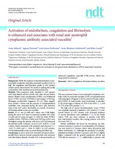
Figure 1 The sequence of events leading to dengue hemorrhage and the possible pathogenic roles of dengue virus (DENV) secreted nonstructural protein 1 (NS1), macrophage migration inhibitory factor (MIF), and coagulation factors cross-reactive autoantibodies in dengue hemorrhage fever (DHF) or dengue shock syndrome (DSS). The timing of clinical symptoms and common complications (upper panel), and possible mechanistic causes (lower panel) following mosquito bite in dengue patients are shown. NS1 protein secreted during early stage of DENV infection binding to prothrombin may inhibit its activation. At later time points, MIF induced by DENV infection may enhance DENV replication through autophagy, which may also contribute to vascular leakage through tight junction disruption. Finally, the production of coagulation factors cross-reactive autoantibodies may further inhibit coagulation and enhance fibrinolysis in dengue patients to cause bleeding.
16
Conclusions Hemorrhage induced by DENV infection may involve both viral factors and host factors. Viral factors such as virus infection, viral sNS1 and DIIIE proteins can affect coagulation in direct or indirect manners. Host factors such as cytokines and autoantibodies induced by DENV may also play a role in the disruption of the balance of coagulation and fibrinolysis as well as in the functions of endothelial cells and platelets. It is the combination of both viral and host factors that may cause the hemorrhage in DHF/DSS. Therefore, besides the direct pathogenic effects of DENV to cells, we propose that viral sNS1, MIF, and coagulation cross-reactive autoantibodies may also play important roles in the pathogenesis of DHF/DSS (Fig. 1). Each of them may play different pathogenic roles in different stages of DENV infection. Blockage of sNS1or MIF may provide alternative approaches to prevent the development of DHF/DSS. Moreover, epitopes mimic to coagulation factors should be avoided in the design of dengue vaccines to prevent possible side effects.
References 1. Davie EW, Fujikawa K, Kisiel W. The coagulation cascade: initiation, maintenance, and regulation. Biochemistry 1991; 30:10363e70. 2. Esmon CT. The protein C pathway. Chest 2003;124:26Se32S. 3. Schoenmakers SH, Reitsma PH, Spek CA. Blood coagulation factors as inflammatory mediators. Blood Cells Mol Dis 2005; 34:30e7. 4. Esmon CT. Crosstalk between inflammation and thrombosis. Maturitas 2004;47:305e14. 5. Esmon CT. Inflammation and thrombosis. J Thromb Haemost 2003;1:1343e8. 6. Cosgriff TM. Viruses and hemostasis. Rev Infect Dis 1989; 11(Suppl. 4):S672e88. 7. Chen JP, Cosgriff TM. Hemorrhagic fever virus-induced changes in hemostasis and vascular biology. Blood Coagul Fibrinolysis 2000;11:461e83. 8. Henchal EA, Putnak JR. The dengue viruses. Clin Microbiol Rev 1990;3:376e96. 9. Chang SF, Huang JH, Shu PY. Characteristics of dengue epidemics in Taiwan. J Formos Med Assoc 2012;111: 297e9. 10. Halstead SB. Antibody, macrophages, dengue virus infection, shock, and hemorrhage: a pathogenetic cascade. Rev Infect Dis 1989;11(Suppl. 4):S830e9. 11. Lei HY. Transient hemophagocytic activity in dengue immunopathogenesis. J Formos Med Assoc 2009;108:595e8. 12. Rothman AL. Dengue: defining protective versus pathologic immunity. J Clin Invest 2004;113:946e51. 13. Lei HY, Yeh TM, Liu HS, Lin YS, Chen SH, Liu CC. Immunopathogenesis of dengue virus infection. J Biomed Sci 2001;8: 377e88. 14. Marchi R, Nagaswami C, Weisel JW. Fibrin formation and lysis studies in dengue virus infection. Blood Coagul Fibrinolysis 2009;20:575e82. 15. Huang YH, Liu CC, Wang ST, Lei HY, Liu HL, Lin YS, et al. Activation of coagulation and fibrinolysis during dengue virus infection. J Med Virol 2001;63:247e51. 16. Sosothikul D, Seksarn P, Pongsewalak S, Thisyakorn U, Lusher J. Activation of endothelial cells, coagulation and fibrinolysis in children with Dengue virus infection. Thromb Haemost 2007; 97:627e34.
Y.-C. Chuang et al. 17. Lin YS, Yeh TM, Lin CF, Wan SW, Chuang YC, Hsu TK, et al. Molecular mimicry between virus and host and its implications for dengue disease pathogenesis. Exp Biol Med (Maywood) 2011;236:515e23. 18. Markoff LJ, Innis BL, Houghten R, Henchal LS. Development of cross-reactive antibodies to plasminogen during the immune response to dengue virus infection. J Infect Dis 1991;164: 294e301. 19. Chungue E, Poli L, Roche C, Gestas P, Glaziou P, Markoff LJ. Correlation between detection of plasminogen cross-reactive antibodies and hemorrhage in dengue virus infection. J Infect Dis 1994;170:1304e7. 20. Huang YH, Chang BI, Lei HY, Liu HS, Liu CC, Wu HL, et al. Antibodies against dengue virus E protein peptide bind to human plasminogen and inhibit plasmin activity. Clin Exp Immunol 1997;110:35e40. 21. Chuang YC, Lei HY, Lin YS, Liu HS, Wu HL, Yeh TM. Dengue virus-induced autoantibodies bind to plasminogen and enhance its activation. J Immunol 2011;187:6483e90. 22. Ames PR, Iannaccone L, Alves JD, Margarita A, Lopez LR, Brancaccio V. Factor XIII in primary antiphospholipid syndrome. J Rheumatol 2005;32:1058e62. 23. Yang YH, Chien D, Wu M, FitzGerald J, Grossman JM, Hahn BH, et al. Novel autoantibodies against the activated coagulation factor IX (FIXa) in the antiphospholipid syndrome that interpose the FIXa regulation by antithrombin. J Immunol 2009; 182:1674e80. 24. Zhao Y, Rumold R, Zhu M, Zhou D, Ahmed AE, Le DT, et al. An IgG antiprothrombin antibody enhances prothrombin binding to damaged endothelial cells and shortens plasma coagulation times. Arthritis Rheum 1999;42:2132e8. 25. Yang CD, Hwang KK, Yan W, Gallagher K, FitzGerald J, Grossman JM, et al. Identification of anti-plasmin antibodies in the antiphospholipid syndrome that inhibit degradation of fibrin. J Immunol 2004;172:5765e73. 26. Bidot CJ, Jy W, Horstman LL, Huisheng H, Jimenez JJ, Yaniz M, et al. Factor VII/VIIa: a new antigen in the anti-phospholipid antibody syndrome. Br J Haematol 2003;120:618e26. 27. Cheng HJ, Lin CF, Lei HY, Liu HS, Yeh TM, Luo YH, et al. Proteomic analysis of endothelial cell autoantigens recognized by anti-dengue virus nonstructural protein 1 antibodies. Exp Biol Med (Maywood) 2009;234:63e73. 28. Lin CF, Lei HY, Shiau AL, Liu HS, Yeh TM, Chen SH, et al. Endothelial cell apoptosis induced by antibodies against dengue virus nonstructural protein 1 via production of nitric oxide. J Immunol 2002;169:657e64. 29. Lin CF, Lei HY, Liu CC, Liu HS, Yeh TM, Wang ST, et al. Generation of IgM anti-platelet autoantibody in dengue patients. J Med Virol 2001;63:143e9. 30. Lin CF, Wan SW, Chen MC, Lin SC, Cheng CC, Chiu SC, et al. Liver injury caused by antibodies against dengue virus nonstructural protein 1 in a murine model. Lab Invest 2008;88: 1079e89. 31. Yen YT, Chen HC, Lin YD, Shieh CC, Wu-Hsieh BA. Enhancement by tumor necrosis factor alpha of dengue virus-induced endothelial cell production of reactive nitrogen and oxygen species is key to hemorrhage development. J Virol 2008;82:12312e24. 32. Bethell DB, Flobbe K, Cao XT, Day NP, Pham TP, Buurman WA, et al. Pathophysiologic and prognostic role of cytokines in dengue hemorrhagic fever. J Infect Dis 1998;177:778e82. 33. Chen LC, Lei HY, Liu CC, Shiesh SC, Chen SH, Liu HS, et al. Correlation of serum levels of macrophage migration inhibitory factor with disease severity and clinical outcome in dengue patients. Am J Trop Med Hyg 2006;74:142e7. 34. Assunc ¸˜ ao-Miranda I, Amaral FA, Bozza FA, Fagundes CT, Sousa LP, Souza DG, et al. Contribution of macrophage migration inhibitory factor to the pathogenesis of dengue virus infection. FASEB J 2010;24:218e28.
Factors contributing to hemorrhage in DENV infection 35. Chuang YC, Lei HY, Liu HS, Lin YS, Fu TF, Yeh TM. Macrophage migration inhibitory factor induced by dengue virus infection increases vascular permeability. Cytokine 2011;54:222e31. 36. Calandra T, Bernhagen J, Mitchell RA, Bucala R. The macrophage is an important and previously unrecognized source of macrophage migration inhibitory factor. J Exp Med 1994;179: 1895e902. 37. Calandra T, Bernhagen J, Metz CN, Spiegel LA, Bacher M, Donnelly T, et al. MIF as a glucocorticoid-induced modulator of cytokine production. Nature 1995;377:68e71. 38. Makita H, Nishimura M, Miyamoto K, Nakano T, Tanino Y, Hirokawa J, et al. Effect of anti-macrophage migration inhibitory factor antibody on lipopolysaccharide-induced pulmonary neutrophil accumulation. Am J Respir Crit Care Med 1998;158: 573e9. 39. Souza DG, Fagundes CT, Sousa LP, Amaral FA, Souza RS, Souza AL, et al. Essential role of platelet-activating factor receptor in the pathogenesis of Dengue virus infection. Proc Natl Acad Sci U S A 2009;106:14138e43. 40. Shyu LY, Yeh TM, Chang HH, Lin DP, Teng YH, Chen LC, et al. Macrophage migration inhibitory factor induces ICAM-1and thrombomobulin expression in vitro. Thromb Res 2012;129:43e9. 41. Chuang YC, Su WH, Lei HY, Lin YS, Liu HS, Chang CP, et al. Macrophage migration inhibitory factor induces autophagy via reactive oxygen species generation. PLoS One 2012;7:e37613. 42. Lee YR, Lei HY, Liu MT, Wang JR, Chen SH, Jiang-Shieh YF, et al. Autophagic machinery activated by dengue virus enhances virus replication. Virology 2008;374:240e8. 43. Mairuhu A, Setiati T, Koraka P, Hack C, Leyte A, Faradz S, et al. Increased PAI-1 plasma levels and risk of death from dengue: no association with the 4G/5G promoter polymorphism. Thromb J 2005;3:17. 44. Huang YH, Lei HY, Liu HS, Lin YS, Chen SH, Liu CC, et al. Tissue plasminogen activator induced by dengue virus infection of human endothelial cells. J Med Virol 2003;70:610e6. 45. Jiang Z, Tang X, Xiao R, Jiang L, Chen X. Dengue virus regulates the expression of hemostasis-related molecules in human vein endothelial cells. J Infect 2007;55:e23e8. 46. Shyu HW, Lin YY, Chen LC, Wang YF, Yeh TM, Su SJ, et al. The dengue virus envelope protein induced PAI-1 gene expression via MEK/ERK pathways. Thromb Haemost 2010;104:1219e27. 47. Chen LC, Yeh TM, Lin YY, Wang YF, Su SJ, Chen CY, et al. The envelope glycoprotein domain III of dengue virus type 2
17
48.
49.
50.
51.
52.
53.
54.
55.
56.
57.
induced the expression of anticoagulant molecules in endothelial cells. Mol Cell Biochem 2010;342:215e21. Chen LC, Shyu HW, Lin HM, Lei HY, Lin YS, Liu HS, et al. Dengue virus induces thrombomodulin expression in human endothelial cells and monocytes in vitro. J Infect 2009;58:368e74. Stearns-Kurosawa DJ, Kurosawa S, Mollica JS, Ferrell GL, Esmon CT. The endothelial cell protein C receptor augments protein C activation by the thrombin-thrombomodulin complex. Proc Natl Acad Sci U S A 1996;93:10212e6. Butthep P, Chunhakan S, Tangnararatchakit K, Yoksan S, Pattanapanyasat K, Chuansumrit A. Elevated soluble thrombomodulin in the febrile stage related to patients at risk for dengue shock syndrome. Pediatr Infect Dis J 2006; 25:894e7. Gutsche I, Coulibaly F, Voss JE, Salmon J, d’Alayer J, Ermonval M, et al. Secreted dengue virus nonstructural protein NS1 is an atypical barrel-shaped high-density lipoprotein. Proc Natl Acad Sci U S A 2011;108:8003e8. Libraty DH, Young PR, Pickering D, Endy TP, Kalayanarooj S, Green S, et al. High circulating levels of the dengue virus nonstructural protein NS1 early in dengue illness correlate with the development of dengue hemorrhagic fever. J Infect Dis 2002;186:1165e8. Avirutnan P, Zhang L, Punyadee N, Manuyakorn A, Puttikhunt C, Kasinrerk W, et al. Secreted NS1 of dengue virus attaches to the surface of cells via interactions with heparan sulfate and chondroitin sulfate E. PLoS Pathog 2007;3:e183. Avirutnan P, Fuchs A, Hauhart RE, Somnuke P, Youn S, Diamond MS, et al. Antagonism of the complement component C4 by flavivirus nonstructural protein NS1. J Exp Med 2010;207: 793e806. Somnuke P, Hauhart RE, Atkinson JP, Diamond MS, Avirutnan P. N-linked glycosylation of dengue virus NS1 protein modulates secretion, cell-surface expression, hexamer stability, and interactions with human complement. Virology 2011;413: 253e64. Lin SW, Chuang YC, Lin YS, Lei HY, Liu HS, Yeh TM. Dengue virus nonstructural protein NS1 binds to prothrombin/thrombin and inhibits prothrombin activation. J Infect 2012;64:325e34. Wills B, Tran VN, Nguyen TH, Truong TT, Tran TN, Nguyen MD, et al. Hemostatic changes in Vietnamese children with mild dengue correlate with the severity of vascular leakage rather than bleeding. Am J Trop Med Hyg 2009;81:638e44.