The Journal of Immunology
Termination of Antigen-Specific Immunity by CD95 Ligand (Fas Ligand) and IL-101 Ramon Barreiro,* Gary Luker,† John Herndon,* and Thomas A. Ferguson2* Following elimination of a foreign invader, the immune system must return to its normal quiescent levels. This process requires removal of reactive immune cells when they are no longer needed. We have explored the role of Fas/Fas ligand (FasL) in terminating immunity and demonstrate that mice defective in these proteins have prolonged immune responses. Studies demonstrate that termination of immunity occurs via the interaction of Fasⴙ lymphoid cells with FasLⴙ nonlymphoid cells at the site of Ag challenge. Our results also show that FasL is absent in quiescent tissue but is rapidly up-regulated during the local immune reaction. This occurs through the production of IL-10. Thus, FasL and IL-10 work in concert to eliminate inflammatory cells and control the duration of an immune response. The Journal of Immunology, 2004, 173: 1519 –1525.
A
properly functioning immune system relies on apoptosis at virtually all stages to control the expansion and expression of immunity. Aberrant deletion of immune cells can lead to lymphoproliferative disorders, autoimmunity and even cancer. This is demonstrated with the lpr and gld mutations affecting Fas (CD95) and its ligand, Fas ligand (FasL,3 CD95L), respectively, which produce several apoptosis defects leading to acceleration of autoimmune responses (1). The finding that Fas/ FasL interactions are required for efficient activation-induced apoptosis in T cells in vitro (2) and for peripheral T cell deletion in vivo (3, 4), suggest that defects in apoptosis contribute to lymphoid cell accumulation and autoimmune dysfunction. In addition, the process of immune privilege in the eye relies to a large degree on FasL expression on resident cells. Here mice without functional FasL cannot control inflammation leading to significant damage following viral (5, 6) or parasitic infection (7). Fas/FasL interactions have also been implicated in other processes including autoimmune thyroiditis (8), tumor progression (9, 10), immune tolerance (5, 11, 12), and immune privilege in the brain (13) and testis (14). Enforced expression of FasL has been shown to protect allografts from rejection in some cases (15, 16) but not others (17). In the skin, tolerance induced by UV irradiation involves Fas/FasL (11, 18) and a number of skin diseases are thought to involve FasL (19). In this tissue, resident keratinocytes (20, 21) and Langerhans cells are (22) capable of expressing FasL, while immigrating T cells (23) and NK cells (24) can also express this death-inducing ligand.
The elimination of cells and termination of an Ag-specific immune response is thought to take place through several mechanisms including apoptosis and the production of inhibitory cytokines such as IL-10. IL-10 was first recognized for its ability to inhibit responses of T cells, monocytes, and macrophages (25). Its primary function seems to be the control of inflammatory responses. Mice without IL-10 cannot control immunity and develop severe colitis. A role for IL-10 in the termination of cutaneous immunity was shown a number of years ago (26, 27), where IL-10 production in the skin inhibited T cell responses to applied Ags. In this paper we have examined the termination of an Ag-specific immune response. Our results demonstrate that up-regulation of FasL through the production of IL-10 induces apoptosis in responding cells. This shows for the first time that ligand-mediated apoptosis is responsible for terminating immunity and helping return the system to homeostasis.
Materials and Methods Mice
*Department of Ophthalmology and Visual Sciences, and †Molecular Imaging Center, Mallinckrodt Institute of Radiology, Washington University School of Medicine, St. Louis, MO 63110
BALB/c were purchased from the National Cancer Institute (Fredrick, MD). The gld and lpr mutations were bred onto the BALB/c background by crossing B6-gld and B6-lpr (originally obtained from The Jackson Labortory, Bar Harbor, ME) to BALB/c for a minimum of 10 generations. Mice were screened by PCR as described (28). Since the mutations were derived from B6 mice these strains are designated C.B6-lpr and C.B6-gld. CD95LP-luciferase reporter mice were obtained from Dr. G. Koretzky (University of Pennsylvania, Philadelphia, PA) and have been described (29). These transgenic mice express luciferase driven by 2.2 kb of the mouse CD95L promoter sequence. This allows quantitative detection of luciferase as a surrogate for endogenous CD95L expression. Groups usually consist of five mice, and experiments were repeated at least twice. All mice were male and 4 – 6 wk of age at the initiation of the experiments. Statistical significance between groups was determined by Student’s t test using a 95% (p ⬍ 0.05) confidence interval.
Received for publication March 16, 2004. Accepted for publication May 10, 2004.
Reagents
The costs of publication of this article were defrayed in part by the payment of page charges. This article must therefore be hereby marked advertisement in accordance with 18 U.S.C. Section 1734 solely to indicate this fact. 1 This work was supported by National Institutes of Health Grants EY12826, EY06765, and EY02687; Research to Prevent Blindness, NY; and Foundation for Fighting Blindness (Owings Mills, MD). 2
Address correspondence and reprint requests to Dr. Thomas A. Ferguson, Department of Ophthalmology and Visual Sciences, Washington University School of Medicine, 660 South Euclid, Box 8096, St. Louis, MO 63110. E-mail address:
[email protected]
3 Abbreviations used in this paper: FasL, Fas ligand; TNCB, 2,4,6-trinitro-1-chlorobenzene; TNBS, 2,4,6-trinitrobenzene sulfonic acid; ROI, regions-of-interest.
Copyright © 2004 by The American Association of Immunologists, Inc.
2,4,6-trinitro-1-chlorobenzene (TNCB) was originally purchased from Eastern Chemical (Smithtown, NY) 2,4,6-trinitrobenzene sulfonic acid (TNBS) was purchased from Sigma-Aldrich (St. Louis, MO). ZVAD-fmk and ZFA-fmk were purchased from Calbiochem (San Diego, CA). Fas-Fc was purchased from R&D Systems (Minneapolis, MN). TRAILR-Fc was obtained from Dr. T. Griffith (University of Iowa, Iowa City, IA). rIL-10 was purchased from BD Biosciences (San Diego, CA).
Immune response Mice were immunized with 0.1 ml of 1% TNCB in acetone/olive oil (3/1) applied to shaved abdominal skin as described (12). Four days later, mice 0022-1767/04/$02.00
1520 were challenged with 0.033 ml of 10 mM TNBS in PBS in the right footpad and 0.033 ml of PBS in the left footpad. Values are expressed in micrometers (⫾ SE) and represent the difference between the right footpad (Ag challenge) and the left footpad (PBS challenge). Measurements were taken 24, 48, 72, and 90 h postinjection by a masked observer. Background values (Bkg) represent the difference between the challenged and unchallenged footpad in unimmunized mice.
Adoptive transfer of immunity Mice were immunized with 0.1 ml of 1% TNCB in acetone/olive oil (3/1) applied to shaved abdominal skin. Four days later spleens were harvested, erythrocyte-free suspensions were prepared (12), and 3–5 ⫻ 107 cells were infused into naive recipients. Mice were immediately challenged and measurements were taken as described above.
Bone marrow chimeras Radiation bone marrow chimeras were prepared as previously described (6) except BALB/c mice are given 8.5 Gy (1 Gy ⫽ 100 rad) of radiation before receiving 107 bone marrow cells. Mice were rested at least 3 wk before use.
FasL expression FasL expression was detected by immunohistochemistry using an antiFasL Ab (N20 from Santa Cruz Biotechnology, Santa Cruz, CA) as described (6). The specificity of the Ab was confirmed by blocking with specific FasL peptide (aa 2–19, 1 g/ml) and staining of mouse eye tissue (6). FasL expression was also detected in CD95LP-luc mice by bioluminescence as described in the next paragraph. Bioluminescence imaging of luciferase on CD95LP-luc mice was performed on a cooled CCD camera (IVIS, Xenogen, Alameda, CA) as described (30). Mice were then given an i.p. injection of 150 mg/kg of Dluciferin (Xenogen) from a 15 mg/ml stock solution in PBS. Approximately 5 min after injection, animals were anesthetized with 2% isoflurane, and imaging began 10 min after administration of D-luciferin. Immediately afterward, imaging of in vivo bioluminescence was performed with animals positioned supine, placing the shaved abdominal skin toward the CCD camera. Bioluminescence images were obtained for acquisition times of 0.5–5 min, depending on amounts of light produced. Anesthesia was maintained during imaging by nose cone delivery of isoflurane. Each mouse was imaged before Ag challenge to establish the background luminescence. Only animals with background photon flux of ⬍100 (see below) were used in the experiments. Mice were then injected on shaved abdominal skin with 0.05 ml PBS or 0.05 ml of 10 mM TNBS. Images were obtained at 12, 24, 36, 48, and 60 h later. In some experiments, 200 ng of IL-10 was injected in the skin and images were obtained 8 and 24 h later.
Quantification of bioluminescence data Corresponding gray-scale photographs and color luciferase images were superimposed using LivingImage (Xenogen) and Igor (Wavemetrics, Lake
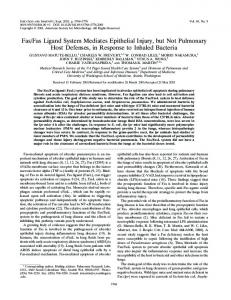
FIGURE 1. Immune responses in BALB/c, C.B-lpr, and C.B6-gld mice. Groups of five mice were immunized with 0.1 ml of 1% TNCB in acetone/olive oil (3/1) applied to shaved abdominal skin. Four days later, mice were challenged with 0.033 ml of 10 mM TNBS in PBS in the right footpad and 0.033 ml of PBS in the left footpad. Values are expressed in micrometers (⫾ SE) and represent the difference between the right footpad (Ag challenge) and the left footpad (PBS challenge). Measurements were taken 24, 48, and 72 h postinjection by a masked observer. Background (Bkg) values represent the difference between the challenged and unchallenged footpad in unimmunized mice. ⴱ, Significantly different from BALB/c immune control (E).
THE TERMINATION OF IMMUNITY Oswego, OR) image analysis software. Signal intensities from regions-ofinterest (ROI) were defined manually, and data were expressed as photon flux (photons per second per cm2) of ROI. Background photon flux was defined from a ROI of the same size drawn over the thorax of each animal, and these data were subtracted from signal intensities measured at sites of injection. Area-under-the-curve analyses were performed with Kaleidagraph (Synergy Software, Reading, PA). Data were reported as mean values for the number of animals indicated in the figures. Pairs were compared with Student’s t test, and values of p ⬍ 0.05 were considered significant.
Results Fas and FasL-deficient mice have prolonged immune responses Using the well characterized system of T cell-mediated cutaneous hypersensitivity (12, 26), we examined the importance of Fas and FasL in the resolution of this response following Ag challenge. Wild-type BALB/c mice along with C.B6-lpr and C.B6-gld mice were sensitized to TNCB. Mice were challenged on day 4, and measurements were obtained 24, 48, and 72 h later. As shown in Fig. 1, wild-type BALB/c had a typical response that peaked at 24 h, but returned to background levels by 48 h. In contrast, C.B6lpr and C.B6-gld mice, which showed a wild-type response at 24 h, still had a significant response 72 h postchallenge. Histological analysis (Fig. 2) confirmed these findings. During the maximal responses at 24 h both BALB (Fig. 2A) and C.B6-gld (Fig. 2B) mice had large numbers of inflammatory cells in the footpads. By 72 h, BALB tissue had returned to normal (Fig. 2C), while C.B6gld skin (fig. 2D) still contained significant inflammatory cell infiltrates. Further confirmation was obtained by demonstrating that local IFN-␥ and TNF production were observed in BALB/c and C.B6-gld mice at 24 h, but by 72 h only C.B6-gld mice expressed these cytokines at the site of antigenic challenge (data not shown). FasL expression in the skin We then localized FasL expression by staining skin sections for FasL protein. As shown in Fig. 3A, significant FasL expression (brown) as noted throughout the skin of TNBS challenged immune mice at 24 h. Challenge with PBS elicited detectable, but significantly less FasL expression (Fig. 3B). To get a more quantitative assessment of FasL expression we used the CD95LP-luc reporter mouse strain (29). These transgenic mice express luciferase driven by 2.2 kb of the mouse CD95L promoter sequence. This allows quantitative detection of luciferase as a surrogate for endogenous CD95L expression. Naive CD95LP-luc mice or CD95LP-luc mice
The Journal of Immunology
1521
FIGURE 2. Histological analysis of challenged skin. Immune BALB/c and C.B6-gld mice we challenged 4 days following immunization. Footpad tissue was obtained 24 and 72 h later. Tissue was fixed overnight with 10% neutral buffered formalin and paraffin embedded sections were stained with H&E. BALB, 24 h (A); C.B6-gld, 24 h (B); BALB/c, 72 h (C); C.B6-gld, 72 h (D). Ep, epidermis; Dm, dermis. Magnification, ⫻100.
given immune T cells from syngeneic nontransgenic (no luciferase) B6 mice were injected s.c. in abdominal skin with 10 mM TNBS. Luciferase expression was monitored in individual mice for 60 h by bioluminescence. Fig. 4 shows that significant luciferase activity was observed at 12 h postinjection of TNBS in immune and nonimmune mice. By 24 h, luciferase activity had dropped significantly in nonimmune mice, but remained high in mice receiving nontransgenic immune T cells. Luciferase activity had fallen by 36 h in immune mice injected with TNBS, but was still higher than the other groups. By 48 h postchallenge, some activity still evident, but by 60 h postchallenge, expression had returned to
FIGURE 3. FasL expression in skin. Footpad tissue was obtained from 4-day immune mice 24 h following challenge with TNBS (A) or PBS (B). Tissues were frozen, embedded, and stained as described (6). Magnification, ⫻100.
background in all groups. We conclude that antigenic challenge in the skin elicits similar FasL expression 12 h postchallenge in immune and nonimmune animals; however, the presence of immune cells (luciferase negative) prolongs FasL expression such that at 24 and 36 h, significant FasL is available. We conclude that skin challenge up-regulates FasL expression. This is sustained by the presence of immune cells.
FIGURE 4. FasL expression in CD95LP-luc mice. Values (photons per second per cm2) obtained from naive CD95LP-luc or CD95LP-luc mice infused with 4-day immune spleen cells from nontransgenic B6 mice. Each point represents the mean of at least three mice. SEs for each point (not shown) were typically ⬍10%.
1522
THE TERMINATION OF IMMUNITY
FIGURE 5. Lymphoid vs nonlymphoid FasL. A, Immune spleen cells from BALB or C.B6-lpr mice were transferred to naive BALB or C.B6-gld recipients. Mice were immediately challenged, and the response was monitored for the next 72 h. Values represent the difference between the right (TNBS) and the left (PBS) footpads. ⴱ, Significantly different from control group BALB/c3 BALB/c (F). B, Bone marrow chimeras constructed by reconstituting BALB/c or C.B6-gld mice with BALB/bone marrow were immunized with TNCB. Four days later, mice were challenged, and the response was monitored for the next 90 h. ⴱ, Significantly different from control group BALB/c3 BALB/c (F).
Lymphoid and nonlymphoid FasL Our results thus far suggest that FasL/Fas interactions play a role in the resolution of this immune response. However, there was significant expression of FasL throughout the skin, including within the inflammatory cell population. Therefore, one explanation for our results is that the response is terminated through autocrine and paracrine interactions among lymphoid cells. We examined this possibility using an adoptive transfer system where sensitized T cells from immune BALB, C.B6-gld, or C.B6-lpr mice were transferred to naive BALB or C.B6-gld recipients (Fig. 5A). Mice were immediately challenged, and the response monitored for the next 72 h. When donor lymphocytes were Fas⫹ and the recipient was FasL⫹ (BALB/c3 BALB/c), the peak 24 h response returned to background level by 48 h. However, if the donor lymphocytes were Fas deficient (C.B6-lpr3 BALB/c; C.B6lpr3 C.B6-gld) or the recipient was FasL deficient (BALB/
c3 C.B6-gld: C.B6-lpr3 C.B6-gld) the immune response persisted to 72 h. When the recipient was FasL⫹ and the lymphocytes were FasL deficient (C.B6-gld3 BALB/c) a wild-type response was observed. Together, this suggested that the relevant FasL was in the recipient animal; however, it was formally possible that the lymphoid cell population in the recipient was involved. To resolve this we constructed radiation bone marrow chimeras (Fig. 5B). BALB/c mice receiving bone marrow from BALB/c mice showed normal development of the immune response at 24 h and typical resolution by 48 h postchallenge. In contrast, C.B6-gld mice reconstituted with wild-type BALB/c lymphoid cells resembled nonchimeric C.B6-gld animals. This was true even at 90 h postchallenge, when a significant response is still evident in animals that did not express functional FasL on nonlymphoid tissue. We conclude that the interaction of lymphoid Fas with nonlymphoid FasL down-regulated the response to TNP in the skin.
The Journal of Immunology
1523 We then examined skin injected with IL-10 for the expression of FasL using the CD95LP-luc mice. As shown in Fig. 7B, peak luciferase activity was observed 8 h following IL-10 injection in nonsensitized animals. These data suggest that one role for IL-10 may be to up-regulate FasL expression. FasL expression creates a barrier to the cutaneous response
FIGURE 6. Role of FasL-induced apoptosis. Groups of five mice were immunized with 0.1 ml of 1% TNCB in acetone/olive oil (3/1) applied to shaved abdominal skin. Four days later mice were challenged with 0.033 ml of 10 mM TNBS in PBS in the right footpad and 0.033 ml of PBS in the left footpad. The first measurement was taken 24 h later. At that time some groups were injected in the challenge site with 100 M ZVAD-fmk, 6 g of Fas-Fc, 6 g of TRAILR-Fc, or 100 M ZFA-fmk indicated by “Inhibitor administration”. Groups were then measured at 48 and 72 h. ⴱ, Significantly different from immune control (F).
The role of apoptosis Results presented thus far are consistent with the idea that FasL expression in the skin regulates the extent of the immune response through the induction of Fas-mediated apoptosis. If this were the case, a direct blockade of either apoptosis or Fas/FasL should prevent the immune reaction from resolving in normal animals. We tested this with the experiment in Fig. 6. Immune BALB/c mice were challenged on day 4 following immunization with TNBS, and at 24 h, when the first measurement was taken, some mice were given the pan-caspase inhibitor, ZVAD-fmk; the cathepsin B inhibitor, ZFA-fmk; the FasLblocking agent, Fas-Fc; or the TRAIL-blocking protein, TRAILR-Fc, directly into the challenge site. The response was then monitored for an additional 2 days. Our results show that the caspase inhibitor and Fas-Fc blocking prevented the response from returning to background levels, while TRAILR-Fc and ZFA-fmk had no effect. This confirms a role for apoptosis via Fas/FasL in the successful resolution of the cutaneous immune response. A role for IL-10 A number of years ago we demonstrated an important role for IL-10 in the resolution of cutaneous immunity (26). In these studies, IL-10 production peaked at 12–14 h postchallenge and was not dependent on the immune status of the animal, as naive and sensitized mice produced the same levels of the cytokine. Recent studies confirm these findings in the present system (data not shown). In addition, injection of IL-10 into the skin of immune mice before challenge prevented the development of immunity (26). In view of our present results showing a role for FasL in termination of immunity, and the well known anti-inflammatory properties of IL-10, we wondered whether there was any relationship between the effect of IL-10 observed in our earlier work and the role of FasL observed here. To address this question, 4-day immune BALB/c or C.B6-gld mice were given IL-10 in the footpad 2 h before Ag challenge, and the response was measured 24, 48, and 72 h later. Data in Fig. 7A show that while IL-10 prevented elicitation of immunity in BALB/c mice, it was ineffective in C.B6-gld animals.
One prediction from this model would be that during peak FasL expression at the site of Ag challenge (12–24 h) it would not be possible to develop an immune response as Fas⫹ cells that enter the site should be killed before they can fully elicit an immune response. To test this, BALB/c or C.B6-gld mice were challenged either 12 h (⫺12 h) before cell transfer or at the time (0 h) of transfer of immune cells. The response was measured 24 h later. As shown in Fig. 8, BALB/c mice challenged 12 h earlier could not make a response. In contrast, there was no barrier to this response in FasL-deficient C.B6-gld mice. Also, if BALB/c mice that were challenged at ⫺12 h received an injection of Fas-Fc into the challenge site, the barrier was overcome and the response was restored. Thus, FasL expression in the skin elicited by Ag challenge can create a barrier whereby immune cells are killed. This confirms the importance of FasL expression to regulating the extent of immune response at the sites of antigenic challenge.
Discussion Ag-directed immune responses develop to specifically combat invaders and remove them before they can be detrimental to the organism. This response provides specificity against the foreign Ag memory to subsequent invasions. A local immune reaction is initiated by attraction of sensitized cells to the site which react to Ag and draw other inflammatory cells to the area. Responding cells are then removed, (presumably by apoptosis) after eliminating the Ag. Using a well-defined system of Ag-specific T cell immunity we report two novel findings concerning removal of cells from a site of antigenic challenge. First, up-regulation of FasL at sites of antigenic challenge eliminates the responding cells via apoptosis. Second, FasL, which is absent in quiescent tissue, is induced in through the production of IL-10. We conclude that these two molecules are critical and work in concert to regulate the duration of an Ag-specific immune response. FasL is expressed on a number of cells and tissues throughout the body (31). Fas/FasL interactions are critical to homeostasis in the immune system and other sites. This was best demonstrated by the aberrant phenotypes observed in Fas and FasL defective lpr and gld mice (23). Our results show that these strains have an additional defect, i.e., they can control the extent of an Ag-specific immune response. Interestingly, while FasL controls the duration of the response, it apparently does not control the magnitude, as gld and lpr animals do not show elevated responses. This suggests that other factors might play a role in controlling the intensity of the response. Inspiration for our present studies came from our work on immune privileged sites. These sites aggressively kill invading lymphoid cells by the constitutive expression of FasL (6, 32). In the eye, for example, constitutive FasL expression on nonlymphoid tissue prevents the entry of activated immune cells as these can cause high levels of nonspecific tissue damage. Similarly, inducible FasL expression in the liver and small intestine deletes the reactive V8⫹ T cells following administration of superantigen (4) and TCR transgenic T cells following administration of peptide (3). The skin functions in a manner similar to inducible immune privileged sites as FasL expression is increased as a result of a local immune response. Whether other factors known to be associated with immune privileged sites (e.g., TGF) are also involved in the skin is not yet known.
1524
THE TERMINATION OF IMMUNITY
FIGURE 7. Role of IL-10. A, Groups of five mice were immunized with 0.1 ml of 1% TNCB in acetone/ olive oil (3/1) applied to shaved abdominal skin. Four days later, mice were challenged with 0.033 ml of 10 mM TNBS in PBS in the right footpad and 0.033 ml of PBS in the left footpad. Two hours before challenge, some mice received 200 ng of rIL-10 directly into the challenge site. Measurements were taken 24, 48, and 72 h postinjection by a masked observer. ⴱ, Significantly different from immune control group (F). B, CD95LP-luc mice were injected with 200 ng of rIL-10 or PBS into abdominal skin. Values were recorded 8 and 24 h later. ⴱ, Significantly different from PBS control group (E).
Histological analysis revealed that there is a very high expression of FasL in the skin that is associated with resident cells and infiltrating inflammatory cells. However, further analysis revealed that FasL expression on nonlymphoid cells was critical. In addition, our studies with the CD95LP-luc mice suggest that the FasL activity on the immune population may not be involved. Although we can’t rule out the possibility that radioresistant lymphoid cells play a role, our results strongly suggest that the important FasL is on resident skin cells. A possible candidate would be the keratinocyte, which appears to be strongly stained in our tissue sections. These cells have been shown to express FasL in vitro in response to cytokine stimulation (20, 21). While our studies are the first to show that FasL expression in the skin regulates the extent of an Ag-specific immune response, the importance of FasL to homeostasis in the skin following UV irradiation is known. For example, following UV irradiation, upregulation of FasL prevented the accumulation of p53 mutations in the epidermis (19). This was shown to be an important defense against the development of cancer. In addition, UV irradiation of skin also increased FasL expression in keratinocytes leading to the elimination of T cells in psoriasis (33). It is also important to note that UV irradiation of skin has been show to induce IL-10 production. Both IL-10 (34) and FasL (11, 18) are thought to be im-
FIGURE 8. FasL expression prevents local immunity. Immune spleen cells from BALB/c were transferred to naive BALB or C.B6-gld recipients. Some groups were challenged with TNBS 12 h before transfer (⫺12 h) and some were challenged at the time of cell transfer (0 h). Measurements were taken 24 h postinjection by a masked observer. ⴱ, Significantly different from control group (BALB/c, 0 h).
The Journal of Immunology portant in systemic tolerance induction observed following UV irradiation of skin. However, a formal relationship among UV, FasL, and IL-10 has not been shown. Both IL-10 and FasL both possess anti-inflammatory capacity, thus, it may not be surprising that they can collaborate. IL-10 is an important inhibitory cytokine, and mice lacking IL-10 have several defects relative to the control of immunity (25, 27). We observed a number of years ago that IL-10 was critical to regulating the extent of a cutaneous immune response (26). In the present studies we demonstrate for the first time a link between IL-10 production, the induction of FasL, and the termination of the immune response. This is supported by several pieces of evidence. First, the kinetics of production of IL-10 and FasL are consistent with a cause and effect relationship. IL-10 production peaked at 12 h (26) while FasL was maximal at 24 h. Second, injection of IL-10 into the challenge site, which suppresses the response in wild-type mice, cannot suppress the response in mice lacking FasL (e.g., gld mice). Third, injection of IL-10 into the skin increased FasL expression in CD95LP-luc mice. Although we don’t yet know whether IL-10 directly induces FasL in skin, there is precedent that this anti-inflammatory cytokine can increase FasL expression. Schmidt et al. (35) showed that IL-10 triggered CD95-mediated apoptosis of human monocytes. It was proposed that this took place through an autocrine and paracrine process. In addition, Georgescu et al. (36) demonstrated that production of IL-10 promoted activation-induced cell death in lymphocytes from patients with systemic lupus erythematosis. It was suggested by these authors that this was part of the pathology of this disease (36). Also, it was recently shown that overexpression of IL-10 in the lacrimal gland led to FasL expression on bystander lymphocytes. This led to tissue destruction and a Sjogren’s-like syndrome (37). In light of our present results, we propose the following series of events leading to termination of a local immune response. Injection of Ag attracts inflammatory cells to the site and leads to increased local IL-10 production. As the immune response ensues FasL expression is increased by the IL-10 and the presence of sensitized cells creating a local “immune privileged” site. As reactive cells become Fas sensitive, they are eliminated by the induction of apoptosis by FasL. It will be interesting to determine what properties of the immune cells are involved in FasL expression and what specific cells are involved in the process. However, even with these unresolved issues, it is clear that the resolution of immunity involves the process of peripheral deletion using both IL-10 and FasL.
References 1. Nagata, S. 1997. Apoptosis by death factor. Cell 88:355. 2. Brunner, T., N. J. Yoo, D. LaFace, C. F. Ware, and D. R. Green. 1996. Activation-induced cell death in murine T cell hybridomas: differential regulation of Fas (CD95) versus Fas ligand expression by cyclosporin A and FK506. Int. Immunol. 8:1017. 3. Pinkoski, M. J., N. M. Droin, T. Lin, L. Genestier, T. A. Ferguson, and D. R. Green. 2002. Nonlymphoid Fas ligand in peptide-induced peripheral lymphocyte deletion. Proc. Natl. Acad. Sci. USA 99:16174. 4. Bonfoco, E., P. M. Stuart, T. Brunner, T. Lin, T. S. Griffith, Y. Gao, H. Nakajima, P. A. Henkart, T. A. Ferguson, and D. R. Green. 1998. Inducible nonlymphoid expression of Fas ligand is responsible for superantigen-induced peripheral deletion of T cells. Immunity 9:711. 5. Griffith, T. S., X. Yu, J. M. Herndon, D. R. Green, and T. A. Ferguson. 1996. CD95-induced apoptosis of lymphocytes in an immune privileged site induces immunological tolerance. Immunity 5:7. 6. Griffith, T. S., T. Brunner, S. M. Fletcher, D. R. Green, and T. A. Ferguson. 1995. Fas ligand-induced apoptosis as a mechanism of immune privilege. Science 270:1189. 7. Hu, M. S., J. D. Schwartzman, G. R. Yeaman, J. Collins, R. Seguin, I. A. Khan, and L. H. Kasper. 1999. Fas-FasL interaction involved in pathogenesis of ocular toxoplasmosis in mice. Infect. Immun. 67:928. 8. Giordano, C., G. Stassi, R. De Maria, M. Todaro, P. Richiusa, G. Papoff, G. Ruberti, M. Bagnasco, R. Testi, and A. Galluzzo. 1997. Potential involvement of Fas and its ligand in the pathogenesis of Hashimoto’s thyroiditis. Science 275:960.
1525 9. Hahne, M., D. Rimoldi, M. Schroter, P. Romero, M. Schreier, L. E. French, P. Schneider, T. Bornand, A. Fontana, D. Lienard, J. Cerottini, and J. Tschopp. 1996. Melanoma cell expression of Fas(Apo-1/CD95) ligand: implications for tumor immune escape. Science 274:1363. 10. O’Connell, J., M. W. Bennett, K. Nally, A. Houston, G. C. O’Sullivan, and F. Shanahan. 2000. Altered mechanisms of apoptosis in colon cancer: Fas resistance and counterattack in the tumor-immune conflict. Ann. NY Acad. Sci. 910:178. 11. Schwarz, A., S. Grabbe, K. Grosse-Heitmeyer, B. Roters, H. Riemann, T. A. Luger, G. Trinchieri, and T. Schwarz. 1998. Ultraviolet light-induced immune tolerance is mediated via the Fas/Fas-ligand system. J. Immunol. 160:4262. 12. Ferguson, T. A., J. Herndon, B. Elzey, T. S. Griffith, S. Schoenberger, and D. R. Green. 2002. Uptake of apoptotic antigen-coupled cells by lymphoid dendritic cells and cross-priming of CD8⫹ T cells produce active immune unresponsiveness. J. Immunol. 168:5589. 13. Flugel, A., F. W. Schwaiger, H. Neumann, I. Medana, M. Willem, H. Wekerle, G. W. Kreutzberg, and M. B. Graeber. 2000. Neuronal FasL induces cell death of encephalitogenic T lymphocytes. Brain Pathol. 10:353. 14. D’Alessio, A., A. Riccioli, P. Lauretti, F. Padula, B. Muciaccia, P. De Cesaris, A. Filippini, S. Nagata, and E. Ziparo. 2001. Testicular FasL is expressed by sperm cells. Proc. Natl. Acad. Sci. USA 98:3316. 15. Li, X. K., T. Okuyama, A. Tamura, S. Enosawa, Y. Kaneda, S. Takahara, N. Funashima, M. Yamada, H. Amemiya, and S. Suzuki. 1998. Prolonged survival of rat liver allografts transfected with Fas ligand-expressing plasmid. Transplantation 66:1416. 16. Swenson, K. M., B. Ke, T. Wang, J. S. Markowitz, M. A. Maggard, G. S. Spear, D. K. Imagawa, J. A. Goss, R. W. Busuttil, and P. Seu. 1998. Fas ligand gene transfer to renal allografts in rats: effects on allograft survival. Transplantation 65:155. 17. Takeuchi, T., T. Ueki, H. Nishimatsu, T. Kajiwara, T. Ishida, K. Jishage, O. Ueda, H. Suzuki, B. Li, N. Moriyama, and T. Kitamura. 1999. Accelerated rejection of Fas ligand-expressing heart grafts. J. Immunol. 162:518. 18. Hill, L. L., V. K. Shreedhar, M. L. Kripke, and L. B. Owen-Schaub. 1999. A critical role for Fas ligand in the active suppression of systemic immune responses by ultraviolet radiation. J. Exp. Med. 189:1285. 19. Hill, L. L., A. Ouhtit, S. M. Loughlin, M. L. Kripke, H. N. Ananthaswamy, and L. B. Owen-Schaub. 1999. Fas ligand: a sensor for DNA damage critical in skin cancer etiology. Science 285:898. 20. Arnold, R., M. Seifert, K. Asadullah, and H. D. Volk. 1999. Crosstalk between keratinocytes and T lymphocytes via Fas/Fas ligand interaction: modulation by cytokines. J. Immunol. 162:7140. 21. Berthou, C., L. Michel, A. Soulie, F. Jean-Louis, B. Flageul, L. Dubertret, F. Sigaux, Y. Zhang, and M. Sasportes. 1997. Acquisition of granzyme B and Fas ligand proteins by human keratinocytes contributes to epidermal cell defense. J. Immunol. 159:5293. 22. Shibaki, A., and S. I. Katz. 2001. Activation through CD40 ligation induces functional Fas ligand expression by Langerhans cells. Eur. J. Immunol. 31:3006. 23. Nagata, S. 1999. Fas ligand-induced apoptosis. Annu. Rev. Genet. 33:29. 24. Johnsen, A. C., J. Haux, B. Steinkjer, U. Nonstad, K. Egeberg, A. Sundan, A. Ashkenazi, and T. Espevik. 1999. Regulation of APO-2 ligand/TRAIL expression in NK cells-involvement in NK cell-mediated cytotoxicity. Cytokine 11:664. 25. Moore, K. W., R. de Waal Malefyt, R. L. Coffman, and A. O’Garra. 2001. Interleukin-10 and the interleukin-10 receptor. Annu. Rev. Immunol. 19:683. 26. Ferguson, T. A., P. Dube, and T. S. Griffith. 1994. Regulation of contact hypersensitivity by interleukin 10. J. Exp. Med. 179:1597. 27. Rennick, D. M., M. M. Fort, and N. J. Davidson. 1997. Studies with IL-10⫺/⫺ mice: an overview. J. Leukocyte Biol. 61:389. 28. Barreiro, R., R. Schadlu, J. Herndon, H. J. Kaplan, and T. A. Ferguson. 2003. The role of Fas-FasL in the development and treatment of ischemic retinopathy. Invest. Ophthalmol. Vis. Sci. 44:1282. 29. Norian, L. A., K. M. Latinis, S. L. Eliason, K. Lyson, C. Yang, T. Ratliff, and G. A. Koretzky. 2000. The regulation of CD95 (Fas) ligand expression in primary T cells: induction of promoter activation in CD95LP-Luc transgenic mice. J. Immunol. 164:4471. 30. Luker, G. D., J. P. Bardill, J. L. Prior, C. M. Pica, D. Piwnica-Worms, and D. A. Leib. 2002. Noninvasive bioluminescence imaging of herpes simplex virus type 1 infection and therapy in living mice. J. Virol. 76:12149. 31. Ferguson, T. A., and D. R. Green. 2001. Fas-ligand and immune privilege: the eyes have it. Cell Death Differ. 8:771. 32. Ferguson, T. A., and T. S. Griffith. 1997. A vision of cell death: insights into immune privilege. Immunol. Rev. 156:167. 33. Gutierrez-Steil, C., T. Wrone-Smith, X. Sun, J. G. Krueger, T. Coven, and B. J. Nickoloff. 1998. Sunlight-induced basal cell carcinoma tumor cells and ultraviolet-B-irradiated psoriatic plaques express Fas ligand (CD95L). J. Clin. Invest. 101:33. 34. Rivas, J. M., and S. E. Ullrich. 1992. Systemic suppression of delayed-type hypersensitivity by supernatants from UV-irradiated keratinocytes: an essential role for keratinocyte-derived IL-10. J. Immunol. 149:3865. 35. Schmidt, M., N. Lugering, H. G. Pauels, K. Schulze-Osthoff, W. Domschke, and T. Kucharzik. 2000. IL-10 induces apoptosis in human monocytes involving the CD95 receptor/ligand pathway. Eur. J. Immunol. 30:1769. 36. Georgescu, L., R. K. Vakkalanka, K. B. Elkon, and M. K. Crow. 1997. Interleukin-10 promotes activation-induced cell death of SLE lymphocytes mediated by Fas ligand. J. Clin. Invest. 100:2622. 37. Saito, I., K. Haruta, M. Shimuta, H. Inoue, H. Sakurai, K. Yamada, N. Ishimaru, H. Higashiyama, T. Sumida, H. Ishida, et al. 1999. Fas ligand-mediated exocrinopathy resembling Sjogren’s syndrome in mice transgenic for IL-10. J. Immunol. 162:2488.