Abbreviations: FasL, Fas ligand; FS, forward scat, HBSS, Hanks' bal- anced salt ..... Ashkenazi A, Dixit VM 1999 Apoptosis control by death and decoy receptors.
0013-7227/04/$15.00/0 Printed in U.S.A.
Endocrinology 145(6):2747–2752 Copyright © 2004 by The Endocrine Society doi: 10.1210/en.2003-0754
Fas Ligand Down-Regulates Cytokine-Induced Fas Receptor Expression on Insulinoma (NIT-1), But Not Islet Cells, from Autoimmune Nonobese Diabetic Mice P. AUGSTEIN, P. HEINKE, E. SALZSIEDER, S. BERG, R. RETTIG, C. SALZSIEDER, L. C. HARRISON
AND
Institute of Diabetes Gerhardt Katsch Karlsburg e. V. (P.A., P.H., E.S., S.B., C.S.), 17495 Karlsburg, Germany; Department of Physiology, Ernst Moritz Arndt University (R.R.), 17489 Greifswald, Germany; and Walter and Eliza Hall Institute of Medical Research (L.C.H.), Parkville 3050, Australia In the pathogenesis of autoimmune type 1 diabetes, the apoptosis receptor Fas appears de novo on the surface of insulinproducing -cells. Fas expression is thought to be induced by proinflammatory cytokines, such as IL-1, interferon-␥ (IFN␥), and TNF␣, released by islet-infiltrating mononuclear cells. To determine whether -cells can modulate their sensitivity to apoptosis at the level of Fas, we investigated the effect of Fas ligand (FasL) on surface expression of Fas in NIT-1 insulinoma cells from nonobese diabetic (NOD) mice prone to autoimmune diabetes and islet cells from NOD and nonautoimmune BALB/c mice. In NIT-1 insulinoma cells, Fas expression induced by the cytokine combination IL-1 and IFN␥ was reduced in the presence of FasL, whereas in islet cells Fas expression was unaffected by FasL. The effect of FasL on
A
POPTOSIS IS A physiological event allowing a multicellular organism to efficiently remove old, superfluous, damaged, or infected cells (1). Apoptotic signals are transduced by cell surface death receptors and their corresponding ligands. Among the best characterized of these is Fas (CD95) and its ligand (FasL), members of the TNF receptor and TNF superfamilies, respectively (2, 3). The Fas/ FasL pathway plays an important role in maintaining homeostatic and cytotoxic effector functions of T cells (4, 5). Whereas Fas is expressed on a variety of cells, FasL is expressed predominantly by activated T cells (5) to trigger a cascade of cytoplasmic events resulting in target cell degradation and death (6). The balance between Fas and FasL expression is a doubleedged sword (3). On the one hand, Fas expression must be sufficient to avert aberrant cell growth; on the other, it must not be so great as to induce uncontrolled apoptosis (7). Moreover, growing evidence suggests that impaired regulation of Fas-mediated apoptosis may be critical in the pathogenesis of autoimmune diseases, including type 1 or insulin-dependent diabetes (8, 9), systemic lupus erythematosus (10), multiple sclerosis (11), Hashimoto’s thyroiditis, and Graves’ disease (12–14).
Abbreviations: FasL, Fas ligand; FS, forward scat, HBSS, Hanks’ balanced salt solution; IFN, interferon; MFI, mean fluorescence intensity; NOD, nonobese diabetic; PI, propidium iodide; SS, side scatter. Endocrinology is published monthly by The Endocrine Society (http:// www.endo-society.org), the foremost professional society serving the endocrine community.
NIT-1 cells was evident during and after the induction of Fas expression by IL-1 and IFN␥. Thus, FasL down-regulates cytokine-induced Fas expression in NOD mouse-derived NIT-1 cells, but not in NOD or BALB/c mouse islets. The ability of NIT-1 cells to down-regulate Fas receptor in response to ligation is similar to that of a variety of tumor cells, which may use this mechanism to escape destruction by cytotoxic T cells. Islets apparently cannot protect themselves against FasL-induced apoptosis by down-regulating the Fas receptor. Understanding how NIT-1 insulinoma cells down-regulate Fas receptor in response to ligation by FasL has therapeutic implications for protecting normal -cells in autoimmune type 1 diabetes. (Endocrinology 145: 2747–2752, 2004)
The regulation of intracellular pathways that transduce FasL-Fas signaling leading to activation of caspases has been extensively investigated (2, 3). Extracellularly, the aggregation state of FasL and the presence of soluble or membrane-bound decoy receptors have important regulatory roles for the outcome of apoptosis (2, 3, 15, 16). In addition, FasL-induced apoptosis might be modulated by Fas endocytosis or hydrolysis of membrane-bound FasL (2, 17, 18). In several cell types, defense mechanisms that modulate the Fas/FasL interaction have been demonstrated. In particular, loss or down-regulation of cell surface Fas protein appears to be a mechanism by which malignant cells can escape from Fas-mediated apoptosis (2, 3, 16). Likewise, down-regulation of Fas protein expression might be an attractive strategy for avoiding cell death associated with T cell-mediated autoimmunity. In autoimmune diabetes the Fas pathway contributes to destruction of pancreatic -cells by autoreactive T cells (19, 20). However, it is unknown whether the -cell can modulate its susceptibility to Fas-mediated apoptosis by regulating the level of Fas expression after FasL binding. By analyzing the effect of FasL on cytokine-induced Fas expression on insulinoma cells from nonobese diabetic (NOD) mice and on islets from NOD and BALB/c mice, the present study examines whether 1) the interaction of Fas receptor with FasL modulates surface Fas expression, and 2) the response of Fas receptor to FasL is equivalent in insulinoma and islet cells.
2747
2748
Endocrinology, June 2004, 145(6):2747–2752
Materials and Methods NIT-1 insulinoma cell line The insulinoma cell line NIT-1 (21) was purchased from American Type Culture Collection (Manassas, VA) and cultured in Ham’s F-12 medium (Sigma-Aldrich Corp., Taufkirchen, Germany) as detailed previously (22). NIT-1 cells were inoculated at a density of 2 ⫻ 105 cells/well in a 24-well tissue culture plate (TPP, Techno Plastic Products AG, Trasadingen, Switzerland) and cultured for 48 h before being exposed to IL-1 (100 U/ml) and interferon-␥ (IFN␥; 1000 U/ml). For induction of apoptosis, Super-FasL (100 ng/ml; Alexis Biochemicals, Grunberg, Germany) was added to NIT-1 cells exposed or not exposed to IL-1 and IFN␥ (TEBU, Frankfurt, Germany). To assess the effects of cytokines on cell morphology, a light microscopic index of NIT-1 cells was determined. All culture plate wells were observed under an inverse microscope, and cells were scored by one observer as 3 (intact colonies of monolayered cells, epithelial morphology of NIT-1 cells, no signs of cell death), 2 (partial defect monolayers, epithelial and round cell morphology, low portion of floating dead cells), or 1 (disturbed monolayer with many round cells on the top, massive presence of floating dead cells and cell debris). After examination by light microscopy, the cells were harvested by treatment with trypsinEDTA solution (Life Technologies, Paisley, Scotland) and washed. They were divided into aliquots, resuspended in culture medium, and kept on ice until analysis of viability, Fas expression, hypodiploidy, and chromatin condensation.
Islet isolation and culture NOD and BALB/c mice were purchased from Møllegaard and Bomholtga˚rd Breeding Center (Ry, Denmark) and Charles River Deutschland GmbH (Sulzfeld, Germany), respectively. Mice were housed at the animal facilities of the Institute of Diabetes Gerhardt Katsch Karlsburg. Islets were isolated from female and male animals according to the method described by Thomas et al. (23). Mice were killed by general anesthesia (Ethane, Abbott GmbH, Wiesbaden, Germany). The abdominal cavity was opened under aseptic conditions, and ice-cold collagenase P (1.5 mg/ml; Roche, Mannheim, Germany) was infused into the ductus choledochus after clamping the duodenal entrance. Pancreata, expanded by the infusion, were removed and collected in a prechilled tube on ice. Collagenase digestion was initiated by the addition of prewarmed (37 C) Hanks’ balanced salt solution (HBSS; Sigma-Aldrich Corp.) and was terminated after 16 min by adding ice-cold HBSS. After washing, only intact islets free from contamination by acinar tissue were hand-picked under a stereomicroscope and washed again (24). This method yielded 129 ⫾ 10 islets/NOD mouse (mean ⫾ sem; n ⫽ 50 preparations) and 222 ⫾ 32 islets/BALB/c mouse (n ⫽ 11). Islets were precultured for 1 d at 37 C under 5% CO2-air in RPMI 1640 with 11 mm glucose and glutamine (BioWhittaker Inc., Verviers, Belgium) supplemented with 7.5% fetal calf serum and Refobacin 40 (50 mg gentamicin/ liter; Merck & Co., Darmstadt, Germany). Islets were exposed to the cytokine combination IL-1 and IFN␥ (50 and 500 U/ml, respectively; TEBU, Frankfurt, Germany) with or without FasL as described above. Islet morphology was assessed after 24 h using a stereomicroscope. The light microscopic index was defined as 3 (well preserved islets of regular shape with no signs of injury or fragmentation), 2 (appearance of a few partially disintegrated islets and low portion of floating islet debris), and 1 (translucent islets, substantial numbers of fragmented islets and islet cell debris). Islets were then dissociated into single cells by fractionated dispase digestion (25), and viability (controls, 95.0 ⫾ 2.9%; IL-1 and IFN␥, 79.2 ⫾ 9.8%; BALB/c mouse islets) was determined by trypan blue exclusion (Sigma-Aldrich Corp.). Flow cytometric analysis of surface Fas expression was performed after a recovery period of 60 min in culture medium at 37 C under 5% CO2-air.
Augstein et al. • Down-Regulation of Fas Expression by FasL in -Cells
binding. Antibody binding was identified by biotinylated antihamster IgG (Dianova, Hamburg, Germany) diluted 1:200 and streptavidin-phycoerythrin diluted 1:500 (BD PharMingen). Mouse thymocytes were used as positive controls (81.20 ⫾ 1.96% Fas⫹ cells). Analysis of Fas antibody Jo2 binding was performed on an EPICS/XL flow cytometer (Coulter, Krefeld, Germany). NIT-1 cells were gated in the first histogram using forward (FS) and side (SS) scatter in the log modus (FS: 2 V, gain 1; SS: 100 V, gain 1). For gating islet cells, the neutral density filter (ND1) was applied to reduce the FS signal by a factor of about 10 (FS: 1 V, gain 1; SS 1 V, gain 1). This gate, representing 100%, was analyzed for phycoerythrin positivity in the FL-2 channel (700 V). Fas expression was quantified by determining the percent binding and mean fluorescence intensity (MFI). To define the specific Jo2 binding of each tissue culture sample, cells were divided and incubated with hamster Ig and Jo2 as described above. Hamster Ig-incubated cells were used to define the region of Fas positivity. Unspecific binding in this sample was subtracted from Jo2 binding. The MFI values were obtained from Jo2-stained samples.
Viability Cell viability was measured by propidium iodide (PI) exclusion. Cells (5 ⫻ 104/test) were stained for 5 min with PI (50 g/ml PBS; 4 C) before flow cytometry. In the first histogram, NIT-1 cells were gated with the same FS and SS settings used for Fas staining. Viable cells were detected in the second histogram as PI negative. PI was identified in the FL3 channel (700 V).
Hypodiploid nuclei Hypodiploid nuclei were determined by flow cytometry according to the protocol described by Thomas et al. (26). Briefly, cells were harvested, centrifuged at 200 ⫻ g for 10 min, washed twice with PBS, resuspended in a hypotonic PI solution containing 0.1% sodium citrate and 0.1% Triton X-100, and incubated at 4 C overnight in the dark. Hypodiploid nuclei were revealed by analysis of the sub-G1 area (22).
Nitric oxide synthesis Nitric oxide production by NIT-1 cells and islets was measured as nitrite accumulation in conditioned medium and determined by the Griess reaction (27). In brief, 150 l cell-free medium were mixed with an equal volume of Griess reagent (one part 0.5% naphthylethylene diamine dihydrochloride and one part 5% sulfanilamide in 25% H3PO4) and incubated in the dark for 30 min at room temperature. The nitrite concentration was determined in triplicate within a concentration range that corresponded to the linear part of the standard curve. Absorption was measured at 550 nm in a microplate reader (ELx800UV, BioTek Instruments, Winooski, VT) (22).
Chromatin condensation NIT-1 cells were fixed at 4 C in 4% phosphate-buffered paraformaldehyde for 30 min and permeabilized (BD FACS Permeabilizing Solution 1, BD PharMingen) as recommended by the supplier, washed again, stained with 4⬘,6⬘-diamidino-2-phenylindole (50 ng/ml) at 37 C for 20 min and examined under a fluorescence microscope (Axioscope, Zeiss, Jena, Germany) (22).
Statistical analysis Results are given as the mean ⫾ sem. ANOVA was performed to compare means among the individual groups analyzed. Cytokine- and FasL-mediated effects were examined by multiple t tests using a Bonferroni-Holm correction to adjust the level of significance.
Surface Fas staining HBSS (Sigma-Aldrich Corp.) supplemented with 2% fetal calf serum and sodium azide (0.1%) was used as buffer for all washing and incubation steps. After washing twice, NIT-1 and islet cells (5 ⫻ 104 cells/ test) were incubated with the hamster anti-Fas antibody Jo2 (2 g/ml; BD PharMingen, Heidelberg, Germany) for 30 min at 4 C. Hamster Ig (2 g/ml; BD PharMingen) was used as a control for unspecific Ig
Results Cytokine- and FasL-induced effects in NIT-1 cells and NOD mouse islets
Exposure of NIT-1 cells to IL-1 and IFN␥ for 24 h increased nitrite accumulation in the culture medium. In ad-
Augstein et al. • Down-Regulation of Fas Expression by FasL in -Cells
Endocrinology, June 2004, 145(6):2747–2752 2749
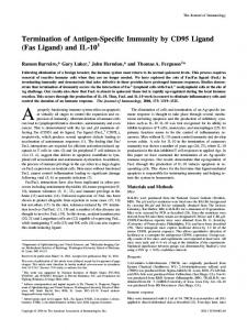
dition, viability and the light microscopy index were reduced in cytokine-treated samples associated with elevated chromatin condensation (Table 1). IL-1 and IFN␥ induced fragmentation of DNA, as revealed by increased percentages of hypodiploid nuclei compared with untreated controls (Fig. 1). After 24 h, the level of hypodiploidy was further enhanced by FasL and was accompanied by a reduced viability and an increased percentage of chromatin condensation. FasL had no effect on cytokine-induced nitrite accumulation (Table 1). Light microscopic assessment of morphology revealed damaged NOD mouse islets after cytokine exposure. The light microscopic index of NOD mouse islets was further lowered in the presence of FasL. Cytokine treatment resulted in increased nitrite accumulation compared with untreated controls. Here, FasL had no additive effect (Table 2). Cytokine-induced Fas receptor expression on NIT-1 cells is down-regulated by FasL
Time course of apoptotic events induced by IL-1 and IFN␥, alone or in the presence of FasL
In IL-1- plus IFN␥-induced apoptosis, surface Fas induction preceded the appearance of hypodiploidy (Fig. 4). Fas was first observed after culture for 6 h with IL-1 and IFN␥ (P ⬍ 0.05 vs. 3 h) and was augmented until 24 h (P ⬍ 0.05). Hypodiploid nuclei were detected after 12 h (P ⬍ 0.01) and remained at a stable level for 24 h of cytokine exposure. If NIT-1 cells were cultured with IL-1, IFN␥, and FasL (Fig. 4), Fas was induced after 6 h (P ⬍ 0.05 vs. 3 h). However, Fas induction waned, and its reduction by FasL was observed after 12 and 24 h (P ⬍ 0.05 vs. IL-1 and IFN␥). The decline in Fas expression preceded the FasL-mediated increase in hypodiploidy. The presence of FasL was associated with significant effects on Fas expression at 24 h (P ⬍ 0.05 vs. IL-1 and IFN␥ treatment). FasL does not alter cytokine-induced Fas expression on NOD and BALB/c mouse islet cells
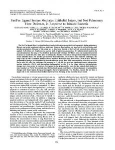
To determine whether FasL could modulate Fas receptor expression, we first examined the induction of Fas in the presence of FasL. The appearance of cytokine-induced Fas expression on NIT-1 cells after exposure to IL-1 and IFN␥ was higher than that after exposure to IL-1, IFN␥, and FasL (Fig. 2). After a 3-h incubation period, IL-1 and IFN␥ induced significant surface Fas expression, with a further increase up to 24 h, whereas in the untreated controls no Fas expression was detectable (P ⬍ 0.01). However, after 12 h in the presence of FasL, cytokine-induced surface Fas expression was significantly diminished (Fig. 2A). To assess the magnitude of the effect of FasL on cytokine-induced Fas expression, a Fas suppression index was defined by setting the level of IL-1- and IFN␥-induced Fas expression at 100%. Accordingly, after 12-h exposure to IL-1, IFN␥, and FasL, the Fas suppression index was 28.4 ⫾ 8.7% (P ⬍ 0.05); it increased to 44.6 ⫾ 11.6% (P ⬍ 0.05) at 24 h and reached a maximum of 58.3 ⫾ 7.4% (P ⬍ 0.01) after 48 h. To investigate the effect of FasL on established surface Fas expression, NIT-1 cells were first exposed to IL-1 and IFN␥ for 24 h to induce Fas surface expression. Thereafter, FasL was added for an additional 24 h. The addition of FasL reduced surface Fas expression (Fig. 2, C and D; and Fig. 3, F–H). After 3 h in the presence of FasL the Fas suppression index was 27.9 ⫾ 7.1% (P ⬍ 0.05); it increased to 35.4 ⫾ 3.0% (P ⬍ 0.01) after 6 h and reached a maximum of 64.1 ⫾ 8.1% (P ⬍ 0.01) after 12 h.
Exposure of NOD mouse islets to IL-1 and IFN␥ for 24 h increased Fas expression. However, addition of FasL had no detectable effect on cytokine-induced surface Fas expression (Fig. 5A). Similarly, Fas expression induced on BALB/c mouse islets by IL-1 and IFN␥ was not affected by FasL (Fig. 5B).
FIG. 1. Induction of hypodiploidy (percentage) in NIT-1 cells after exposure to IL-1 and IFN␥ without (E) or with (F) FasL. Controls were untreated (䡺) or supplemented with FasL (f). Data are the mean ⫾ SEM from at least six individual experiments. **, P ⬍ 0.01; *, P ⬍ 0.05 (vs. untreated control). 2⫹, P ⬍ 0.01; ⫹, P ⬍ 0.05 (vs. IL-1 and IFN␥ alone).
TABLE 1. Induction of apoptosis in IL-1 plus IFN␥-treated NIT-1 cells by coculture with FasL for 24 h
Control Control ⫹ FasL IL-1 ⫹ IFN␥ IL-1 ⫹ IFN␥ ⫹ FasL
Viability (%)
Light microscopic index
Chromatin condensation (%)
Nitrite (mol/liter)
82.71 ⫾ 2.13 82.72 ⫾ 3.24 69.86 ⫾ 2.85b 58.78 ⫾ 2.96a
2.79 ⫾ 0.11 2.35 ⫾ 0.18a 1.71 ⫾ 0.13b 1.50 ⫾ 0.15
1.10 ⫾ 0.56 1.08 ⫾ 0.58 7.28 ⫾ 2.07c 14.82 ⫾ 3.98a
0.81 ⫾ 0.19 0.89 ⫾ 0.43 2.39 ⫾ 0.40b 1.94 ⫾ 0.34
Data are the mean ⫾ SEM of at least five individual experiments. a P ⬍ 0.05 vs. treatment without FasL. b P ⬍ 0.01 vs. untreated control. c P ⬍ 0.05 vs. untreated control.
2750
Augstein et al. • Down-Regulation of Fas Expression by FasL in -Cells
Endocrinology, June 2004, 145(6):2747–2752
TABLE 2. NO production and morphological effects observed after exposure of NOD mouse islets to IL-1 plus IFN-␥ and/or FasL for 24 h
Light microscopic index Nitrite (mol/liter)
Control
Control ⫹ FasL
IL-1 ⫹ IFN␥
IL-1 ⫹ IFN␥ ⫹ FasL
2.69 ⫾ 0.04 0.92 ⫾ 0.16
2.61 ⫾ 0.16a 1.28 ⫾ 0.32
1.85 ⫾ 0.10b 1.64 ⫾ 0.28b
1.56 ⫾ 0.15a 1.60 ⫾ 0.20
Data are given for 200 islets/ml culture medium and are the mean ⫾ a P ⬍ 0.05 vs. treatment without FasL. b P ⬍ 0.01 vs. untreated control.
SEM
of at least four individual experiments.
FIG. 2. Effects of FasL on induction of surface Fas expression (A and B) and on established Fas expression (C and D) monitored as the percent binding (A and C) and MFI (B and D) of Fas antibody Jo2. Fas expression was induced (A and B) by IL-1 and IFN␥ alone (E) or the combination of IL-1, IFN␥, and FasL (F) and analyzed by flow cytometry at the indicated time points. FasL was added to cytokine-treated (24 h) NIT-1 cells with established surface Fas expression (C and D), and the effect on Fas expression was analyzed for up to another 24 h. Controls were untreated (䡺) or supplemented with FasL (f). Data are the mean ⫾ SEM from at least four individual experiments. *, P ⬍ 0.05; **, P ⬍ 0.01 (vs. untreated control). ⫹, P ⬍ 0.05 (vs. IL-1 and IFN␥ alone).
Discussion
Apoptotic cell death mediated by the Fas receptor is recognized as one mechanism of -cell loss in autoimmune diabetes (19, 20, 28). Cytokines secreted by pancreatic isletfiltrating mononuclear cells have been attributed -cell apoptotic effects (29). In accordance with the data presented in this study, proinflammatory (IL-1 and TNF␣) and type 1 (IFN␥) cytokines have been shown to induce Fas expression on -cells (8, 30 –32) and render -cells susceptible to Fasmediated apoptosis (33, 34). The interaction of cytokine-induced Fas with FasL⫹ on islet-infiltrating T cells (33) is thought to activate signal transduction pathways (2) leading to -cell death (19, 20, 29). We observed an increase in apoptosis of IL-1/IFN␥-treated NIT-1 cells in the presence of FasL. Similarly, transgenic expression of FasL on -cells themselves increased susceptibility of NOD mouse islets to cytokine-induced apoptosis (35) and accelerated diabetes development (36). Based on this evidence for Fas-mediated -cell death, we asked whether Fas⫹ NOD mouse islet and
insulinoma cells could counter autoreactive FasL⫹ T cells by down-regulating Fas in response to ligation with FasL. The experimental work of this study was designed in two stages. Initially screening experiments were performed with NIT-1 cells, an insulinoma cell line derived from NOD mice, and key experiments were then performed on freshly isolated islets from NOD and BALB/c mice. In NIT-1 cells, cytokine-induced Fas expression was reduced in the presence of FasL. This effect of FasL was evident during induction as well as after establishing surface Fas expression. A trivial explanation for this observation might be that FasL blocks binding of the Jo2 antibody used to detect Fas. This is highly unlikely because the effect of FasL was not observed with islets. Our findings with NIT-1 cells could be explained by inducible Fas internalization similar to that observed in a variety of tumor cells that escape attack by activated cytotoxic T cells (7, 37). Tumor cells can avoid apoptosis by modulation of Fas and/or FasL expression, Fas-associated death domain-like IL-1-converting enzyme inhibitory pro-
Augstein et al. • Down-Regulation of Fas Expression by FasL in -Cells
Endocrinology, June 2004, 145(6):2747–2752 2751
FIG. 3. Assessment of FasL effects on established surface Fas expression demonstrated by representative flow cytometric histograms. NIT-1 cells were cultured with IL-1 and IFN␥ for 24 h to induce Fas detected by Jo2 binding (A). Controls were untreated (B). Addition of FasL for 3 h (F), 6 h (G), and 12 h (H) to IL-1- and IFN␥-treated NIT-1 cells resulted in reduction of surface Fas expression compared with samples maintained with cytokine treatment (C–E). Controls were unaffected by addition of FasL (not shown). The given regions for Jo2 positivity were obtained by measuring the unspecific hamster Ig binding for each sample.
FIG. 4. Time course of surface Fas expression (䡺 and f) and hypodiploidy (E and F) induced by IL-1 and IFN␥ alone (䡺 and E) or combined with FasL (f and F). NIT-1 cells were exposed for 24 h to IL-1 and IFN␥ or IL-1, IFN␥, and FasL. Data are the mean ⫾ SEM from at least four individual experiments. *, P ⬍ 0.05; **, P ⬍ 0.01 (vs. preceding time point). ⫹, P ⬍ 0.05 (vs. treatment with IL-1 and IFN␥).
tein-mediated inhibition of Fas signaling or expression of decoy receptors in response to an immune attack or chemotherapy. Moreover, the ability to internalize Fas and thereby escape immune attack has been correlated with malignancy (38). Alternatively, ongoing apoptosis that results in suppression of protein synthesis including Fas or in reduction of the number of Fas⫹ cells could account for diminished Fas expression. To evaluate these possibilities we compared the time course of cytokine- and FasL-induced apoptosis and determined what comes first after the addition of FasL, a decrease in Fas expression or an increase in hypodiploidy. A decrease in Fas expression preceded hypodiploidy during induction as well as after establishing Fas expression. Nevertheless, hypodiploidy is a late apoptotic event and we cannot entirely exclude the possibility that early mechanisms in apoptosis contributed to diminished Fas expression. Algeciras-Schimnich et al. (17) suggested that FasL first induces Fas microaggregates at the cell surface, followed by formation of the death-inducing signaling complex. Thereafter, large Fas surface clusters are formed and are positively regulated by caspase-8 (17). As described also for the TNF re-
FIG. 5. Effects of FasL on cytokine-induced surface Fas expression of islet cells from NOD (A) and BALB/c (B) mice. Islets were cultured for 24 h with IL-1 and IFN␥ (50 and 500 U/ml, respectively; ). Controls (䡺) were untreated. To reveal the effects of FasL on induction of Fas expression, samples were simultaneously exposed to IL-1, IFN␥, and FasL (o). After 24 h, cytokine-induced Fas expression was analyzed by flow cytometry and quantified as the percent binding and MFI. Data are the mean ⫾ SEM from at least four individual experiments. *, P ⬍ 0.05; **, P ⬍ 0.01 (vs. controls).
ceptor pathway (18), Fas-FasL complexes are internalized through an endosomal pathway controlled by actin filaments. Interestingly, Elsner et al. (39) reported that the activity of endocytosis-associated genes, such as megalin, clathrin, and calcineurin, is increased in rat insulinoma cells (RIN5mF) after exposure to IL-1, TNF␣, and IFN␥. This cytokine-activated endocytosis machinery might contribute to the Fas receptor down-regulation that we observed.
2752
Augstein et al. • Down-Regulation of Fas Expression by FasL in -Cells
Endocrinology, June 2004, 145(6):2747–2752
Islets of NOD mice have been shown to be susceptible to Fas-mediated cell death in vitro and in vivo (9, 20, 26, 36). However, in contrast to NIT-1 cells, Fas expression on cytokine-treated islets from NOD mice was not affected by FasL. To investigate whether there was a relation to autoimmune diabetes susceptibility, we also performed experiments using islets from diabetes-resistant BALB/c mice. Similarly, these islets responded to IL-1 and IFN␥ with upregulation of Fas expression, but Fas was maintained at a high level even in the presence of FasL. Thus, in contrast to NOD mouse NIT-1 insulinoma cells, FasL did not downregulate cytokine-induced Fas expression on islet cells from NOD or BALB/c mice. Apparently, in contrast to immortalized NIT-1 cells, primary mouse islet cells lack the downregulation mechanism that could protect them from Fasmediated apoptosis. Understanding what accounts for this discrepancy could lead to therapeutic approaches that protect -cells in autoimmune diabetes.
14.
15. 16. 17. 18. 19.
20. 21. 22.
Acknowledgments 23.
Received June 16, 2003. Accepted March 8, 2004. Address all correspondence and requests for reprints to: Petra Augstein, Ph.D., Institute of Diabetes Gerhardt Katsch Karlsburg e.V, Greifswalder Strasse 11e, 17495 Karlsburg, Germany. E-mail: augstein@mail. uni-greifswald.de. This work was supported by grants from Deutsche Forschungsgemeinschaft (AU 151/1-1 and 1-2) and Ministerium fu¨ r Bildung, Wissenschaft und Kultur Mecklenburg-Vorpommern (IDK 97 007 80/SOM and IDK 97 007 80/HSP III).
References 1. Hengartner MO 2001 Apoptosis. DNA destroyers. Nature 412:27–29 2. Schmitz I, Kirchhoff S, Krammer PH 2000 Regulation of death receptormediated apoptosis pathways. Int J Biochem Cell Biol 32:1123–1136 3. Sharma K, Wang RX, Zhang LY, Yin DL, Luo XY, Solomon JC, Jiang RF, Markos K, Davidson W, Scott DW, Shi YF 2000 Death the Fas way: regulation and pathophysiology of CD95 and its ligand. Pharmacol Ther 88:333–347 4. Wallach D, Varfolomeev EE, Malinin NL, Goltsev YV, Kovalenko AV, Boldin MP 1999 Tumor necrosis factor receptor and Fas signaling mechanisms. Annu Rev Immunol 17331– 67:67 5. Li-Weber M, Krammer PH 2002 The death of a T-cell: expression of the CD95 ligand. Cell Death Differ 9:101–103 6. Hengartner MO 2000 The biochemistry of apoptosis. Nature 407:770 –776 7. Daniel PT, Wieder T, Sturm I, Schulze-Osthoff K 2001 The kiss of death: promises and failures of death receptors and ligands in cancer therapy. Leukemia 15:1022–1032 8. Suarez-Pinzon W, Sorensen O, Bleackley RC, Elliott JF, Rajotte RV, Rabinovitch A 1999 -Cell destruction in NOD mice correlates with Fas (CD95) expression on -cells and proinflammatory cytokine expression in islets. Diabetes 48:21–28 9. Suarez-Pinzon WL, Power RF, Rabinovitch A 2000 Fas ligand-mediated mechanisms are involved in autoimmune destruction of islet  cells in nonobese diabetic mice. Diabetologia 43:1149 –1156 10. Georgescu L, Vakkalanka RK, Elkon KB, Crow MK 1997 Interleukin-10 promotes activation-induced cell death of SLE lymphocytes mediated by Fas ligand. J Clin Invest 100:2622–2633 11. Ouallet J, Baumann N, Marie Y, Villarroya H 1999 Fas system up-regulation in experimental autoimmune encephalomyelitis. J Neurol Sci 170:96 –104 12. Mysler E, Bini P, Drappa J, Ramos P, Friedman SM, Krammer PH, Elkon KB 1994 The apoptosis-1/Fas protein in human systemic lupus erythematosus. J Clin Invest 93:1029 –1034 13. Giordano C, Richiusa P, Bagnasco M, Pizzolanti G, Di Blasi F, Sbriglia MS, Mattina A, Pesce G, Montagna P, Capone F, Misiano G, Scorsone A, Pugliese A, Galluzzo A 2001 Differential regulation of Fas-mediated apoptosis in both thyrocyte and lymphocyte cellular compartments correlates with opposite
24. 25. 26. 27. 28.
29. 30. 31. 32. 33.
34. 35. 36. 37. 38. 39.
phenotypic manifestations of autoimmune thyroid disease. Thyroid 11:233– 244 Stassi G, Todaro M, Bucchieri F, Stoppacciaro A, Farina F, Zummo G, Testi R, De Maria R 1999 Fas/Fas ligand-driven T cell apoptosis as a consequence of ineffective thyroid immunoprivilege in Hashimoto’s thyroiditis. J Immunol 162:263–267 Ashkenazi A, Dixit VM 1999 Apoptosis control by death and decoy receptors. Curr Opin Cell Biol 11:255–260 Schneider P, Tschopp J 2000 Apoptosis induced by death receptors. Pharm Acta Helv 74:281–286 Algeciras-Schimnich A, Shen L, Barnhart BC, Murmann AE, Burkhardt JK, Peter ME 2002 Molecular ordering of the initial signaling events of CD95. Mol Cell Biol 22:207–220 Idriss HT, Naismith JH 2000 TNF␣ and the TNF receptor superfamily: structure-function relationship(s). Microsc Res Tech 50:184 –195 Nakayama M, Nagata M, Yasuda H, Arisawa K, Kotani R, Yamada K, Chowdhury SA, Chakrabarty S, Jin ZZ, Yagita H, Yokono K, Kasuga M 2002 Fas/Fas ligand interactions play an essential role in the initiation of murine autoimmune diabetes. Diabetes 51:1391–1397 Savinov AY, Tcherepanov A, Green EA, Flavell RA, Chervonsky AV 2003 Contribution of Fas to diabetes development. Proc Natl Acad Sci USA 100: 628 – 632 Hamaguchi K, Gaskins HR, Leiter EH 1991 NIT-1, a pancreatic -cell line established from a transgenic NOD/Lt mouse. Diabetes 40:842– 849 Augstein P, Dunger A, Salzsieder C, Heinke P, Kubernath R, Bahr J, Fischer U, Rettig R, Salzsieder E 2002 Cell surface trafficking of Fas in NIT-1 cells and dissection of surface and total Fas expression. Biochem Biophys Res Commun 290:443– 451 Thomas HE, Parker JL, Schreiber RD, Kay TW 1998 IFN-␥ action on pancreatic  cells causes class I MHC upregulation but not diabetes. J Clin Invest 102:1249 –1257 Hahn HJ, Jahr H, Kohnert KD, Zuhlke H 1975 Investigations on isolated islets of Langerhans in vitro. Influence of temperature changes during preparation on some parameters of insulin metabolism. Horm Res 6:169 –176 Witt S, Ziegler B, Ziegler M 1989 Detection of islet cell specificity of monoclonal islet cell surface antibodies by means of double-staining immunofluorescence. Acta Histochem 86:111–115 Thomas HE, Darwiche R, Corbett JA, Kay TW 1999 Evidence that  cell death in the nonobese diabetic mouse is Fas independent. J Immunol 163:1562–1569 Green LC, Wagner DA, Glogowski J, Skipper PL, Wishnok JS, Tannenbaum SR 1982 Analysis of nitrate, nitrite, and [15N]nitrate in biological fluids. Anal Biochem 126:131–138 Maedler K, Fontana A, Ris F, Sergeev P, Toso C, Oberholzer J, Lehmann R, Bachmann F, Tasinato A, Spinas GA, Halban PA, Donath MY 2002 FLIP switches Fas-mediated glucose signaling in human pancreatic  cells from apoptosis to cell replication. Proc Natl Acad Sci USA 99:8236 – 8241 Eizirik DL, Mandrup-Poulsen T 2001 A choice of death: the signal-transduction of immune-mediated  cell apoptosis. Diabetologia 44:2115–2133 Liu D, Pavlovic D, Chen MC, Flodstrom M, Sandler S, Eizirik DL 2000 Cytokines induce apoptosis in -cells isolated from mice lacking the inducible isoform of nitric oxide synthase (iNOS⫺/⫺). Diabetes 49:1116 –1122 Liu D, Darville M, Eizirik DL 2001 Double-stranded ribonucleic acid (RNA) induces -cell Fas messenger RNA expression and increases cytokine-induced -cell apoptosis. Endocrinology 142:2593–2599 Zumsteg U, Frigerio S, Hollander GA 2000 Nitric oxide production and Fas surface expression mediate two independent pathways of cytokine-induced murine -cell damage. Diabetes 49:39 – 47 Moriwaki M, Itoh N, Miyagawa J, Yamamoto K, Imagawa A, Yamagata K, Iwahashi H, Nakajima H, Namba M, Nagata S, Hanafusa T, Matsuzawa Y 1999 Fas and Fas ligand expression in inflamed islets in pancreas sections of patients with recent-onset type I diabetes mellitus. Diabetologia 42:1332–1340 Stassi G, De Maria R, Trucco G, Rudert W, Testi R, Galluzzo A, Giordano C, Trucco M 1997 Nitric oxide primes pancreatic  cells for Fas-mediated destruction in insulin-dependent diabetes mellitus. J Exp Med 186:1193–1200 Petrovsky N, Silva D, Socha L, Slattery R, Charlton B 2002 The role of Fas ligand in  cell destruction in autoimmune diabetes of NOD mice. Ann NY Acad Sci 958:204 –208 Silva DG, Petrovsky N, Socha L, Slattery R, Gatenby P, Charlton B 2003 Mechanisms of accelerated immune-mediated diabetes resulting from islet  cell expression of a fas ligand transgene. J Immunol 170:4996 –5002 Muschen M, Warskulat U, Beckmann MW 2000 Defining CD95 as a tumor suppressor gene. J Mol Med 78:312–325 French LE, Tschopp J 2002 Defective death receptor signaling as a cause of tumor immune escape. Semin Cancer Biol 12:51–55 Elsner M, Souza K., Lenzen S, Tiedge M 2002 Expressionsanalysen von Zytokin-induzierten Genen in insulinproduzierenden RINm5F Zellen durch die Real-Time-Polymerasekettenreaktion. Diabetes Stoffwechsel 11(Suppl 1):59
Endocrinology is published monthly by The Endocrine Society (http://www.endo-society.org), the foremost professional society serving the endocrine community.