Behavioral Neuroscience 2004, Vol. 118, No. 5, 1062–1070
Copyright 2004 by the American Psychological Association 0735-7044/04/$12.00 DOI: 10.1037/0735-7044.118.5.1062
Fast Positive Feedback Between the Adrenocortical Stress Response and a Brain Mechanism Involved in Aggressive Behavior Menno R. Kruk
Jo´zsef Hala´sz
Leiden/Amsterdam Center for Drug Research
Institute of Experimental Medicine
Wout Meelis
Jo´zsef Haller
Leiden/Amsterdam Center for Drug Research
Institute of Experimental Medicine
Aggressive behavior induces an adrenocortical stress response, and sudden stressors often precipitate violent behavior. Experiments in rats revealed a fast, mutual, positive feedback between the adrenocortical stress response and a brain mechanism controlling aggression. Stimulation of the aggressive area in the hypothalamus rapidly activated the adrenocortical response, even in the absence of an opponent and fighting. Hypothalamic aggression, in turn, was rapidly facilitated by a corticosterone injection in rats in which the natural adrenocortical stress response was prevented by adrenalectomy. The rapidity of both effects points to a fast, mutual, positive feedback of the controlling mechanisms within the time frame of a single conflict. Such a mutual facilitation may contribute to the precipitation and escalation of violent behavior under stressful conditions.
metyrapone-treated rats rapidly reinstates aggressive behavior (Mikics et al., 2004). Also, injecting corticosteroids into the hypothalamus of the golden hamster rapidly facilitates aggression (Hayden-Hixson & Ferris, 1991). These findings suggest that aggressive behavior is facilitated by the corticosteroids that are secreted in an anticipatory response to the social challenge itself, even before actual fighting starts. This concept implies a mutual stimulatory interaction between mechanisms that control brain areas involved in aggression and the stress response. However, the contribution of aggression-related brain mechanisms to the adrenocortical stress response cannot be assessed in a paradigm such as “spontaneous” territorial conflict, because the hypothalamic– pituitary–adrenal (HPA) axis is activated by behavioral activation per se. Moreover, the activation of the HPA axis strongly depends on the behavior of the opponent (Haller et al., 1996). Aggression can also be evoked by direct electrical activation of the hypothalamic attack area of the rat (Hala´sz, Liposits, Meelis, Kruk, & Haller, 2002; King & Hoebel, 1968; Koolhaas, 1978; Koolhaas & Wiepkema, 1976; Kruk, 1991; Kruk et al., 1983, 1984, 1998; Kruk, Meelis, Van der Poel, & Mos, 1981; Kruk, Van der Poel, & de Vos-Frerichs, 1979; Lammers, Kruk, Meelis, & Van der Poel, 1988a; Mos et al., 1983; Panksepp, 1971; Panksepp & Trowill, 1969; Roberts & Nagel, 1996; Siegel, Roeling, Gregg, & Kruk, 1999; Vergnes & Karli, 1969, 1970; Woodworth, 1971). Hypothalamic aggression has been used to trace the network involved in the control of aggression (Hala´sz et al., 2002; Roberts & Nagel, 1996; Roeling et al., 1994). A similar type of neural organization seems to be involved in different species (see, e.g., Kruk, 1991 and Siegel et al., 1999, for a review of earlier studies in the cat and rat). The paradigm of hypothalamic attack has several useful characteristics for the study of the adrenocortical stress response in aggressive behavior. It has successfully been used to quantify
Stress is a major factor promoting aggression and violence in humans (Barnett, Fagan, & Booker, 1991; Tardiff, 1992), and aggression has been convincingly correlated with stress in several human situations (Guerra, Huesmann, Tolan, Van Acker, & Eron, 1995; Sanson, Smart, Prior, & Oberklaid, 1993; Vaux & Ruggiero, 1983). Also, control of stress and violent behavior is a priority objective among health authorities (Stone & Kelner, 2000). However, how stress mechanisms and mechanisms involved in aggression interact is only partly understood. Plasma corticosteroids rise fast and early in the course of an agonistic encounter between rats (Schuurman, 1980), even before actual aggressive behavior is observed (Haller, Barna, & Baranyi, 1995). There are several reasons to believe that the surge in plasma glucocorticoids caused by the confrontation with a potential adversary plays an important role in the subsequent aggressive conflict. Inhibiting corticosterone synthesis by adrenocorticotropic hormone (ACTH) antiserum or the corticosterone synthesis blocker metyrapone inhibits aggressive behavior (Haller, Kiem, & Makara, 1996; Mikics, Kruk, & Haller, 2004), whereas corticosterone treatment administered to
Menno R. Kruk and Wout Meelis, Section of Medical Pharmacology, Leiden/Amsterdam Center for Drug Research, Leiden, the Netherlands; Jo´zsef Hala´sz and Jo´zsef Haller, Institute of Experimental Medicine, Budapest, Hungary. This project was supported by a grant from the Harry Frank Guggenheim Foundation in New York to Menno R. Kruk, and an international cooperation grant from the Netherlands Organization for Scientific Research and its Hungarian counterpart Orsza´gos Tudoma´nyos Kutata´ti Alapprogramok (Grant 048.011.025). We greatly appreciate the encouragement of E. R. de Kloet. Correspondence concerning this article should be addressed to Menno R. Kruk, Section of Medical Pharmacology, Leiden/Amsterdam Center for Drug Research, P.O. Box 9502, RA 2300 Leiden, the Netherlands. E-mail:
[email protected] 1062
STRESS AND AGGRESSION
effects of hormones and drugs on brain mechanisms involved in aggression as changes in the threshold current intensity required to evoke attacks (Bermond, Mos, Meelis, Van der Poel, & Kruk, 1982; Katz & Thomas, 1976; Kruk, 1991; Kruk et al., 1984; MacDonnell & Fessock, 1969; Mos, Olivier, Van Oorschot, & Dijkstra, 1984; Olivier, Mos, & Rasmussen, 1990; Siegel et al., 1999; Van der Poel et al., 1982). Stimulation-evoked aggression in this paradigm requires the presence of an opponent (Kruk, 1991; Kruk et al., 1979; Levinson & Flynn, 1965; Siegel et al., 1999). This characteristic was used here to study the adrenocortical response of activating an aggressive brain mechanism in fighting and not-fighting rats. In addition, the hypothalamic paradigm is well suited to study fast effects of hormones because it allows precise control over the timing of attacks. Aggression is elicited within seconds after stimulation onset, and it stops immediately when stimulation stops. Moreover, it is relatively insensitive to changes in opponent behavior (Kruk, 1991; Kruk et al., 1979; Siegel et al., 1999). The impact of the behavior of the opponent can be further minimized by anesthetizing them with a high dose of morphine, as hypothalamically stimulated rats—in contrast to spontaneously attacking ones (Kruk, Ha´ lasz, Haller, & Bot, 2002)—readily attack such anesthetized opponents (Siegel et al., 1999). Recently, it was shown that such stimulation basically activates the same brain areas as territorial aggression (Hala´ sz, Liposits, Kruk, & Haller, 2002; Hala´ sz, Liposits, Meelis, et al., 2002). The aim of the present experiments was to test the hypothesis of a mutual stimulatory interaction between brain mechanisms controlling aggressive behavior and the stress response. We stimulated the hypothalamic attack area of rats in both the presence and the absence of an opponent, and assessed whether HPA axis activation was due to behavioral activation or to the activation of aggressionrelated brain mechanisms. We also studied the effects of acute corticosterone treatments on the threshold of attacks, that is, on the responsiveness of the hypothalamic attack area to stimulation. The behavioral specificity of adrenalectomy and corticosterone injection effects was assessed by determining their effects on thresholds for hypothalamic teeth-chattering evoked in the hypothalamic aggressive area. Teeth-chattering is evoked from a medial hypothalamic area that partially overlaps with the hypothalamic attack area (Lammers et al., 1988a). It has a similar behavioral pharmacology (Kruk, 1991; Van der Poel et al., 1982) and is a clear sign of distress in the rat.
Method Subjects Experimental subjects, male Wistar rats weighing 350 – 400 g at the start of the experiments, were obtained from Charles River Laboratories (via Broekman, Veldhoven, the Netherlands). Each rat was allowed to recover from transportation for at least 1 week, fed on standard laboratory food, and given free access to tap water. Temperature was maintained at 22 ⫾ 1 °C, and humidity was 60 ⫾ 10%. Rats were housed in groups of 10 before electrode implantation and in individual cages thereafter. Opponents were male rats from the same supplier and were maintained under similar conditions. Their weight was 250 –300 g. Opponents were used only once, and they received an intraperitoneal injection of morphine (10 mg/kg) 20 min before encounters to produce profound sedation and analgesia during attacks. The experiments were all performed in the active (dark) phase of
1063
both stimulated rats and their opponents. A 12:12-hr inverted day–night schedule was imposed on the rats, with lights on at 2000. All experiments were approved by the Ethical Committee on Animal Experimentation of Leiden University, in accordance with Dutch laws on animal experimentation.
Electrode Implantation Electrode implantation and stimulation were performed as described in Kruk et al. (1979). In brief, male adult Wistar rats were deeply anesthetized by an intraperitoneal injection of a mixture of midazolam, atropine, and Hypnorm (0.5 mg/kg, 1.0 mg/kg, and 1 ml/kg body weight, respectively). Bipolar electrodes were implanted at the coordinates RC ⫺1.9, ML 1.0, DV 8.2 from bregma. Electrodes and connectors were kept in place by dental carboxylate cement covered by acrylate dental cement and anchored to the skull by stainless steel screws. After electrode implantation, the rats were singly housed in Macrolon cages. They were allowed to recover for at least 1 week. Electrodes for teeth-chattering were implanted at the same coordinates as used for attack, by the same surgical procedures.
Threshold Determinations Behavioral testing took place outside the home cage in a test cage (50 cm long ⫻ 60 cm wide ⫻ 100 cm high) with a glass front wall. Subjects could move around freely, in no way hampered by the connecting wires. An opponent anesthetized with morphine (10 mg/kg) was introduced into the cage before stimulation. Each opponent was used only once and was killed with an overdose of Nembutal immediately after a threshold determination. The attack threshold current intensity was determined by means of the up-and-down method of Wetherill (1966). In brief, electrical stimulations with a train duration of 10 s were delivered. Trains were separated by 50-s pauses. Forty-hertz biphasic pulses with phase duration of 0.2 ms and a phase interval of 12.3 ms were used. If attack behavior was observed within the 10 s of stimulation, the intensity of the next train was lowered by 20 A. If no attack occurred, stimulation intensity of the next train was increased by 20 A. A threshold determination was completed when six current intensity-related changes from attack to no attack, or from no attack to attack, were obtained in response to stimulation. This procedure lasted less than 20 min. From the current intensities at which these six response changes occurred, the threshold for attack was estimated according to the method of Wetherill (1966). After the completion of experiments, the position of the electrode tip was verified by histological procedures as described elsewhere (Lammers, Meelis, Kruk, & Van der Poel, 1987). Only rats having the electrode tip located within the hypothalamic attack area showed attacks (Kruk et al., 1983; Lammers et al., 1988a). Stable baseline thresholds of a specific electrode in an individual rat can be obtained after three threshold determinations on subsequent days (Kruk et al., 1979). Attack thresholds stabilize somewhere between 30 and 120 A, depending on the precise electrode position within the attack area. Teeth-chattering thresholds were determined by the same procedures and stimulation parameters as used for the attack thresholds. However, no opponent was present, and hence no aggressive behavior was evoked. Teeth-chatter thresholds stabilize between 20 and 30 A.
Corticosterone Manipulations Adrenalectomy was performed under ether anesthesia between 0900 and 1200, via the dorsal approach. A corticosterone pellet (25 mg corticosterone and 75 mg cholesterol) was implanted subcutaneous immediately after adrenalectomy to avoid neuronal death that is observed in nonreplaced adrenalectomized rats (MacLennan, Smith, & Darlington, 1998). As previously shown, such pellets maintain plasma corticosterone levels of about 90 –100 nmol/L for 3 weeks (Haller, Van de Schraaf, & Kruk, 2001). This level corresponds to approximately 30% of the normal levels observed in
1064
´ SZ, MEELIS, AND HALLER KRUK, HALA
the active (dark) period. Acute changes in corticosterone levels were produced by injecting intraperitoneal corticosterone-HBC (2-hydroxypropyl--cyclodextrin) complex (RBI, Natick, MA). The dose injected (0.25 mg/kg body weight) rapidly increases plasma levels of corticosterone to stress levels of about 1,000 nmol/L (Haller et al., 2001). Controls received HBC in equimolar concentrations. Preliminary experiments showed that HBC has no behavioral effects of its own.
Hormone Measurements Levels of ACTH and corticosterone were assessed by specific and direct radioimmunoassays (RIAs) as described previously (Zelena et al., 2003). In brief, ACTH and corticosterone antibodies were raised in rabbits in our laboratory. Tracers were iodinated by the chloramine-T method, and 50 l or 10 l plasma aliquots were assayed in the ACTH or corticosterone RIA, respectively. In this study, 25-l aliquots of medium were assayed after appropriate dilution by RIA buffer.
Experimental Design Experiment 1 determined the effects of hypothalamic stimulation on HPA axis activation (n ⫽ 11). Rats were implanted with electrodes aimed at the hypothalamic attack area. After 1 week of recovery, rats were submitted to three threshold determinations, which were separated by 2 days. In each of these threshold determinations, a naive opponent was present. Blood was sampled immediately before and after threshold determination by the tail incision technique (Fluttert, Dalm, & Oitzl, 2000). Plasma was separated by centrifugation and was kept at ⫺20 °C in EDTA-coated vials until hormone measurements. Experiment 2 determined the effect of fighting on the attack area stimulation-induced activation of the HPA axis (n ⫽ 6). Rats were implanted with electrodes as described above. After 1 week of recovery, they were confronted with an opponent in the stimulation cage, and the thresholds of attack were determined. Two days later, the rats were reintroduced into the stimulation cage and stimulated in the absence of an opponent, in accordance with the same stimulation protocol used to determine the previous attack threshold in the same rats, when there was an opponent present to attack. That is, for each rat, the precise pattern of its stimulation during the first threshold determination was repeated during the second threshold determination, but the second time no opponent was present and no attack or other overt behavioral effects where observed during stimulation. Two days later, the same rats were again reintroduced to the stimulation cage, but in this case, they were not stimulated and there was no opponent present. They stayed in the empty stimulation cage for a duration equal to that of the previous threshold determination. In this experiment rats served as their own controls; the design—stimulating the rat in absence of an opponent with exactly the same stimulations that were used to determine the thresholds in the presence of an opponent— did not allow for a design with a balanced order of treatments. Plasma was sampled and stored before and after each session, as in Experiment 1. Experiment 3 assessed the effects of a surge in plasma corticosterone on attack thresholds (n ⫽ 14). Rats were implanted with electrodes, and after a recovery of 1 week, three attack thresholds were determined at 2-day intervals. As shown by Experiments 1 and 2, attack area stimulation produced high stress levels of plasma glucocorticoids. Therefore, rats were adrenalectomized and implanted with low-release corticosterone pellets, immediately after the last threshold determination, to avoid interference from endogenous corticosterone production. After another week of recovery, three attack thresholds were determined at 2-day intervals. No treatment was applied before the first threshold determination; an acute corticosterone treatment was applied 10 min before the second threshold determination, whereas the third threshold determination was preceded by a vehicle injection. An injection of 0.25 mg/kg HBC corticosterone produces a transient increase in circulating corticostersone similar to the natural corticosterone response to a stressor (Haller et al., 2001).
Experiment 4 assessed the duration of corticosterone effects on attack thresholds (n ⫽ 15). Rats were implanted with electrodes aimed at the hypothalamic attack area, and after 1 week of recovery, their attack thresholds were assessed in three tests performed 2 days apart. Rats were adrenalectomized and, after another week of recovery, were submitted to four threshold determinations. Each threshold was preceded by a control or corticosterone injection. Corticosterone injections were administered 10, 60, or 240 min before, whereas control (vehicle HBC) injections were administered 10 min before, attack threshold determinations. Treatment order was randomized over the group. Experiment 5 assessed the effects of adrenalectomy and mimicking the adrenocortical stress response by an acute injection of corticosterone on hypothalamic teeth-chattering thresholds (n ⫽ 7). Teeth-chattering was evoked within the hypothalamic attack area at the same coordinates and with same stimulation parameters as used in the experiments on hypothalamic attack. However, there was no opponent present during teethchattering threshold determinations, and no aggressive behavior was observed. The conditions HBC– corticosterone and HBC alone were presented in balanced order. Rats served as their own controls. One day after the completion of the experiment, blood samples were taken by tail incision (Fluttert et al., 2000) to confirm the success of the adrenalectomy procedure.
Statistics Corticosterone data were expressed as mean (⫾ SEM). Changes in attack thresholds (Experiment 3 and 4) were expressed as percentage of baseline, that is, the last preadrenalectomy level. This approach was used because attack thresholds show slight individual variation that is probably due to slight individual differences in the location of the electrode within the attack area (Kruk et al., 1983; Lammers, 1988a). Therefore, values obtained in experimental tests were compared with values obtained in the same rat in preliminary tests. STATISTICA (2001) Version 6.0 was used for a repeated measures analysis of corticosterone levels, ACTH concentrations, and behavioral threshold changes. Dunnett’s test was used for post hoc comparisons.
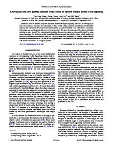
Results Adrenocortical Response to Stimulation in the Hypothalamic Attack Area In Experiment 1, stimulation of the attack area in intact rats increased circulating ACTH, F(1, 30) ⫽ 89.10, p ⬍ .0001 (see Figure 1A). The response did not change over three subsequent attack threshold determinations, F(2, 30) ⫽ 0.80, p ⬍ .4; interaction, F(2, 30) ⫽ 0.33, p ⬍ .70. Stimulation of the attack area also rapidly increased circulating corticosterone, F(1, 30) ⫽ 249.20, p ⬍ .0001 (see Figure 1B). The level after stimulation did not change over three subsequent attack threshold determinations, F(2, 30) ⫽ 2.11, p ⬍ .20. However, there was an interaction between the treatment order and the response, as a result of an increased corticosterone level before the third threshold determination, F(2, 30) ⫽ 4.93, p ⬍ .02, whereas there was no corresponding increase in ACTH in the same rats (Figure 1A). In Experiment 2, the adrenocortical response of another group of rats was determined under three different conditions to assess the importance of the presence or absence of an opponent on the stimulation-induced stress response (see Figures 1C and 1D). Placement of these rats in the stimulation cage with neither stimulation nor opponent served as a control. Levels of ACTH changed significantly before and after experimental treatments, F(1, 15) ⫽
STRESS AND AGGRESSION
1065
Figure 1. Electrical stimulation of the hypothalamic attack area increases plasma corticosterone irrespective of fighting. A and B: Plasma adrenocorticotropic hormone (ACTH) and corticosterone concentrations before and after the stimulation of the hypothalamic attack area during three threshold determinations performed every 2nd day. C and D: Plasma ACTH and corticosterone concentrations before and after tests performed in the presence or absence of an opponent. Asterisks indicate a significant difference from nonstimulated controls ( p ⬍ .05).
91.50, p ⬍ .0001. Treatment condition was also a significant factor, F(2, 15) ⫽ 7.95, p ⬍ .005. There was a significant interaction between treatment and response, F(2, 15) ⫽ 13.28, p ⬍ .0005. Figure 1C and post hoc testing show that stimulation in the presence of an opponent produced the same ACTH response as stimulation in the absence of an opponent (Dunnett’s p ⫽ .90). Placement in the stimulation cage without stimulation produced a much smaller ACTH response. This response differs significantly from the stimulation-with-opponent and stimulation-withoutopponent treatments (Dunnett’s p ⬍ .001, p ⬍ .003, respectively). The effects on corticosterone responses in the same rats were similar (Figure 1D). Corticosterone levels changed significantly before and after experimental treatments, F(1, 15) ⫽ 234.10, p ⬍ .0001. Treatment
condition was also significant, F(2, 15) ⫽ 11.77, p ⬍ .0008. There was a significant interaction between treatment and response, F(2, 15) ⫽ 14.07, p ⬍ .0003. Figure 1D and post hoc testing show that stimulation in the presence of an opponent produced the same corticosterone response as stimulation in the absence of an opponent (Dunnett’s p ⫽ .90). Placement in the stimulation cage without stimulation produced a much smaller corticosterone response. This response differs significantly from the stimulation-with-opponent and stimulation-without-opponent treatments (Dunnett’s p ⬍ .0008, p ⬍ .0007, respectively). These results demonstrate that activating the hypothalamic aggressive area is in itself a sufficient condition to obtain a considerable adrenocortical response. The confrontation with an oppo-
1066
´ SZ, MEELIS, AND HALLER KRUK, HALA
nent apparently is not required. The small increase observed in the absence of stimulation and opponent is probably due to an anticipatory stress response to the introduction into the test cage, a setting where these rats had fought before.
Rapid Facilitation of Attack In Experiment 3 (see Figure 2A), the rapid increase in plasma corticosterone observed after stimulation of the hypothalamic attack area was mimicked by the intraperitoneal injection of corticosterone to adrenalectomized rats. Peak levels of plasma corticosterone obtained by injection corresponded with the levels reached in stimulated rats with intact adrenals (basal levels in uninjected rats: 121.4 ⫾ 14.3 nmol/L; 10 min after injection: 1,139.2 ⫾ 88.3 nmol/L; 30 min after injection: 198.8 ⫾ 35.9 nmol/L). Such injections given 10 min before the start of the determination of an attack threshold facilitated hypothalamic attack behavior by 30% compared with the threshold after a vehicle injection in the same rats: repeated measures analysis of variance (ANOVA), F(2, 26) ⫽ 6.31, p ⬍ .0058. Thresholds following corticosterone injection differ significantly from no treatment and vehicle treatment at p ⬍ .024 and p ⬍ .0018, respectively. In Experiment 4 (see Figure 2B), adrenalectomized rats were assigned to four treatment conditions in a random order, after which attack thresholds were determined. There is a significant effect of treatment: repeated measures ANOVA, F(3, 18) ⫽ 4.01, p ⬍ .023. Only corticosterone injections given 10 min before the attack threshold determination differed significantly from vehicle injections ( p ⬍ .0037). Post hoc analysis showed that only corticosterone injected 10 min before encounters induced a significant decrease in attack thresholds compared with vehicle injections. These data show that an acute surge in plasma corticosterone
Figure 2. Rapid facilitation of hypothalamic aggression by corticosterone in adrenalectomized rats. A: Electrical thresholds for attack in noninjected, corticosterone-injected, and vehicle-injected rats. Average attack threshold control ⫽ 90.5 ⫾ 6.9 A. B: Electrical thresholds for attack in rats treated with vehicle (10 min before stimulation) or corticosterone 10, 60, and 240 min before stimulation. Average attack threshold for vehicle control ⫽ 97.5 ⫾ 15.1 A. Asterisks indicate a significant difference from thresholds obtained after vehicle injections ( p ⬍ .05).
facilitated attack behavior in this model by reducing the current intensity necessary for the induction of attack. In Experiment 5, teeth-chattering thresholds (22.6 A on average) were generally lower than attack thresholds (approximately 90 A). Adrenalectomy successfully reduced circulating corticosterone to 32.1 ⫾ 9.3 nmol/L. Figure 3 shows that neither adrenalectomy nor an acute injection of corticosterone had any effect on teeth-chattering thresholds evoked by electrical stimulation of the hypothalamic attack area: repeated measures ANOVA, F(3, 18) ⫽ 1.34, p ⬍ .29. There was no evidence of any order of treatment (i.e., vehicle or vehicle ⫹ corticosterone first) effects.
Discussion The results show that the stimulation of the hypothalamic attack area in rats induces strong HPA axis activation. This response is due to the stimulation per se, and not to the associated fighting, as it is preserved in the absence of an opponent, that is, when rats do not show overt signs of behavioral activation. On the other hand, an experimentally induced acute surge in corticosterone facilitates the aggressive response to hypothalamic stimulation. The effect lasts less than 1 hr. That is, it seems related to the presence of corticosterone, because corticosterone levels decreased sharply within 30 min after injection. Taken together, these results strongly suggest mutual stimulatory interaction between brain mechanisms involved in attack and the stress response. The activation of the brain mechanism controlling attack induces the activation of the HPA axis per se, whereas the activation of the HPA axis increases the sensitivity to stimulation of the mechanism activated via the hypothalamic attack area. See Figure 4 for a schematic presentation of this concept. The proaggressive effect of acute corticosteroids surges has been demonstrated before in mice (Brain & Haug, 1992), rats (Haller et al., 1997; Mikics et al., 2004), and hamsters (HaydenHixson & Ferris, 1991). Increased HPA axis reactivity was shown to correlate with certain types of human aggressive behavior as well (Guerra et al., 1995; Sanson et al., 1993; Vaux & Ruggiero, 1983). Conversely, aggressive behavior was shown to induce HPA axis activation (Haller et al., 1995; Schuurman, 1980). However, the relationship between aggressive behavior and the adrenocortical stress response remains poorly understood, because in experiments on spontaneous aggression it is difficult to distinguish between the distinct contributions of neuronal and behavioral consequences of social challenges to the adrenocortical stress response. In spontaneous aggression, the effect on the HPA axis could be due either to behavioral activation (fights) or to the activation of brain mechanisms involved in aggression. The hypothalamic attack paradigm allows distinguishing between these different contributions to adrenocortical activation. The effect of electrical stimulation on the HPA axis in the presence and absence of an opponent was virtually the same, demonstrating that fighting is not required to activate the HPA axis. Such a mechanism may well function in spontaneously attacking rats, as we have shown earlier that the hypothalamic attack area is strongly activated during territorial conflicts (Hala´ sz, Liposits, Kruk, et al., 2002). Classical stimulation and lesion studies (see Siegel et al., 1999, for a review) suggest the existence of an attack-stimulating axis, the main elements of which are the medial amygdala, the hypothalamic centers controlling affective aggression, and the periaque-
STRESS AND AGGRESSION
Figure 3. Absence of effects of adrenalectomy (ADX) and acute corticosteroid (Cort) injections on mean (⫾ SE) teeth-chattering evoked from the hypothalamic attack area. Veh ⫽ vehicle.
ductal gray. During spontaneous aggression (e.g., territorial conflict) the hypothalamic attack area is also activated (Hala´ sz, Liposits, Kruk, & Haller, 2002; Haller, Liposits, Meelis, et al., 2002). During such conflicts, the increased activity in the hypothalamic attack area probably also facilitates the adrenocortical stress response. The precise neuronal and molecular mechanisms underlying the attack area-mediated corticosterone response are still unknown. The projections from the hypothalamic attack area to the paraventricular nucleus of the hypothalamus (Roeling et al., 1994) could possibly mediate the interaction. But whether these sparse projections are sufficient to induce the activation of the corticotropinreleasing factor neurons in the paraventricular nucleus is not known. However, the dense varicosities on fibers projecting from the hypothalamic attack area in many directions (Roeling et al., 1994), and the equally dense varicosities on corticotropin-releasing hormone (CRH) fibers projecting from the paraventricular nucleus through the attack area (Makara, 1985), suggest that the mechanism mediating aggressive responses may interact, directly at the
1067
level of the hypothalamus, with the mechanism regulating the adrenocortical stress response. Alternatively, both the adrenocortical stress response to stimulation and the facilitation of the aggressive response could be caused by other intrahypothalamic mechanisms. Central vasopressin facilitates the adrenocortical stress response, and vasopressin receptor blockade in the anterior hypothalamus suppresses aggression in golden hamster (Ferris & Potegal, 1988). However, the observed interaction may also be caused by extrahypothalamic mechanisms. Efferent fibers of the attack area reach the main aminergic nuclei (Roeling et al., 1994). These nuclei in turn project to neuroendocrine stress mechanisms (Grino, Paulmyer-Lacroix, Faudon, Renard, & Anglade, 1994; Szafarczyk, Alonso, Ixart, Malaval, & Assenmacher, 1985) and may mediate the observed stress response. Hypothalamic aggression is selectively sensitive to serotonergic drugs and the betablocker propranolol (Kruk, 1991). The stimulation-induced adrenocortical stress response may be solely due to the classical neuroendocrine pathway via CRH and ACTH. However, there is evidence suggesting that the response may be facilitated by other pathways. Direct stimulation of the adrenocortical stress response in the paraventricular nucleus of the hypothalamus produces the same corticosterone response as stimulation of the attack area. The ACTH response to attack area stimulation was much smaller, suggesting the involvement of another facilitating mechanism in the case of the attack area (Kruk et al., 1998). Vasopressinmediated facilitation of the adrenocortical response may be involved (Ferris & Potegal, 1988), or a direct neural pathway that changes the responsiveness of the adrenals to stimulation by ACTH. The observation that rapid changes in circulating corticosteroids exert a fast facilitating feedback control on a brain mechanism directly mediating aggressive behavior has several interesting implications. The high stress levels of glucocorticoids, rapidly produced by the challenge-induced activation of brain mechanisms controlling aggression, may in turn produce a fast facilitation of
Figure 4. Graphic representation depicting proposed hypothetical mechanisms involved in the relationship between attack-controlling brain mechanisms and the adrenocortical stress response. HPA ⫽ hypothalamic– pituitary–adrenal; PVN ⫽ paraventricular nucleus of the hypothalamus.
1068
´ SZ, MEELIS, AND HALLER KRUK, HALA
the very same brain mechanisms (see Figure 4). Such mutual facilitation could constitute a vicious circle, which would explain why aggressive behavior escalates so easily, and why it is so difficult to stop once it has started, especially because corticosteroids rapidly pass the blood– brain barrier. Short-lasting facilitation of aggression by previous hypothalamic stimulation has been reported in the cat (Sledjesky & Flynn, 1972) and in the rat (Kruk et al., 1981), with a half-life of approximately 12 and 8 s, respectively. Longer lasting facilitation, called priming, has also been demonstrated in territorial aggression—a more “natural” paradigm than hypothalamic aggression—in the golden hamster and the rat (Potegal, 1992; Potegal & Coombes, 1995). This longer lasting facilitation is associated with c-Fos activation of the amygdala (Potegal, Ferris, Hebert, Meyerhof, & Skaredoff, 1996). The amygdala is strongly activated by stimulation of the hypothalamic attack area. Moreover, c-Fos activity in the amygdala of the rat during aggression is under corticosteroid control (Hala´ sz, Liposits, Kruk, & Haller, 2002; Hala´ sz, Liposits, Meelis, et al., 2002). Our findings also suggest that rapid increases in corticosteroids caused by stressors unrelated to fighting may precipitate violent behavior by lowering thresholds for attack. This concept also implies that an anticipatory increase in corticosterone in environments previously associated with aggression could possibly lead to place-dependent violent habits in individuals who are nonviolent in other settings. Previous aggression is also known to facilitate subsequent aggressive behavior in humans (Tardiff, 1992). Also, persons hospitalized for hostility have deviant stress responses to serotonergic challenges (Coccaro, Kavoussi, & Hauger, 1995; Rinne, Westerberg, Den Boer, & Van den Brink, 2000). In our opinion, behavioral constructs such as “general arousal” caused by corticosteroids or changes in “aversive or hedonic quality” of the stimulation do not provide plausible explanations for the observed facilitation of the behaviorally distinct and directly observable response to hypothalamic attack. First, corticosterone does not change the threshold for the concomitant behavioral response of teeth-chattering (Figure 3). Teeth-chattering is a clear sign of distress that is experienced by the rat in many different aversive settings. Corticosterone inhibits stimulationinduced hypothalamic flight (Kruk et al., 2002). Second, corticosterone also facilitates territorial fighting in rats in a very specific way (Hala´ sz, Liposits, Kruk, & Haller, 2002; Haller et al., 1995). Last, whether the effects of corticosterone on hypothalamic aggression are mediated by an effect on the hedonic properties of stimulation is still an open question. However, neither the aversive nor the rewarding properties of hypothalamic stimulation predict the ability to elicit aggressive behavior by means of an electrode (Herndon, Adrian, & McCoy, 1979; Kruk, 1991; Kruk et al., 1984; Roberts & Kiess, 1964), although the rewarding properties of stimulation seem to be associated with the more “complete” form of hypothalamic aggression (Koolhaas & Wiepkema, 1976). The fast onset and the short duration of the effects of corticosterone are intriguing and may be mediated by similar nongenomic mechanisms that mediate other fast behavioral effects of glucocorticoids (Makara & Haller, 2001; Rose, Moore, & Orchinik, 1993; Sandi, Venero, & Guaza, 1996; Wehling, 1997). Alternatively, they may be caused by very fast genomic effects (see, e.g., Joe¨ ls & De Kloet, 1994; Mikics et al., 2004). Whether the results reported here are just an interesting characteristic of the hypothalamic paradigm used and whether they are also valid under more naturalistic conditions and in other species
are important questions that require additional study. However, the adrenocortical stress response is a phylogenetically old, wellconserved mechanism in mammals that prepares the organism for fight or flight. The hypothalamic areas involved in these responses also seem to have a similar role in many different species. These mechanisms have been studied in rats (Adams, Boudreau, Kokonowski, Oberteuffer, & Yohay, 1993; Albert, Nanji, Brayley, & Madryga, 1979; Hala´ sz, Liposits, Meelis, et al., 2002; Haller et al. 1998; King & Hoebel, 1968; Koolhaas, 1978; Koolhaas & Wiepkema, 1976; Kruk, 1991; Kruk et al., 1979, 1981, 1984, 1998; Lammers, 1988a; Olivier et al., 1990; Panksepp, 1971; Panksepp & Trowill, 1969; Roberts & Nagel, 1996; Roeling, Schuurmans, & Veening, 1993; Vergnes & Karli, 1969, 1970; Woodworth, 1971), golden hamsters (Ferris & Potegal, 1988; Hayden-Hixon & Ferris, 1991), opossums (Adamec, 1990; Roberts, Steinberg, & Means, 1967), cats (Hess, 1928; Hess & Bru¨ gger, 1943; Hunsperger, 1956; Katz & Thomas, 1976; Levinson & Flynn, 1965; MacDonnell & Fessock, 1969; Roberts & Bergquist, 1968; Roberts & Kiess, 1964; Siegel et al., 1999; Sledjeski & Flynn, 1972), monkeys (Herndon et al., 1979; Lipp & Hunsperger, 1978), and even domestic fowl (Phillips & Youngren, 1971). Moreover, the same basic principles in hypothalamic behavioral organization seem to apply in these species. Mechanisms involved in aggression and flight behavior are found in close association, in an area adjacent to the mechanism controlling the endocrine, adrenocortical flight or fight response (Adamec, 1990; Albert et al., 1979; Hala´ sz, Liposits, Kruk, & Haller, 2002; Hala´ sz, Liposits, Meelis, et al., 2002; Lammers et al., 1998a, 1988b; Lipp & Hunsperger, 1978; Roberts & Nagel, 1996). Such a similarity in organization and function across species suggests that a rapid, mutual, positive feedback between the adrenocortical stress response and an “aggressive brain mechanism” may also operate under more naturalistic conditions in other animal species and in humans. Treatment of pathological violence and lack of impulse control in humans is notoriously difficult. The same applies to hypothalamic attack behavior, which was used as a psychopharmacological and behavioral paradigm to study pathological violence (Kruk, 1991; Olivier et al., 1990). The results presented here indicate that the adrenocortical stress response that accompanies conflict may effectively cancel out the effect of therapies intended to reduce violent behavior. Therefore, regulation of the adrenocortical stress response may offer a novel approach to the understanding and control of violent behavior. Agents, which have an anti-stress action (e.g., neurosteroids, Reddy & Kulkarni, 1996; corticotropinreleasing factor antagonists, Heinrichs et al., 1994), and certain anxiolytics, which reduce different stress-induced behaviors (Korte, Koolhaas, Schuurman, Traber, & Bohus, 1990), may also be effective in counteracting acute stress-precipitated violence.
References Adamec, R. E. (1990). Role of the amygdala and medial hypothalamus in spontaneous feline aggression and defense. Aggressive Behavior, 16, 207–222. Adams, D. B., Boudreau, W., Kokonowski, C., Oberteuffer, K., & Yohay, K. (1993). Offense produced by chemical stimulation of the anterior hypothalamus of the rat. Physiology & Behavior, 53, 1127–1132. Albert, D. J., Nanji, N., Brayley, K. N., & Madryga, F. J. (1979). Hyperactivity as well as mouse killing is induced by electrical stimulation of the lateral hypothalamus in the rat. Behavioural and Neural Biology, 27, 59 –71.
STRESS AND AGGRESSION Barnett, O. W., Fagan, R. W., & Booker, J. M. (1991). Hostility and stress as mediators of aggression in violent men. Journal of Family Violence, 6, 217–241. Bermond, B., Mos, J., Meelis, W., Van der Poel, A. M., & Kruk, M. R. (1982). Aggression induced by stimulation of the hypothalamus: Effects of androgens. Pharmacology Biochemistry and Behavior, 16, 41– 45. Brain, P. F., & Haug, M. (1992). Hormonal and neurochemical correlates of various forms of animal “aggression.” Psychoneuroendocrinology, 17, 537–551. Coccaro, E. F., Kavoussi, R. J., & Hauger, R. L. (1995). Physiological responses to d-fenfluramine and ipsapirone challenge correlate with indices of aggression in males with personality disorder. International Clinical Psychopharmacology, 10, 177–179. Ferris, C. F., & Potegal, M. (1988). Vasopressin receptor blockade in the anterior hypothalamus suppresses aggression in hamsters. Physiology & Behavior, 44, 235–239. Fluttert, M., Dalm, S., & Oitzl, M. S. (2000). A refined method for sequential blood sampling by tail incision in rats, Laboratory Animals, 34, 372–378. Grino, M., Paulmyer-Lacroix, O., Faudon, M., Renard, M., & Anglade, G. (1994). Blockade of alpha 2-adrenoceptors stimulates basal and stressinduced adrenocorticotropin secretion in the developing rat through a central mechanism independent from corticotropin-releasing factor and arginine vasopressin. Endocrinology, 135, 2549 –2557. Guerra, N. G., Huesmann, L. R., Tolan, P. H., Van Acker, R., & Eron, L. D. (1995). Stressful events and individual beliefs as correlates of economic disadvantage and aggression among urban children. Journal of Consulting and Clinical Psychology, 63, 518 –528. Hala´ sz, J., Liposits, A., Kruk, M. R., & Haller, J. (2002). Neural background of glucocorticoid dysfunction-induced abnormal aggression in rats: Involvement of fear- and stress-related structures. European Journal of Neuroscience, 15, 561–569. Hala´ sz, J., Liposits, Z., Meelis, W., Kruk, M. R., & Haller, J. (2002). Hypothalamic attack area-mediated activation of the forebrain in aggression. NeuroReport, 13, 1267–1270. ´ braham, I., Zelena, D., Juha´ sz, G., Makara, G. B., & Kruk, Haller, J., A M. R. (1998). Aggressive experience affects the sensitivity of neurons towards pharmacological treatment in the hypothalamic attack area. Behavioural Pharmacology, 9, 1469 –1475. Haller, J., Albert, I., & Makara, G. B. (1997). Acute behavioral effects of corticosterone lack specificity but show marked context-dependency. Journal of Neuroendocrinology, 9, 515–518. Haller, J., Barna, I., & Baranyi, M. (1995). Hormonal and metabolic responses during psychosocial stimulation in aggressive and nonaggressive rats. Psychoneuroendocrinology, 20, 65–74. Haller, J., Kiem, D. T., & Makara, G. B. (1996). The physiology of social conflict in rats: What is particularly stressful? Behavioral Neuroscience, 110, 353–359. Haller, J., Van de Schraaf, J., & Kruk, M. R. (2001). Deviant forms of aggression in glucocorticoid hyporeactive rats: A model for ‘pathological’ aggression? Journal of Neuroendocrinology, 13, 102–107. Hayden-Hixon, D. M., & Ferris, C. F. (1991). Steroid specific regulation of agonistic responding in the anterior hypothalamus of male hamsters. Physiology & Behavior, 50, 793–799. Heinrichs, S. C., Menzhagi, F., Pich, E. M., Baldwin, H. A., Rassnich, S., Britton, K. T., & Koob, G. F. (1994). Anti-stress action of a corticotropin-releasing factor antagonist on behavioral reactivity to stressors of varying type and intensity. Neuropsychopharmacology, 11, 179 –186. Herndon, J. G., Adrian, A. P., & McCoy, M. (1979). Orthogonal relationship between electrically elicited social aggression and self-stimulation from the same brain sites. Brain Research, 171, 374 –380. Hess, W. R. (1928). Stammganglien-reizversuche [Brainstem stimulation explorations]. Berichte de Gesammten Physiologie, 47, 554. Hess, W. R., & Bru¨ gger, M. (1943). Das subkortikale Zentrum de affec-
1069
tiven Abwehrreaktion [The subcortical center of the affective defense reaction]. Helvetica Physiologica et Pharmacologica Acta, 1, 33–52. Hunsperger, R. W. (1956). Affectreactionen auf elektrische Reizung der Affectiven Abwehr-reaktion [Affective reactions on electrical stimulation of the affective defense reaction]. Helvetica Physiologica et Pharmacologica Acta, X, 70 –92. Joe¨ ls, M., & De Kloet, R. E. (1994). Mineralocorticoid and glucocorticoid receptors in the brain: Implications for ion permeability and transmitter systems. Progress in Neurobiology, 43, 1–36. Katz, R. J., & Thomas, E. (1976). Effects of a novel anti-aggressive agent upon two types of brain stimulated behavior. Psychopharmacology, 4, 79 – 82. King, M. B., & Hoebel, B. G. (1968). Killing elicited by brain stimulation in rats. Communications in Behavioural Biology, 173–177. Koolhaas, J. M., (1978). Hypothalamically induced intraspecific aggressive behaviour in the rat. Experimental Brain Research, 32, 365–375. Koolhaas, J. M., & Wiepkema, P. R. (1976). Aspects of ICSS that intrigue an ethologist. In A. Wauqier & E. T. Rolls (Eds.), Brain-stimulation reward (pp. 400 – 401). Amsterdam: North-Holland Publishing Company. Korte, S. M., Koolhaas, J. M., Schuurman, T., Traber, J., & Bohus, B. (1990). Anxiolytics and stress-induced behavioral and cardiac responses: A study of diazepam and ipsapirone. European Journal of Pharmacology, 179, 393– 401. Kruk, M. R. (1991). Ethology and pharmacology of hypothalamic aggression in the rat. Neuroscience and Biobehavioral Reviews, 15, 527–538. Kruk, M. R., Ha´ lasz, J., Haller, J., & Bot, M. [Morphinized rats intruding into the territory of a naive resident are not attacked: Role of the intruder in resident–intruder social conflict]. Unpublished raw data. Kruk, M. R., Meelis, W., Van der Poel, A. M., & Mos, J. (1981). Electrical stimulation as a tool to trace physiological properties of the hypothalamic network in aggression. In P. F. Brain & D. Benton (Eds.), NATOAdvanced Study Institute Sub-Series D: The biology of aggression (pp. 383–395). Alphen aan den Rijn, the Netherlands: Sijthoff and Noordhoff. Kruk, M. R., Van der Laan, C. E., Meelis, W., Phillips, R. E., Mos, J., & Van der Poel, A. M. (1984). Brain stimulation induced agonistic behaviour: A novel paradigm in ethopharmacological aggression research. In K. A. Miczek, M. R. Kruk, & B. Olivier (Eds.), Progress in Clinical and Biological Research Series: Vol. 167. Ethopharmacological aggression research (pp. 157–177). New York: Alan Liss. Kruk, M. R., Van der Poel, A. M., & de Vos-Frerichs, T. P. (1979). The induction of aggressive behaviour by electrical stimulation in the hypothalamus of male rats. Behaviour, 70, 292–322. Kruk, M. R., Van der Poel, A. M., Meelis, W., Hermans, J., Mostert, P. G., & Lohman, A. H. M. (1983). Discriminant analysis of the localization of aggression-inducing electrode placements in the hypothalamus of male rats. Brain Research, 260, 61–97. Kruk, M. R., Westphal, K. G. C., Van Erp, A. M. M., Van Asperen, J., Cave, B. J., Slater, E., de Koning, J., & Haller, J. (1998). The hypothalamus: Cross-roads of endocrine and behavioural regulation in grooming and aggression. Neuroscience & Biobehavioral Reviews, 23, 163–177. Lammers, J. H., Kruk, M. R., Meelis, W., & Van der Poel, A. M. (1988a). Hypothalamic substrates for brain stimulation-induced attack, teethchattering and social grooming in the rat. Brain Research, 449, 311–327. Lammers, J. H., Kruk, M. R., Meelis, W., & Van der Poel, A. M. (1988b). Hypothalamic substrates for brain stimulation-induced patterns of locomotion and escape jumps in the rat. Brain Research, 449, 294 –310. Lammers, J. H. C. M., Meelis, W., Kruk, M. R., & Van der Poel, A. M. (1987). Hypothalamic substrates for brain stimulation-induced grooming, digging and circling in the rat. Brain Research, 418, 1–19. Levinson, P. K., & Flynn, J. P. (1965). The objects attacked by cats during stimulation of the hypothalamus. Animal Behaviour, 13, 217–220. Lipp, H. P., & Hunsperger, R. W. (1978). Threat, attack and flight elicited by electrical stimulation of the ventromedial hypothalamus of the mar-
1070
´ SZ, MEELIS, AND HALLER KRUK, HALA
moset monkey Callithrix jacchus. Brain, Behavior and Evolution, 15, 260 –293. MacDonnell, M. F., & Fessock, L. (1969). Disulfiram and brain-stimulated aggressive behavior. Quarterly Journal of Studies on Alcohol, 30, 719 – 723. MacLennan, K. M., Smith, P. F., & Darlington, C. L. (1998). Adrenalectomy-induced neuronal degeneration. Progress in Neurobiology, 54, 481– 498. Makara, G. B. (1985). Mechanisms by which stressful stimuli activate the pituitary-adrenal system. Federation Proceedings, 44, 149 –153. Makara, G. B., & Haller, J. (2001). Non-genomic effects of glucocorticoids in the neural system. Evidence, mechanisms and implications. Progress in Neurobiology, 65, 367–390. Mikics, E., Kruk, M. R., & Haller, J. (2004). Genomic and nongenomic effects of glucocorticoids on aggressive behavior in male rats. Psychoneuroendocrinology, 29, 618 – 635. Mos, J., Lammers, J. H. C. M., Van der Poel, A. M., Bermond, B., Meelis, W. & Kruk, M. R. (1983). Effects of midbrain ventral gray lesions on spontaneous and electrically induced aggression in the rat. Aggressive Behavior, 9, 13–155. Mos, J., Olivier, B., Van Oorschot, R., & Dijkstra, H. (1984). Postpartum aggression in rats does not influence threshold currents for EBS-induced aggression. Brain Research, 404, 263–266. Olivier, B., Mos, J., & Rasmussen, D. (1990). Behavioural pharmacology of the serenic, eltoprazine. Drug Metabolism and Drug Interactions, 8, 31– 83. Panksepp, J. (1971). Aggression elicited by electrical stimulation of the hypothalamus in albino rats. Physiology & Behavior, 6, 321–329. Panksepp, J., & Trowill, J. (1969). Electrically induced affective attack from the hypothalamus of the albino rat. Psychonomic Sciences, 16(3), 118 –119. Phillips, R. E., & Youngren, O. M. (1971). Brain stimulation and species typical behaviour: Activities evoked by electrical stimulation of the brains of chickens. Animal Behaviour, 19, 757–779. Potegal, M. (1992). Time course of aggressive arousal in female hamsters and male rats. Behavioral and Neural Biology, 58(2), 120 –124. Potegal, M., & Coombes, K. (1995). Attack priming and aggressive arousal in female Syrian golden hamsters. Animal Behaviour, 49, 931–947. Potegal, M., Ferris, C. F., Hebert, M., Meyerhof, J., & Skaredoff, L. (1996). Attack priming in female Syrian golden hamsters is associated with a c-fos-coupled process within the corticomedial amygdala. Neuroscience, 75, 869 – 880. Reddy, D. S., & Kulkarni, S. K. (1996). Role of GABA-A and mitochondrial diazepam binding inhibitor receptors in the antistress activity of neurosteroids in mice. Psychopharmacology, 128, 280 –292. Rinne, T., Westerberg, H. M. G, Den Boer, J. A., & Van den Brink, W. (2000). Serotonergic blunting to meta-chlorophenylpiperazine (m-CPP) highly correlates with sustained childhood abuse in impulsive and autoaggressive female borderline patients. Biological Psychiatry, 47, 531– 539. Roberts, W. W., & Bergquist, E. H. (1968). Attack elicited by hypothalamic stimulation in cats raised in social isolation. Journal of Comparative and Physiological Psychology, 66, 590 –595. Roberts, W. W., & Kiess, H. O. (1964). Motivational properties of hypothalamic aggression in cats. Journal of Comparative and Physiological Psychology, 58, 187–193. Roberts, W. W., & Nagel, J. (1996). First order projections of sites eliciting attack and flight in rats. Behavioral Neuroscience, 110, 509 –527. Roberts, W. W., Steinberg, M. L., & Means, L. W. (1967). Hypothalamic mechanisms for sexual, aggressive, and other motivational behaviors in the opossum, Didilphis virginiana. Journal of Comparative Physiology & Behavior, 3, 563–566. Roeling, T. A. P., Schuurmans, R., & Veening, J. G. (1993). Behavioural
responses of bicuculline methiodide into the ventral hypothalamus in freely moving socially interacting rats. Brain Research, 615, 121–127. Roeling, T. A., Veening, J. G., Kruk, M. R., Peters, J. P., Vermelis, M. E., & Niewenhuys, R. (1994). Efferent connections of the hypothalamic “aggression area” in the rat. Neuroscience, 59, 1001–1024. Rose, J. D., Moore, F. L., & Orchinik, M. (1993). Rapid neurophysiological effects of corticosterone on medullary neurons: Relationship to stress-induced suppression of courtship clasping in an amphibian. Neuroendocrinology, 57, 815– 824. Sandi, C., Venero, C., & Guaza, C. (1996). Novelty-related rapid locomotor effects of corticosterone in rats. European Journal of Neurosciences, 8, 794 – 800. Sanson, A., Smart, D., Prior, M., & Oberklaid, F. (1993). Precursors of hyperactivity and aggression. Journal of the American Academy of Child and Adolescent Psychiatry, 32, 1207–1216. Schuurman, T. (1980). Hormonal correlates of agonistic behavior in adult male rats. Progress in Brain Research, 53, 415– 420. Siegel, A., Roeling, T. A., Gregg, T. R., & Kruk, M. R. (1999). Neuropharmacology of brain-stimulation-evoked aggression. Neuroscience & Biobehavioral Reviews, 23, 359 –389. Sledjeski, M. B., & Flynn, J. P. (1972). Post-stimulus excitability at attack sites in the cat’s hypothalamus. Brain Research, 40, 516 –522. STATISTICA (Version 6.0) [Computer software]. (2001). Tulsa, OK: StatSoft. Stone, R., & Kelner, K. (2000, July 28). Violence: No silver bullet. Science, 289, 569. Szafarczyk, A., Alonso, G., Ixart, G., Malaval, F., & Assenmacher, I. (1985). Diurnal stimulated and stress-induced ACTH release in rats is mediated by ventral noradrenergic bundle. American Journal of Physiology, 249, E219 –E226. Tardiff, K. (1992). The current state of psychiatry in the treatment of violent patients. Archives of General Psychiatry, 49, 493– 499. Van der Poel, A. M., Olivier, B., Mos, J., Kruk, M. R., Meelis, W., & Van Aken, J. H. M. (1982). Anti-aggressive effect of a new phenylpiperazine compound (DU27716) on hypothalamically induced behavioural activities. Pharmacology Biochemistry and Behavior, 17, 147–153. Vaux, A., & Ruggiero, M. (1983). Stressful life change and delinquent behavior. American Journal of Community Psychology, 11, 169 –183. Vergnes, M., & Karli, P. (1969). Effets de la stimulation de l’hypothalamus late´ ral, de l’amygdale et de l’hippocampe sur le comportement d’agression interspe´ cefique rat-souris [Effects of stimulation of the lateral hypothalamus, amygdala, and hippocampus on rodent interspecies aggressive behavior]. Physiology & Behavior, 4, 889 – 894. Vergnes, M., & Karli, P. (1970). De´ clenchement d’un comportement d’agression par stimulation e´ lectrique de l’hypothalamus me´ dian chez le rat [Elicitation of aggressive behavior by electric stimulation of the medial hypothalamus in the rat]. Physiology & Behavior, 5, 1427–1430. Wehling, M. (1997). Specific, nongenomic actions of steroid hormones. Annual Review of Physiology, 59, 365–393. Wetherill, G. B. (1966). Sequential estimation of points on quantal response curves. In G. Barrie & G. B. Wetherill (Eds.), Sequential methods in statistics (pp. 162–179). London: Methuen & Company, Ltd. Woodworth, C. H. (1971). Attack elicited in rats by electrical stimulation of the lateral hypothalamus. Physiology & Behavior, 6, 345–353. Zelena, D., Mergl, Z., Foldes, A., Kovacs, K. J., Toth, Z., & Makara, G. B. (2003). Role of hypothalamic inputs in maintaining pituitary-adrenal responsiveness in repeated restraint. American Journal of Physiology, Endocrinology and Metabolism, 285, E1110 –E1117.
Received December 26, 2003 Revision received April 12, 2004 Accepted April 15, 2004 䡲