bruit over the mass is suggestive of pseudoaneurysm, this is not present in all cases. (2). Thus, angiography is commonly necessary for definitive diagnosis,.
Donald
G. Mitchell,
MD
Laurence Needleman, Alfred B. Kurtz, MD
MD
#{149} .
Artery Conventional
Pseudoaneurysm: Duplex and
Three patients with clinically suspected pseudoaneurysm as a complication of femoral puncture were referred for ultrasound (US) evaluation with both conventional duplex Doppler US and color Doppler imaging. Pseudoaneurysm (n = 2) and simple hematoma (n 2) were depicted with both Doppler systems, and a separate pseudoaneurysm and a hematoma were found in one patient. These diagnoses were confirmed surgically. Distinctive Doppler spectral waveforms and color Doppler findings enabled confident diagnoses. Color Doppler imaging allowed faster detection of intraaneurysmal flow, and the track between the injured artery and the pseudoaneurysm was identified only with color Doppler imaging. Duplex Doppler US with color Doppler imaging allows for the rapid, unequivocal diagnosis of pseudoaneurysm, thus enabling prompt treatment without the need for invasive diagnostic modalities. Index ies,
terms:
Aneurysm,
femoral,
92.12984
femoral,
92.12984
92.73
#{149} Arteries,
#{149} Ultrasound
(US),
US
#{149} Arter-
F
artery
EMORAL
an
femoral cy
of
studies
1987;
puncture,
165:687-690
of
the
is increased
frequen-
by
antico-
agulation, hypertension, or improper technique (1-5). It is a pulsatile hematoma that results from leakage of blood into the soft tissues anterior to the femoral artery, with subsequent fibrous encapsulation and failure of the defect in the vessel wall to heal (1-3, 6, 7). A patent channel between the vessel and the fluid space is thus maintained, and blood flows into and out of the pseudoaneurysm during the cardiac cycle (6-8). Since it is not a true aneurysm, in that it is not lined by a complete arterial wall (2, 7, 9), a pseudoaneurysm requires prompt surgery to prevent subsequent expansion or rupture (2, 4, 10, 1 1). Pseudoaneurysms as
focal
usually
masses
near
the
of
ficult
to
tions
from
differentiate
intrinsic
transmitted
of
ic clinical ble systolic suggestive is not
the
present
mass
in
all
cases
(2).
diagnosis, findings may
although be subtle
an-
Duplex proved
many
Doppler useful
vascular
ultrasound (US) in the diagnosis abnormalities
From
sion
the Department
of Ultrasound,
ty Hospital,
phia,
PA
revision
and
0 RSNA,
also
683-685)
June
Universi-
Streets, April
May
Divi-
Jefferson
Walnut
Received
requested
June 8; accepted quests to 0CM.
See
Thomas
10th 19107.
of Radiology.
27;
8, 1987;
revision
9. Address
Philadelreceived
reprint
re-
1987 the
article
by
in this issue.
Shimamoto
et al.
(pp.
of
its use in the diagnosis of femoral artery pseudoaneumysm has not, to our knowledge, been reported. A mecently developed advance in Doppler technology, color Doppler imaging, promises to extend the utility of Doppler imaging by displaying flow and structure in a single two-dimensional
image
(15,
16).
We
present
bruit
Three
pulsatile
was
noted
and US
not
unhad
near
after an au-
to 5 days
mass
with
the
site
of ante-
pseudoaneurysm diagnosis confirmed
angiography was
had and
anticoagulation cardiopulmonary
was in each surgically.
to confirm
performed
case
the
in any
diag-
case.
Technique
All patients were initially examined with conventional B-mode and Doppler US imaging with a commercially available duplex unit (Ultramark 4; Advanced Technology Laboratories, Bellevue, Wash.) equipped with a 5-MHz mechanical
sector
duplex
transducer.
Following
duplex scanning, patients with a color Doppler unit Medical Systems, Issaquah,
(12-14),
but 1
patients
surgery.
a focal
US
(6-8).
has
graft
femoral referred for
catheterization
subsequently received therapy for immediate
nosis
Thus,
suspected were
These
cardiac
Repeat
necessary
with
US examination. dergone
suspected. The was subsequently
audiis this
METHODS
Material
nial puncture,
pulsa-
an mass
AND
Three patients artery pseudoaneurysm
is a nonspecif-
is commonly
for definitive giographic
Case
be obscured by Thus, palpable
sign (2, 7). While bruit over the of pseudoaneurysm,
angiography
MATERIALS
dible
pulsations,
with US’
cussed.
surgery,
arteni-
MD MD MD
three cases of suspected femoral antery pseudoaneurysm in which the differentiation of pseudoaneurysm from hematoma was possible with conventional duplex Doppler US supplemented by color Doppler imaging. A typical image and spectral waveform for a pseudoaneurysm are described, and easily avoidable potential diagnostic pitfalls are dis-
bypass
present
site
a! puncture. They must be differentiated from simple hematomas, which do not require surgery. It can be dif-
pulsatility Radiology
is
complication
artery which
MD #{149} Barry B. Goldberg, MD #{149} Mathew D. Rifkin, #{149} Oksana H. Baltarowich,
Diagnosis Color Doppler
pseudoaneurysm
uncommon
and pulsations may overlying hematoma.
studies,
Doppler
Bezzi, G. Pennell, Vilaro, MD
Maria
.
Femoral
#{149} Mario
#{149} Rebecca
the use of both 5- and phased-array transducers. tion
was
with
recorded
7.5-MHz This
digitally
a frame-by-frame
A color
Doppler
were examined (Quantum Wash.) with
and
linear examinareviewed
playback. image
is generated
by
processing the reflected ultrasound echoes not only for amplitude, as is done for conventional
B-mode
also for phase in frequency
and frequency (15). between transmitted
ceived therefore
ultrasound, or be calculated
US
imaging,
but
A shift and re-
Doppler shift, for each pixel
can in
687
an image. to the played or
The
in
as either
Doppler
played words,
this
relative is dis-
With
the
study,
col-
veloci-
which
frequency
as saturation deep shades relatively
in
transducer,
to the
creasing increased
pixel
or blue.
used
to the
proportional
of flow each
red
unit
ty relative
sent
direction
transducer
is
shift,
is dis-
of color. In other of red on blue repre-
slow
velocities,
flow velocity “whiteness”
while
in-
is represented of the colon.
as
RESULTS Diagnoses In two patients, pseudoaneurysm was identified at both duplex and colon Doppler examination, while only a simple hematoma was found in
the
third
patient.
In
one
of
the
pa-
tients with a pseudoaneurysm, an additional simple hematoma was noted several centimeters away from the pseudoaneumysm. These diagnoses correlated with the surgical findings in all three patients.
Duplex
US
Findings
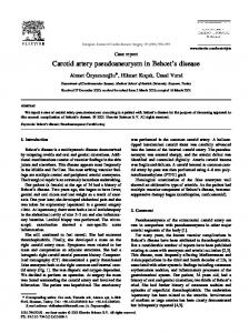
Pseudoaneurysm and simple hematomas could not be distinguished on the basis of B-mode findings alone. Both entities appeared as complex collections with anechoic portions, and unequivocal pulsatility could not be demonstrated in any collection. Pulsed Doppler evaluation mevealed turbulent pulsatile flow within both pseudoaneurysms, establishing the diagnosis. In one of these patients, a distinctive “to-and-fro” Doppler waveform was observed at the neck of the pseudoaneurysm, consisting of high-frequency flow (5-8 kHz) toward the transducer throughout systole and intermediatefrequency flow (2-4 kHz) away from the transducer throughout diastole (Fig. 1). The distance between the nearest artery and the pseudoaneurysm
was
approximately
patient. In neither pseudoaneurysm necting it to the onstrated.
Color Doppler Findings
1 cm
in
each
patient with a was the track conartery clearly dem-
Imaging
In both patients in whom diagnoses of pseudoaneunysm were made on the basis of Doppler findings, a high-velocity flow jet was noted entening the pseudoaneurysm at its neck during systole, with eddy currents of flow away from the transducer noted on either side of the jet 688
Radiology
#{149}
Figure quency
frequency
1. Distinctive waveform (>5 kHz) systolic flow
flow
in the opposite
into
from the neck of a pseudoaneunysm, the pseudoaneunysm (small arrows)
direction
throughout
(Figs. 2, 3). During diastole, swirls of color filled the collection, and continuous flow leaving the pseudoaneumysm was seen at the neck. In both patients, the tracks connecting the pseudoaneurysm with the artery were easily detected, appearing as straight lines with high-velocity flow toward the pseudoaneurysm throughout systole and away from the pseudoaneurysm throughout diastole. The range gate of the color Doppler unit allowed for duplex scanning of the neck of the pseudoaneumysm and the track leading to it. In these sites, the distinctive to-and-fro waveform was seen.
DISCUSSION The three cases in this series demonstrate the ability of both conventional duplex Doppler US and color Doppler imaging to allow differentiation of pseudoaneurysm secondary to arterial puncture from hematoma. This is an important differential diagnosis, because a pseudoaneunysm must be resected promptly to prevent expansion or rupture (2, 4, 10, 11), while a simple hematoma is usually managed conservatively. B-mode US has been reported as useful for differentiating pseudoaneurysm from nonpulsatile hematoma, because of the capability of demonstrating visible pulsatile expansion of the pseudoaneurysm and echoes within hematoma (4). In our series, echogenicity was not useful, since both hematomas and pseudoaneumysms had anechoic portions. We were not able to identify pulsatile ex-
diastole
(large
consisting of high-freand intermediate-
arrows).
pansion reliably, perhaps because of surrounding clot or fibrous tissue. Other modalities have been suggested as alternatives to arteniography, such as intravenous digital subtraction angiography (11) and dynamic computed tomography (4). These modalities share the disadvantage of requiming intravascular administration of contrast material, however, and are thus more invasive than Doppler examination.
Conventional
duplex
Doppler
and
colon Doppler imaging are both capab!e of depicting pseudoaneurysm of the femoral artery by demonstrating arterial pulsations within an anechoic space in proximity to an injured vessel. If the neck of the pseudoaneurysm or the track leading to it is examined with pulsed Doppler, a distinctive spectral waveform can be obtamed with either Doppler system. This waveform, which has not to our knowledge been previously described in the literature, consists of holosystolic high-velocity flow toward the transducer followed by ho-
lodiastolic
flow
of intermediate
ye-
locity in the opposite direction, a pattern that is typical of pseudoaneumysm. During systole, blood flows from the injured artery into the pseudoaneurysm. As arterial pressure drops during diastole, blood leaves the pseudoaneurysm and reenters the artery gradient
(1). Because the pressure between the pseudoaneurysm and the artery is lower during diastole than during systole, a lower velocity of flow is noted during diastole. The contribution of color Doppler
December
1987
a.
b.
Figure 2. Color Doppler images of same patient as in Figure 1. Flow toward the transducer is red, and flow away from the transducer is blue. Flow with a frequency shift higher than 2 kHz toward (a) or away from (b) the transducer is displayed in green. (a) During systole, blood flows from the femoral artery (FA) into the pseudoaneurysm through the track (black arrows), which is red. The flow of highest velocity
flow
(white
is within the center of the jet, depicted in green. Eddy currents of reversed arrows). The remainder of the pseudoaneurysm is filled with thrombus tent shape of the color, defining the edge of the thrombus and differentiating
(T). Delineation
the direction
a. Figure
of blood
flow
in the pseudoaneurysm
and
the track
is reversed,
are
it from
seen
seen
to either
churning,
in blue.
side
of the
of the thrombus The
slowly
most
flowing
rapid
flow
systolic
jet,
depicted
was enhanced blood.
(b)
in blue
by a consis-
During
is in the center
diastole,
of the jet.
b.
Color Doppler images of a pseudoaneurysm. Flow with a frequency shift higher than 1.5 kHz toward (a) or away from (b) the transducer is displayed in green. (a) During systole, forward flow (red) is seen in the femoral artery (FA), in the track leading to the pseudoaneurysm (black arrows), and in the jet entering the pseudoaneurysm. Reversed flow (blue) in the pseudoaneurysm is seen to the left of forward flow. Velocity in the cephalic portion of the pseudoaneurysm is too low to allow depiction by color Doppler imaging, and color does
3.
not
extend to the edge of the thrombus and at the neck of the pseudoaneunysm diastole, except for the continuous exiting
(white
the track, out
imaging
in our
faster and more intraaneurysmal same information
series
was
to allow
complete analysis flow. While the might have been
of
obtained with conventional duplex Doppler US, more skill and effort would have been required. With colon Doppler imaging, the neck of the pseudoaneumysm
fied,
Volume
was
as was
165
the
track
Number
readily
connecting 3
identi-
the
arrows). (arrow).
flow
at the
(b)
During
The
complex
neck.
Color
early
diastole,
pattern
of
extends
red
to the
reversed
flow
and
within
edge
blue
of the
injured artery with the pseudoaneurysm. This anatomic information, if communicated to the surgical team preoperatively, can facilitate repair of the pseudoaneurysm. An easily avoidable potential pitfall of color Doppler imaging is antifactual color noise, which can fill anechoic spaces. Colon noise can easily be differentiated from flow because
(blue) the
is seen
in the
pseudoaneurysni
femoral
artery,
varied
in
through-
thrombus.
(a) it is a homogeneous mixture of med and blue pixels that demonstrate random change only, (b) a comesponding Doppler audible or spectral waveform cannot be obtained, and (c) it can be eliminated if the colon threshold and gain are set properly. In conclusion, we have described distinctive Doppler spectral waveform that reflects the characteristic
Radiology
.
a
689
pattern of flow within a pseudoaneurysm. When observed in the groin following arterial trauma, we considen this waveform to be strongly suggestive of pseudoaneurysm. While conventional duplex Doppler US can
depict
flow
mysm sis,
and color
within thus
Doppler
a pseudoaneu-
establish imaging
the
Doppler
examination.
use of invasive ities. U
diagnostic
rysms. 3.
Perl
WR,
Yao
JST,
of peripheral Clin
5, Wener
Am
JJ.
Bergan
arterial
North
4.
L, Lyon
after
1973;
12.
aneu-
1979;
59:693-
Hessel
Clin
DC
and
Radiol
SJ, Adams
DF,
Radiology
15.
RW,
Wass
JL.
Two
10.
vascular I, Aakhus
Acta 199. Marx
Radiol
truth
trauma.
AJR
T, Evensen
about
11.
injuries
[Diagn]
M, Cardiner
of pe1968;
A.
(Stockh) CA,
Angiogra-
of the extremities.
Miller
false aneurysms.
1975;
from iatrogenic need for early
16:193-
disease.
16.
Merritt
US. for low-
In: Zwiebel
to vascular
1986; 333-350. RS, van Merode
APC.
of multi-gate
Doppler
ultraso-
CRB,
color
T, Hick
Cardiovascular pulsed
Med
Biol
Bluth
P,
applications
Doppler 1986;
&
systems.
Ul-
12:357-370.
El, Sullivan
flow mapping
MA.
of peripheral
vessels: comparison of angiodynography with conventional duplex US. Presented at the 72nd Scientific Assembly and An-
nual Meeting of North
of the Radiological
America,
30-December
Chicago,
Society
November
5, 1986.
RH. The AJR 1985;
145:193-194. Mills JL, Wiedeman JE, Robison lett JW Jr. Minimizing mortality morbidity nies: the
154:529-530.
venous
trasound
Radiolo-
diagnosis
2d ed. New York: Crone
Stratton, Reneman
Hoeks
signs of pseudoaneunysm:
phy in vascular
9.
pseudoaneurysm:
WJ, ed. Introduction
Corn-
102:431-440.
modal-
artery
with real-time and pulsed Doppler Radiology 1986; 158:55-56. Barnes RW. Doppler techniques
er-extremity
HL.
systolic jet and diastolic washout. gy 1982; 144:79-82. Love L, Braun T. Arteriography
Enge
14.
37:585-
Abrams
of angiography.
1981; 138:273-281. Kneipke DL, Holden
ripheral 8.
patic
ultrasound
1986;
Zwiebel WJ. Duplex carotid sonography. In: Zwiebel WJ, ed. Introduction to vascular ultrasonography. 2d ed. New York: Crone & Stratton, 1986; 139-216. Falkoff CE, Taylor KJW, Morse 55. He-
nography.
angiographic
7.
Ann
WC, Ruttley MST. femoral artery:
tomographic
plications 6.
13.
Pseudoaneu-
Med
Fitzgerald EJ, Bowsher False aneurysm of the appearances. 588.
5.
WS.
angiography.
42:173-175.
computed
Duplex
Rapoport 5, Sniderman KW, Morse SS, Proto MH, Ross CR. Pseudoaneurysm: complication of faulty technique in femoral arterial puncture. Radiology 1985;
Flinn
Surg
rysms
References 1.
LA,
706.
facilitates
Doppler US with color Doppler imaging allows the rapid, unequivocal diagnosis of pseudoaneurysm to be made, thus permitting its prompt treatment without the need for the
Queral
Management
diagno-
detection of the neck of the pseudoaneurysm and the track connecting it with the injured artery, and helps localize the mange gate for
pulsed
2.
arterial recognition
JC, Haland injuand
prompt repair. J Vasc Sung 1986; 4:22-27. Holland BA, Jeffrey RB, Brant-Zawadzki M. Intravenous digital subtraction angiography in the demonstration of femoral pseudo-aneurysms. AJR 1983; 141:607-
608.
690
Radiology
#{149}
December
1987