RESEARCH ARTICLE 4375
Development 140, 4375-4385 (2013) doi:10.1242/dev.097733 © 2013. Published by The Company of Biologists Ltd
FGF signaling sustains the odontogenic fate of dental mesenchyme by suppressing β-catenin signaling Chao Liu1, Shuping Gu1, Cheng Sun1, Wenduo Ye1, Zhongchen Song1, Yanding Zhang2 and YiPing Chen1,2,* SUMMARY Odontoblasts and osteoblasts develop from multipotent craniofacial neural crest cells during tooth and jawbone development, but the mechanisms that specify and sustain their respective fates remain largely unknown. In this study we used early mouse molar and incisor tooth germs that possess distinct tooth-forming capability after dissociation and reaggregation in vitro to investigate the mechanism that sustains odontogenic fate of dental mesenchyme during tooth development. We found that after dissociation and reaggregation, incisor, but not molar, mesenchyme exhibits a strong osteogenic potency associated with robustly elevated β-catenin signaling activity in a cell-autonomous manner, leading to failed tooth formation in the reaggregates. Application of FGF3 to incisor reaggregates inhibits β-catenin signaling activity and rescues tooth formation. The lack of FGF retention on the cell surface of incisor mesenchyme appears to account for the differential osteogenic potency between incisor and molar, which can be further attributed to the differential expression of syndecan 1 and NDST genes. We further demonstrate that FGF signaling inhibits intracellular βcatenin signaling by activating the PI3K/Akt pathway to regulate the subcellular localization of active GSK3β in dental mesenchymal cells. Our results reveal a novel function for FGF signaling in ensuring the proper fate of dental mesenchyme by regulating β-catenin signaling activity during tooth development.
INTRODUCTION A variety of craniofacial organs and tissues, such as the Meckel’s cartilage, maxillary and mandible bone, trigeminal ganglion and dentin-producing odontoblasts, derive from craniofacial neural crest cells (Chai et al., 2000; Chung et al., 2009). Despite originating from the same progenitor population, craniofacial bone and tooth exhibit distinct developmental, morphological and histological characteristics (Lumsden, 1988; D’Souza et al., 1999; Chai et al., 2000; Zhang et al., 2005; James et al., 2006). It is well established that multiple signaling pathways, including Wnt, TGFβ/BMP, Hh and FGF signaling, are involved in regulating every step of tooth development (Thesleff and Mikkola, 2002; Tummers and Thesleff, 2009), but the mechanisms that specify and ensure the odontogenic fate in dental mesenchyme remain largely unknown. FGF signaling has been implicated in regulating tooth development at several distinct steps. FGF signaling might be involved in the specification of odontogenic fate in both dental epithelial and dental mesenchyme, as evidenced by Fgf8 expression in the presumptive dental epithelium and its induction of Pitx2 and Pax9, the earliest molecular markers of the dental epithelium and mesenchyme, respectively, to determine the tooth-forming site (Neubüser et al., 1997; Trumpp et al., 1999; St Amand et al., 2000). At the bud stage, epithelial FGF4 and FGF8 are likely to activate Fgf3 in the dental mesenchyme through the mediation of Msx1 and Runx2, and FGF3 in turn, possibly together with FGF10, acts back on the dental epithelium to induce/maintain Shh expression in the enamel knot (Bei and Maas, 1998; Kettunen et al., 2000; Aberg et
1
Department of Cell and Molecular Biology, Tulane University, New Orleans, LA 70118, USA. 2College of Life Science, Fujian Normal University, Fuzhou, Fujian Province 350108, P.R. China. *Author for correspondence (
[email protected])
Accepted 19 August 2013
al., 2004). At the cap stage, expression of several FGFs in the enamel knot stimulates cell proliferation in the dental epithelium, leading to epithelial folding and cusp patterning (Jernvall et al., 1994; Jernvall and Thesleff, 2000). Furthermore, releasing FGF signaling from suppression by Sprouty factors leads to tooth formation in the diastema region, indicating a potential role for FGF signaling in the regulation of odontogenic fate (Klein et al., 2006; Li et al., 2011b). The essential role of canonical Wnt (Wnt/β-catenin) signaling in tooth development has been well documented (Liu and Millar, 2010). Many Wnt ligands are expressed in the developing tooth, predominantly in the epithelial component, with WNT5A, a noncanonical Wnt, in the mesenchyme (Dassule and McMahon, 1998; Sarkar and Sharpe, 1999). These Wnt ligands appear to act in both intra- and intertissue manners to regulate tooth development. Epithelial deletion of Catnb (Ctnnb1 – Mouse Genome Informatics), the gene encoding β-catenin, or Gpr177 (Wls – Mouse Genome Informatics), the product of which is required for secretion of Wnts, leads to an arrest of tooth development at the bud or early cap stage (Liu et al., 2008; Zhu et al., 2013). A similar developmental defect was also observed in mice lacking Catnb in the dental mesenchyme (Chen et al., 2009). Conversely, constitutive activation of β-catenin signaling in oral epithelium induces ectopic tooth formation (Järvinen et al., 2006; Liu et al., 2008). Although βcatenin signaling activity is present in the dental mesenchyme of the E12.5 incisor (Fujimori et al., 2010), such activity has never been reported in the incisor mesenchyme beyond E12.5 and was not detected in developing molar mesenchyme using several Wnt/βcatenin signaling reporter mouse lines, including BATGAL, TOPGAL and TCF/Lef-lacZ mice (Liu et al., 2008), suggesting that Wnt/β-catenin activity is maintained at a very low level, if any, in the dental mesenchyme. Elevated Wnt/β-catenin signaling results in the formation of bone-like tissues in the dental pulp (Chen et al., 2009; Li et al., 2011a). Thus, a finely tuned level of Wnt/β-catenin signaling is essential for proper tooth development.
Development
KEY WORDS: β-catenin signaling, FGF, Tooth development
Development 140 (21)
4376 RESEARCH ARTICLE
In this study, we investigated the mechanisms underlying our previous finding that early molar and incisor tooth germs exhibit distinct tooth-forming capability after dissociation and reaggregation in vitro (Song et al., 2006).
through 15% and 30% sucrose series, embedded in O.C.T. compound (Tissue-Tek) and cryosectioned at 10 μm. Sections were then subjected to standard X-Gal staining (Chai et al., 2000).
MATERIALS AND METHODS
For immunohistochemical staining, samples were fixed in Z-Fix (Anatech) at room temperature for 2 hours, dehydrated with 15% and 30% sucrose series, embedded in O.C.T. and cryosectioned at 10 μm. Immunohistochemical staining was conducted according to the manufacturer’s instruction using the following antibodies: mouse monoclonal anti-FGF3 antibody (Santa Cruz), rat anti-mouse syndecan 1 antibody (BD Pharmingen), rat monoclonal antibodies against heparan chondroitin sulfate, heparan dermatan sulfate and heparan keratan sulfate, respectively (Antibodies-online), biotinylated rabbit anti-mouse antibody (Vector Laboratories), horseradish peroxidase-coupled goat anti-rabbit IgG (Sigma), and Alexa Fluor 488 goat anti-rat antibody (Molecular Probes). For immunocytochemical staining, lower incisor and molar germs were treated with dispase and epithelia removed. Dental mesenchyme was collected and treated with trypsin to make a single-cell suspension as described above. Suspended dental mesenchymal cells were placed onto cell culture dishes and cultured in DMEM supplemented with 20% FBS for 6-12 hours, and then fixed with Z-Fix for 20 minutes. Immunocytofluorescence was performed using primary antibodies against β-catenin (Millipore), GSK3βY216 (Abcam) and GSK3βser9 (Abcam). Alexa Fluor 488 and Alexa Fluor 546 goat anti-rabbit IgG (Molecular Probes) were used as the secondary antibodies. Immunoblotting was performed as described previously (Iwata et al., 2006). Rabbit polyclonal antibodies against P-AktSer473 and total Akt (Cell Signaling) and mouse monoclonal antibodies against β-actin and FGF3 (Santa Cruz) were used as primary antibodies. IRDye 800cw goat anti-rabbit IgG and IRDye 800cw donkey anti-mouse IgG were used as secondary antibodies (Li-Cor).
BATGAL mice (Maretto et al., 2003) were obtained from Jackson Laboratories and were crossed onto the CD-1 background. All wild-type mice were CD-1 background and purchased from Charles River. Animals and procedures used in this study were approved by the Institutional Animal Care and Use Committee of Tulane University. Tissue recombination, organ culture, bead implantation and subrenal culture
Embryonic day (E) 13.5 or E14.5 embryos were collected from timed pregnant mice. To prepare tooth reaggregates, mandibular incisor or molar germs from one litter of embryos were isolated and pooled, respectively, then treated with 0.25% trypsin in 1 mM EDTA at 37°C for 5 minutes, and then dispersed into a single-cell suspension by mechanical aspiration with a micropipette. About 1×10⁶ cells from either the incisor or molar pool were added to a 1.5-ml Eppendorf tube, centrifuged at 3000 rpm (550 g) for 5 minutes, and incubated at 37°C and 5% CO2 for 1 hour to allow the formation of a firm cell pellet. Cell pellets were removed from Eppendorf tubes, placed in Trowell type organ culture in DMEM supplemented with 20% FBS overnight prior to being subjected to subrenal culture as described previously (Zhang et al., 2003; Song et al., 2006). To prepare tooth reaggregates with exchanged dental epithelial cells, isolated incisor and molar germs were treated with 2 mg/ml dispase at 37°C for 30 minutes, washed with DMEM containing 20% FBS, and then dental epithelia were separated from dental mesenchyme with the aid of fine forceps. Incisor mesenchyme was pooled together with molar epithelia and vice versa, and pooled dental tissues were further treated with 0.25% trypsin in 1 mM EDTA at 37°C for 2 minutes to generate the single-cell suspension and tooth reaggregates as described above. Protein-soaked bead preparation and implantation in tooth reaggregates or intact dental mesenchyme were performed as reported previously (Li et al., 2011b). Affi-Gel Blue agarose beads or heparin beads (100-200 μm in diameter; Bio-Rad) were used as carriers for FGF8 (0.5 mg/ml), FGF4 (0.4 mg/ml), FGF3 (0.5 mg/ml), FGF9 (0.5 mg/ml), FGF10 (0.5 mg/ml), DKK1 (0.4 mg/ml) and WNT10B (0.1 mg/ml) (all from R&D Systems). For the heparinase treatment experiment, E14.5 BATGAL molar germs were isolated and treated with dispase, and the epithelia were removed, as described above. The remaining dental mesenchyme was dispersed into single-cell suspension and pelleted. Cell pellet containing ~1×10⁶ cells was resuspended in 0.5 ml heparinase buffer (New England Biolabs). The final concentration of heparinases was adjusted as follows: heparinase I (150 unit/ml), heparinase II (10 unit/ml) and heparinase III (40 unit/ml). The cell suspension was incubated at 37°C for 1 hour before reaggregation and organ culture. Histology, in situ hybridization and X-Gal staining
Samples for histological analysis were harvested and fixed in 4% paraformaldehyde (PFA)/PBS. Ossified samples were subjected to Decalcifier I (Leica Biosystems) for demineralization for a week, and then dehydrated through graded ethanol, cleared with xylene, embedded in paraffin, and sectioned at 10 µm for standard Hematoxylin/Eosin (H&E) staining (Presnell and Schreibman, 1997). For in situ hybridization, samples were harvested in ice-cold PBS and fixed in 4% PFA/PBS at 4°C overnight prior to dehydration through graded ethanol and embedding in paraffin. Samples were sectioned at 10 μm and subjected to non-radioactive in situ hybridization as described (Yu et al., 2005b). At least three samples were used for each probe. For whole-mount X-Gal staining, samples were fixed with 4% PFA/PBS at room temperature for 20 minutes and then stained for β-galactosidase activity according to a standard procedure (Chai et al., 2000). For section XGal staining, samples were fixed in 4% PFA/PBS at 4°C overnight, passed
Quantitative RT-PCR
For quantitative (q) RT-PCR analysis, samples were subjected to RNA extraction using the RNAqueous-4PCR Kit (Ambion). The high capacity cDNA Reverse Transcription Kit (Applied Biosystems) was used for cDNA synthesis. qPCR was carried out on the 7500 Fast Real-Time PCR System (Applied Biosystems) with gene-specific primers and SYBR Green. Values were normalized to Gapdh using the 2–ΔΔCt method. Data from at least three independent experiments or samples for each gene were used for analysis.
RESULTS Incisor mesenchymal cells adopt an osteogenic fate in tooth germ reaggregates We reported previously that E13.5 mouse molar germ, after dissociation and reaggregation, is able to form a well differentiated tooth organ, whereas incisor germ fails (Song et al., 2006) (Fig. 1A,B). We followed up on this observation to investigate the underlying mechanism. We first tested if failed tooth formation in incisor reaggregates results from a loss of odontogenic competence in the dental epithelial cells after dispersion by exchanging dissociated epithelial cells and mesenchymal cells between E13.5 incisor and molar germ. Reaggregates composed of incisor epithelial and molar mesenchymal cells formed teeth (n=9/10), but reaggregates constituted by incisor mesenchymal and molar epithelial cells failed (n=0/10) and generated bony structures and keratinized cysts after 2 weeks in subrenal culture (Fig. 1C,D). These observations indicate that the incisor mesenchyme loses its odontogenic capability to instruct dispersed dental epithelial cells to form teeth. Gene expression assays demonstrate the expression of mesenchymal odontogenic markers, including Pax9, Bmp4 and Msx1, in mesenchymal cells surrounding the reorganized dental epithelial structures in molar reaggregates but not in incisor
Development
Animals
Immunostaining and immunoblotting
FGF sustains odontogenic fate
RESEARCH ARTICLE 4377
reaggregates after 3 days in culture (Fig. 1E-J). By contrast, osteogenic markers, including Runx2, osteocalcin (Bglap – Mouse Genome Informatics) and osterix (Sp7 – Mouse Genome Informatics), were activated in mesenchymal cells of incisor reaggregates but not in molar reaggregates (Fig. 1K-N; data not shown), which was further confirmed by qPCR assay (Fig. 1Q). These expression patterns persisted in incisor and molar reaggregates after 5 days in subrenal culture (data not shown). The retained expression of Pitx2, a dental epithelial molecular marker, in the reorganized dental epithelial masses in both incisor and molar reaggregates indicates their odontogenic fate (Fig. 1O,P). These results suggest a deviation of odontogenic fate and the rapid adoption of osteogenic fate in the incisor mesenchyme after dissociation and reaggregation. Wnt/β-catenin signaling is robustly activated in mesenchymal cells of incisor reaggregates We next investigated the molecular basis for the adoption of osteogenic fate in mesenchymal cells of incisor reaggregates. Wnt/β-catenin signaling plays a crucial role in promoting osteogenic fate and the maturation of osteoblasts (Clément-Lacroix et al., 2005; Gaur et al., 2005). We suspected that dissociation and reaggregation
of incisor tooth germ led to the activation of β-catenin signaling and subsequent osteogenesis. β-catenin signaling activity is restricted to the dental epithelium of E13.5 incisor and molar, as detected by the BATGAL transgenic reporter (Fig. 2A,B). Using the BATGAL reporter mice, we found that after 3 days in subrenal culture, robust BATGAL activity could be detected in both epithelial and mesenchymal cells in E13.5 incisor reaggregates (Fig. 2D; n=14/14). By contrast, BATGAL activity was detected only in the reorganized dental epithelial structures in molar reaggregates (Fig. 2C; n=20/20). However, in molar reaggregates after 5 days in subrenal culture, BATGAL activity was detected in some mesenchymal cells that were not associated with the forming tooth but also expressed osteocalcin (Fig. 2E,F), indicating an association of active β-catenin signaling with osteogenic fate. We examined whether molar epithelial cells played an inhibitory role in suppressing β-catenin signaling in molar reaggregates. E13.5 BATGAL incisor and molar mesenchyme without dental epithelium were dissociated and reaggregated. Robust BATGAL activity was detected in the incisor but not in the molar mesenchymal reaggregate as early as 2 hours in organ culture (Fig. 2E,F), suggesting the existence of a mechanism to inhibit β-catenin signaling in the molar mesenchyme.
Development
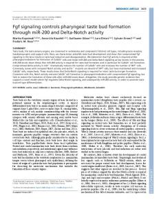
Fig. 1. Adoption of osteogenic fate by mesenchymal cells in E13.5 incisor reaggregates. (A-D) H&E staining of mouse tooth reaggregates after 2 weeks in subrenal culture shows tooth formation (arrows) in E13.5 molar reaggregate (A) and in the reaggregate of E13.5 molar mesenchymal cells and incisor epithelial cells (C). Tooth formation failed in E13.5 incisor reaggregate (B) and reaggregate of E13.5 incisor mesenchymal cells and molar epithelial cells (D). (B,D) Arrows point to keratinized cysts. (E-N) In situ hybridization of tooth reaggregates after 3 days in subrenal culture shows expression of Pax9, Bmp4 and Msx1 but absence of Runx2 and osteocalcin expression in E13.5 molar reaggregates (E,G,I,K,M), and opposite expression patterns of these genes in E13.5 incisor reaggregates (F,H,J,L,N). Arrows point to gene expression sites. (O,P) Pitx2 expression is seen in the reorganized epithelial structures in E13.5 molar (O) and incisor (P) reaggregates. (Q) Realtime RT-PCR results show dramatically elevated expression of osteogenic markers in incisor (In) reaggregates as compared with molar (Mol) reaggregates after 3 days in subrenal culture. **P