United States Department of Agriculture / Forest Service Rocky Mountain Region, Forest Health Protection Rocky Mountain Research Station General Technical Report RMRS-GTR-241 December 2010
Rocky Mountain Region, Forest Health Protection. 2010. Field guide to diseases & insects of the Rocky Mountain Region. Gen. Tech. Rep. RMRS-GTR-241 Fort Collins, CO: U.S. Department of Agriculture, Forest Service, Rocky Mountain Research Station. 336 p.
Abstract This field guide is a forest management tool for field identification of biotic and abiotic agents that damage native trees in Colorado, Kansas, Nebraska, South Dakota, and Wyoming, which constitute the USDA Forest Service’s Rocky Mountain Region. The guide focuses only on tree diseases and forest insects that have significant economic, ecological, and/ or aesthetic impacts; this guide does not necessarily cover all possible damage agents. Management suggestions are provided where available. The field guide is divided into two sections: one describes both diseases and damage caused by animals and abiotic factors, and the other describes insects. Agents are presented by the type and/or location of the injury on the tree. Written descriptions, color photographs, a general index, an index by host tree species, descriptive tables, and line drawings are all provided to assist users in identifying damaging agents.
You may order additional copies of this publication by sending your mailing information in label form through one of the following media. Please specify the publication title and series number. Publishing Services Telephone FAX E-mail Web site Mailing address
(970) 498-1392 (970) 498-1122
[email protected] http://www.fs.fed.us/rm/publications Publications Distribution Rocky Mountain Research Station 240 West Prospect Road Fort Collins, CO 80526
Authors Kurt K. Allen James T. Blodgett Kelly S. Burns Robert J. Cain Sheryl L. Costello Tom J. Eager Jeri Lyn Harris Brian E. Howell Roy A. Mask Willis C. Schaupp, Jr. Jeffrey J. Witcosky James J. Worrall
Acknowledgments Great appreciation is extended to the entomologists and pathologists who have studied and published on the often complicated life cycles, subtle taxonomic variations, and effective management strategies of the diverse organisms presented in this field guide. References are cited at the end of this guide. The following individuals have all provided invaluable observations through personal communication and published materials: Whitney Cranshaw and William Jacobi with Colorado State University; David Leatherman and Ingrid Aguayo, formerly with Colorado State Forest Service; Mark Harrell with Nebraska Forest Service; John Ball with South Dakota State University; Robert Mathiasen from Northern Arizona University; Les Koch with the Wyoming State Forestry Division; Jose Negron with the USDA Forest Service, Rocky Mountain Research Station (RMRS); and John Schmid, formerly with RMRS. Field guides produced in the Pacific Northwest, Northern and Intermountain, and Southwestern Regions of the USDA Forest Service provided insights for layout and design. Many of the photographs presented here were taken by Federal, State, and university pathologists and entomologists from the Rocky Mountain Region or adjacent USDA Forest Service regions and were made available by the University of Georgia through www.bugwood.org. Thanks to MaryLou Fairweather with USDA Forest Service, Southwestern Region, for providing scanned photographs from the Field Guide to Insects and Diseases of Arizona and New Mexico. Special thanks also to Jose Negron and Bill Jacobi for their reviews and comments, to Justin Backsen and Diane Hildebrand for their help during manuscript preparation, and to Lindy Myers, Lane Eskew, and Connie Lemos for editing, layout, and publication of the field guide.
ii
Contents About This Field Guide.............................................................................1 Role of Diseases and Insects in Forest Ecosystems..................................2 Diagnosing Tree Problems........................................................................3
Diseases Dwarf and True Mistletoes Introduction to Dwarf Mistletoes.........................................................6 Douglas-Fir Dwarf Mistletoe.............................................................10 Limber Pine Dwarf Mistletoe............................................................12 Lodgepole Pine Dwarf Mistletoe.......................................................13 Pinyon Dwarf Mistletoe.....................................................................15 Southwestern Dwarf Mistletoe...........................................................16 Juniper Mistletoe.................................................................................18 Root Diseases and Stem Decays Introduction to Decay Diseases..........................................................19 Annosus Root Disease........................................................................25 Armillaria Root Disease......................................................................28 Black Stain Root Disease....................................................................31 Coniophora Root and Butt Rot..........................................................35 Root Diseases with White Pocket Rots..............................................38 Schweinitzii Root and Butt Rot.........................................................40 White Mottled Rot.............................................................................42 Aspen Trunk Rot................................................................................44 Brown Crumbly Rot...........................................................................46 Gray-Brown Saprot............................................................................48 Red Ray Rot........................................................................................49 Red Ring Rot......................................................................................51 Rust-Red Stringy Rot and Red Heart Rot.........................................53 Stem Decays of Hardwoods in the Plains..........................................56 Cankers Introduction to Cankers and Overview of Aspen Cankers................60 Black Canker.......................................................................................64 Black Knot of Cherry and Plum.........................................................66 Botryodiplodia Canker.......................................................................67 Cryptosphaeria Canker.......................................................................69 Cytospora Canker of Aspen...............................................................71 Cytospora Canker of Conifers............................................................73 Hypoxylon Canker..............................................................................75 Sooty-Bark Canker.............................................................................77 iii
Wilts Introduction to Wilt Diseases.............................................................79 Dutch Elm Disease.............................................................................81 Oak Wilt.............................................................................................84 Pine Wilt.............................................................................................87 Verticillium Wilt.................................................................................89 Rusts Introduction to Rust Diseases.............................................................91 Broom Rusts of Spruce and Fir..........................................................94 Comandra and Stalactiform Blister Rusts..........................................97 Gymnosporangium Rusts.................................................................100 Western Gall Rust............................................................................103 White Pine Blister Rust....................................................................105 Foliage Diseases Introduction to Foliage Diseases.......................................................107 Anthracnose......................................................................................110 Brown Felt Blight.............................................................................113 Cercospora Blight of Junipers...........................................................114 Davisomycella and Lophodermella Needle Casts............................115 Elytroderma Needle Cast.................................................................118 Ink Spot............................................................................................120 Marssonina Leaf Blight....................................................................121 Melampsora Rusts............................................................................124 Red Band and Brown Spot Needle Blights......................................125 Septoria Leaf Spot and Canker........................................................128 Shoot Blights Pine Shoot Blight and Canker..........................................................129 Shepherd’s Crook..............................................................................132 Abiotic Injuries and Miscellaneous Burls, Galls, and Tumors...................................................................134 Animal Damage................................................................................135 Abiotic Foliage Damage...................................................................138 Abiotic Stem Damage......................................................................145 Wetwood...........................................................................................149
Insects Bark Beetles Introduction to Bark Beetles.............................................................154 Ash Bark Beetles...............................................................................156 Aspen bark beetles............................................................................157
iv
Blue Spruce Engraver.......................................................................159 Cedar Bark Beetles............................................................................161 Douglas-Fir Beetle............................................................................162 Douglas-Fir Pole and Engraver Beetles............................................163 Elm Bark Beetles..............................................................................165 Fir Engraver......................................................................................167 Limber Pine Engraver......................................................................169 Lodgepole Pine Beetle......................................................................170 Mountain Pine Beetle.......................................................................172 Pine Ips Species (Engraver Beetles)..................................................174 Pinyon Ips.........................................................................................177 Pinyon Twig Beetles..........................................................................179 Red Turpentine Beetle......................................................................181 Roundheaded Pine Beetle.................................................................183 Spruce Beetle....................................................................................184 Spruce Engraver Beetles...................................................................187 Twig Beetles......................................................................................188 Western Balsam Bark Beetle.............................................................190 Western Pine Beetle..........................................................................193 Defoliators Introduction to Defoliating Insects..................................................195 Aspen Leafminer..............................................................................197 Bagworm...........................................................................................199 Cottonwood Leaf Beetle...................................................................201 Douglas-Fir Tussock Moth..............................................................202 Elm Leaf Beetle................................................................................204 Fall Webworm...................................................................................207 Forest Tent Caterpillar......................................................................209 Large Aspen Tortrix..........................................................................212 Needleminers....................................................................................214 Oak Leafroller...................................................................................216 Pandora Moth...................................................................................218 Pine Butterfly....................................................................................220 Pine Sawflies.....................................................................................222 Tiger Moth.......................................................................................225 Western Pine Budworm...................................................................227 Western Pine Tussock Moth............................................................229 Western Spruce Budworm................................................................231 Western Tent Caterpillar..................................................................233 Wood Borers Introduction to Wood Borers............................................................236 Agrilus quercicola................................................................................239
v
Ambrosia Beetles..............................................................................240 Bronze Poplar Borer..........................................................................242 Carpenter Ants.................................................................................244 Carpenterworm.................................................................................245 Cottonwood Borer............................................................................247 Elm Borer.........................................................................................248 Flatheaded Wood Borers (Metallic Wood Borers)...........................250 Juniper Borers...................................................................................252 Lilac (Ash) Borer..............................................................................253 Poplar Borer......................................................................................255 Roundheaded Wood Borers (Longhorned Beetles).........................256 Twolined Chestnut Borer.................................................................258 Wood Wasps (Horntails)..................................................................260 Sap-Sucking Insects, Gall Formers, and Mites Introduction to Sap-Sucking Insects, Gall Formers, and Mites.......262 Cooley Spruce Gall Adelgid.............................................................265 European Elm Scale.........................................................................266 Gall (Eriophyid) Mites.....................................................................267 Giant Conifer Aphids.......................................................................269 Oystershell Scale...............................................................................270 Petiolegall Aphids.............................................................................272 Pine Needle Scale.............................................................................274 Pinyon Needle Scale.........................................................................276 Spider Mites......................................................................................278 Bud and Shoot Insects Introduction to Bud, Shoot, Branch, and Terminal Insects..............280 Pine Tip Moth..................................................................................281 Pitch Moths......................................................................................282 Pitch Nodule Moths.........................................................................283 White Pine Weevil and Lodgepole Pine Terminal Weevil..............285 Glossary................................................................................................287 References.............................................................................................297 Host-Pest Index....................................................................................312 General Index.......................................................................................322 Instructions for Submitting Insect and Disease Specimens for Identification..............................................................................335 Technical Assistance Sources..............................................................336
vi
About This Field Guide This field guide details the most commonly encountered diseases and insects of forest trees in the Rocky Mountain Region. Descriptions of diseases, insects, and physical injuries focus on the most diagnostic features of each. Color photographs, line drawings, and tables are used to illustrate and emphasize characteristics described in the text. Diseases and insects in plains hardwood trees are not covered in depth. Ornamental trees are sometimes affected by the diseases or insects included in this guide but may not be specifically mentioned as hosts. This guide presents diseases and then insects. Entries are arranged according to the part of trees typically damaged by the agent described. The disease section describes dwarf and true mistletoes, decays (including root diseases and stem decays), cankers, wilts, rusts, foliage diseases, shoot blights, and abiotic injuries and miscellaneous diseases. The insect section describes bark beetles, defoliators, wood borers, sap-sucking insects, gall formers, mites, and bud and shoot insects. At the end of the guide is a subject index, a host-pest index to damaging agents by tree species and part of the tree affected, and a glossary of terms. The host-pest index provides a rapid means of assessing the number and variety of agents described for each tree species. This field guide applies to the USDA Forest Service’s Rocky Mountain Region, which includes Colorado, Wyoming, South Dakota, Kansas, and Nebraska (fig. 1). Additional hosts, diseases, and insect pests may be encountered outside this Region that are not included here, and a few of the diseases and insects included in this guide may not be seen in other areas.
Figure 1. Rocky Mountain Region area covered by this Field Guide. Image: Sheryl Costello, USDA Forest Service.
USDA Forest Service RMRS-GTR-241. 2010.
1
Plant pathologists and entomologists are available to assist resource managers with identification of insects and pathogens encountered in the forests. A list of offices that provide this assistance is included at the back of this field guide along with instructions for specimen collection and shipping.
Role of Diseases and Insects in Forest Ecosystems Most forest insects and diseases are native to and important players in the forest ecosystems of the Rocky Mountain Region. Some insects and diseases reduce tree growth, cause mortality, reduce timber productivity, create hazardous situations in developed sites, and change wildlife habitat, fire hazard conditions, and overall watershed quality. In terms of mortality and growth loss, the impacts of diseases and insects on forests are far greater than those of fire or any other disturbance. Along with fire, insects and diseases are the major disturbance agents for changing forest age, density, composition, and structure on a stand or landscape level. They provide food and habitat for other wildlife, increase structural diversity of forests, and are important forest recyclers in the Rocky Mountains. Typically, forest insects are at low or endemic levels. From time to time, significant outbreaks cause rapid changes across the forest. For example, bark beetles can build to high populations relatively fast and can cause widespread tree mortality, significantly altering forest conditions. Diseases are more likely to increase gradually or remain at similar levels over time, depending on forest conditions. Dwarf mistletoes and root diseases are ever-present in the forest and are some of the main contributors to growth loss, reduced vigor, and mortality in their hosts. Insects and diseases often interact, such as when root diseases predispose trees to bark beetle attack. At times, natural or human-made events or conditions can cause insects or diseases to become more active. This can occur from the high-elevation subalpine forests to the urban forests that line the streets of our cities and towns. Fire, wind, and drought can weaken host trees and create favorable environments for insects and diseases to attack. Altered age, density, composition, and structure can also lead to greater susceptibility and increased disease and insect activity. For example, through past harvesting practices and fire exclusion, many ponderosa pine stands in southern Colorado now have dense understories of shade-tolerant white fir, with consequent increased activity of root diseases to which it is susceptible. Forest insect and disease management often involves changing the conditions that have created the pest situation. Reducing stand density in conifer stands can lower bark beetle impact. Removing mistletoe-infected overstory trees from a regenerating pine stand or converting to even-aged management where that is consistent with the natural disturbance regime can protect regeneration and 2
USDA Forest Service RMRS-GTR-241. 2010.
reduce mistletoe impacts in future stands. Increasing the diversity of cultivars that are planted along our city streets and windbreaks can also reduce pest impacts. The main goal of most forest managers is to maintain a healthy forest that is capable of producing a variety of resources. Knowledge of the role that forest insects and diseases play is critical to achieving the desired conditions from the forest. This manual provides users with a tool to begin identifying and understanding forest insects and diseases that may be detrimental to management goals. Please refer to your State Department of Agriculture or your local pest control specialist for the rules, regulations, and applicable allowances concerning specific control measures.
Diagnosing Tree Problems Several steps must be taken in order to effectively diagnose tree problems. The following are general guidelines: • Properly identify the tree. It is important to know exactly what species you are looking at. It is also important to know what the leaves, bark, trunk, and roots should look like under “normal” conditions. • Carefully observe the symptoms expressed. Look for patterns on the tree and in the surrounding vegetation. ○○ Check for host specificity. Biotic agents tend to affect one species, are clumpy in distribution, show progressive symptoms, and usually impact specific plant parts. Abiotic agents tend to affect many species relatively uniformly. ○○ Carefully examine the types of symptoms and the part of the plant impacted. ○○ Typical symptoms include: underdevelopment of tissues or organs (stunting and malformed leaves); overdevelopment of tissues or organs (galls, brooms, and stress cones); necrosis (death) of plant parts (wilting, dieback, and leaf spots); and alteration of normal appearance (chewing and chlorosis [pale coloration in leaves and flowers]). ○○ Examine how the symptoms are distributed. If the entire tree is impacted, there is likely something wrong with the roots or stem, or there may be an environmental cause. Single or randomly scattered affected branches are often associated with insects or diseases. • Determine the history of the tree and the site. Has the root system been disturbed? Have chemicals been used? Has there been any harvesting? Other site factors, such as changing water relations, extreme temperatures, or wind, may affect tree vigor. • Look for signs of biotic agents such as fungal fruiting bodies, parasitic plants, larvae, or adult insects. • Identify agents. Laboratory tests may be necessary in some circumstances.
USDA Forest Service RMRS-GTR-241. 2010.
3
Sources of Additional Information Additional information on insects and diseases can be found online at: • USDA Forest Service, Rocky Mountain Region: http://www.fs.usda.gov/ goto/r2/fh; • USDA Forest Service, Northern and Intermountain Region: http://www. fs.fed.us/r1-r4/spf/fhp/mgt_guide/; • USDA Forest Service, Southwestern Region: http://www.fs.fed.us/r3/resources/health/field-guide/index.shtml; • Colorado State University Extension: http://www.ext.colostate.edu/pubs/ pubs.html#insects; and • State Forestry Agencies and County Extension Offices. Updated Forest Insect and Disease Leaflets can be found online at: • USDA Forest Service, Forest Health Protection: http://www.fs.fed.us/r6/nr/ fid/wo-fidls/index.shtml. Images and publications can also be found online at: • University of Georgia and USDA Forest Service online database: http:// www.bugwood.org/.
4
USDA Forest Service RMRS-GTR-241. 2010.
USDA Forest Service RMRS-GTR-241. 2010.
5
D WA R F A N D T R U E M I S T L E T O E S
Introduction to Dwarf Mistletoes Parasitic vascular plants with conifer hosts
Pathogens—Dwarf mistletoes (Arceuthobium spp.) are parasitic plants of co-
nifers that obtain almost all of their needs, including water, mineral, and carbon nutrients, from their hosts. Fulfilling these resource requirements stresses infected trees, causing reductions in growth, cone, and seed production and, with high infection levels, mortality. Dwarf mistletoes are some of the most common and easily identified disease agents where they occur in the coniferous forests of the Rocky Mountain Region. Hosts—Five species of dwarf mistletoes occur in this Region, each with a specific set of susceptible hosts (table 1). Signs and Symptoms—Dwarf mistletoes produce aerial shoots on branches or stems of infected trees (fig. 2). Shoots are nearly leafless and vary in color; yellow, brown, purple, or green shoots are common. Plants size also varies within and among species. Douglas-fir dwarf mistletoe shoots are often shorter than the host’s leaves, while southwestern dwarf mistletoe shoots are typically 3-6 inches (7-15 cm) long. When shoots are shed, characteristic basal cups remain (fig. 3). Infection with dwarf mistletoe also causes characteristic deformities in the host. Witches’ brooms are areas of profuse, dense branching often induced by dwarf mistletoe infection (fig. 4). Branch swellings are often found in the immediate vicinity of local infections (fig. 2). Cankers (areas of dead cambium) are often associated with older infections. Dieback of the host’s foliage from the top-down and eventual mortality is often observed in trees that have been infected for many Table 1. Dwarf mistletoes and their hosts in the Rocky Mountain Region. Dwarf mistletoe (DM)
Primary hosta
Other hostsa
Lodgepole pine DM Lodgepole pine Secondary: ponderosa pine Arceuthobium americanum Occasional: whitebark and limber pines Rare: Engelmann and blue spruce, bristlecone pine Limber pine DM Limber pine, Rare: ponderosa pine, lodgepole pine A. cyanocarpum Whitebark pine, Bristlecone pine Pinyon DM Pinyon pine None A. divaricatum Douglas-fir DM Douglas-fir Rare: subalpine fir, Englemann and blue spruce A. douglasii Southwestern DM Ponderosa pine Occasional: bristlecone pine, lodgepole pine A. vaginatum subsp. Rare: limber and southwestern white pine, blue spruce cryptopodium a Hosts are in the following categories: Primary: more than 90% infection when close to heavily infected trees. Secondary: frequently infected (50-90% infection) when close to heavily infected principal hosts. Occasional: occasionally infected (5-50% infection) when close to heavily infected principal hosts. Rare: rarely infected (≤5% infection), even when close to heavily infected principal hosts.
6
USDA Forest Service RMRS-GTR-241. 2010.
D WA R F A N D T R U E M I S T L E T O E S
Figure 2. Aerial shoots of American dwarf mistletoe plant on lodgepole pine. Note the swelling in the branch associated with the aerial shoots. Photo: Brian Howell, USDA Forest Service. Figure 3. After aerial shoots are shed and basal cups remain as signs of dwarf mistletoe infection. Photo: Kelly Burns, USDA Forest Service.
Figure 4. Large witches’ brooms formed on a ponderosa pine that is heavily infected with southwestern dwarf mistletoe. The top of the tree is dying back. Photo: Bob Cain, USDA Forest Service.
Figure 5. Lodgepole pine killed as a result of lodgepole pine mistletoe infection. Note the typical witches’ brooms. Photo: Jim Worrall, USDA Forest Service.
years (fig. 5), depending on the species of dwarf mistletoe and host, the level of infection, and site factors. These symptoms on the hosts are often associated with mistletoe infection but may also be caused by other agents. Plants or basal cups should be present for positive identification of dwarf mistletoe infection. Disease Cycle—Dwarf mistletoes have separate male and female plants (figs. 6-7). Seeds are produced annually on mature female plants. These are explosively released and typically fly less than 33 ft (10 m). Upon germination, the dwarf mistletoe plant produces a specialized root-like structure that contacts the phloem and xylem of the host, from which the parasite obtains nutrients and water. Aerial shoots appear 3-5 or more years after infection; the time period before shoots are visible is known as the latent period (fig. 8). Dwarf mistletoes spread both within and between tree crowns. As a result of the explosive seed-dispersal mechanism, infections tend to build up initially in the lower portion of the crown and spread gradually upward. Lateral spread USDA Forest Service RMRS-GTR-241. 2010.
7
D WA R F A N D T R U E M I S T L E T O E S
Figure 6. Male flowers on shoots of southwestern dwarf Figure 7. Immature female berries on shoots of southwestern dwarf mistletoe. Photo: Brian mistletoe. Photo: Brian Howell, USDA Forest Service. Howell, USDA Forest Service.
Figure 8. Generalized life cycle of dwarf mistletoes, as exemplified by lodgepole pine dwarf mistletoe on lodgepole pine (from Hawksworth and Dooling 1984).
of dwarf mistletoe through single-storied stands averages about 1.5 ft (0.5 m) per year. Spread is most rapid from infected overstory to adjacent regeneration. Long-distance seed dispersal by birds is not common but can introduce dwarf mistletoe to new areas. Impact—As parasites, dwarf mistletoes cause significant changes in physiological processes and structural characteristics of infected trees, resulting in changes 8
USDA Forest Service RMRS-GTR-241. 2010.
D WA R F A N D T R U E M I S T L E T O E S
Figure 9. Evidence of squirrel or porcupine feeding on sugar-rich phloem found near southwestern dwarf mistletoe infections. Photo: Brian Howell, USDA Forest Service.
in the structure and function of forest communities. Tree growth and vigor usually decline when more than half of the crown is parasitized. Most trees survive infection for decades, but small trees tend to decline and die more quickly than large ones. Tree mortality in areas with extensive infection is often three to four times higher than in uninfected areas. Bark beetles frequently attack heavily infected trees, especially during drought. Extensive dwarf mistletoe infection greatly reduces forest productivity. However, infection has some benefits for wildlife. Large witches’ brooms provide nesting and seclusion habitat for birds and small mammals. Snags created by dwarf mistletoe infection offer habitat for cavity-nesting birds. A few species are known to feed on shoots of dwarf mistletoes and the sugar-rich phloem found in and around infection sites (fig. 9). Management—The first step when making management decisions in stands infected with dwarf mistletoe is to quantify the incidence and severity of infection. Although many systems have been used to rate levels of infection by dwarf mistletoe, one is now used almost universally: Hawksworth’s 6-class dwarf mistletoe rating (DMR) system (fig. 10). Many disease parameters and management recommendations are provided in terms of DMR because this system has been used for many years. A tree’s DMR ranges from 0 (uninfected) to 6 (over half the branches infected throughout the crown). Rate each third of the crown on a scale from 0 to 2, then sum the thirds for the tree rating (fig. 10). Binoculars should be used to enFigure 10. The 6-class Hawksworth dwarf mistletoe rating system (from hance detection. Hawksworth and Wiens 1996). USDA Forest Service RMRS-GTR-241. 2010.
9
D WA R F A N D T R U E M I S T L E T O E S Silvicultural control of dwarf mistletoes can be effective and should be considered for use in a variety of stand conditions and dwarf mistletoe infection levels. Because dwarf mistletoes require a living susceptible host, silvicultural options include pruning, harvesting, and favoring non-host species. Due to the explosive seed dispersal mechanism, implementing buffer strips around infection centers or around sanitized patches can also be effective. A thorough discussion of management options based on stand conditions and objectives is outside the scope of this manual but can be found on the Region 2 Forest Health Management website at: http://www.fs.fed.us/r2/fhm/bugcrud/DM_MgmtGuide_R2.pdf. References: 66, 70, 158
Douglas-Fir Dwarf Mistletoe Large brooms, small aerial shoots
Pathogen—Douglas-fir dwarf mistletoe (Arceuthobium douglasii) causes large
brooms but has very small shoots. Aerial shoots are olive green, average 4/5 inch (20 mm) long (maximum 3 inches or 8 cm) and 1/25-1/16 inch (1-1.5 mm) in diameter, and have fan-shaped branching (fig. 11). Douglas-fir dwarf mistletoe occurs throughout the range of Douglas-fir in the central and southern mountains of Colorado. It is absent from northern Colorado (except for the extreme northwest) and from the portion of Wyoming in the Rocky Mountain Region (fig. 12). Hosts—Douglas-fir dwarf mistletoe primarily infects its namesake, although several true firs and spruces are occasional or rare hosts.
Figure 11. Douglas-fir dwarf mistletoe parasitizing Douglas-fir. Note that the mistletoe plants are growing with the branch (systemic infection). Photo: Robert Mathiasen, Northern Arizona University.
10
Figure 12. Distribution of Douglas-fir dwarf mistletoe in the Rocky Mountain Region (from Hawksworth and Wiens 1996).
USDA Forest Service RMRS-GTR-241. 2010.
D WA R F A N D T R U E M I S T L E T O E S
Figure 13. Douglasfir dwarf mistletoe plants, which are approximately the same length as the host’s leaves. Photo: USDA Forest Service.
Figure 14. Large brooms are typical in Douglas-fir heavily infected with Douglas-fir dwarf mistletoe. Photo: USDA Forest Service.
Figure 15. Trees heavily infected with Douglas-fir dwarf mistletoe typically die from the top-down, causing dead (spike) tops. Photo: USDA Forest Service.
Signs and Symptoms—Douglas-fir dwarf mistletoe has the smallest shoots
(fig. 13) of all mistletoes in the Region, but it can form the largest witches’ brooms (fig. 14). Douglas-fir infections grow along with the infected host branches (systemic infection). Mistletoe shoots may be spread along young host branches or be aggregated near the annual bud scars. Because shoots are so small, they are normally detectable only in branches close to the ground. Witches’ brooms, which are used for detection and rating, become noticeable about 10 years after infection and develop best in direct sunlight. Brooms occur mostly in the lower half of tree crowns. They can weigh hundreds of pounds, can break off of the tree, and are considered hazards in developed sites. Impact—Dwarf mistletoe is the most detrimental disease of Douglas-fir. Damages typically associated with dwarf mistletoe infection are: growth reduction, spike tops (fig. 15), reduced cone and seed production, and mortality. Infections have reportedly increased in abundance since the late 1800s. In northern Idaho and western Montana, Douglas-fir stands have become more widespread due to fire suppression, a history of selective harvesting that removed pines and encouraged USDA Forest Service RMRS-GTR-241. 2010.
11
D WA R F A N D T R U E M I S T L E T O E S shade-tolerant species, and white pine blister rust, which largely eliminated western white pine. Data on growth effects from western Montana indicate that light, medium, and severe infections caused decreases in basal area growth rate of 14, 41, and 69%, respectively. Effects on height growth were similar. Horizontal spread in single-storied stands is estimated at 1.5-2 ft (45-61 cm) per year. Upward spread in crowns is about 4-6 inches (10-15 cm) per year. Please see the Introduction to Dwarf Mistletoes entry for disease cycle and management information. References: 59, 70
Limber Pine Dwarf Mistletoe Infects five-needle pines
Pathogen—Aerial shoots of limber pine dwarf mistletoe (Arceuthobium cyano-
carpum) are yellow-green, 1 1/4-2 3/4 inches (3-7 cm) long, and up to 1/13 inch (2 mm) diameter. Branching is fan-shaped and shoots are densely clustered (fig. 16). Limber pine dwarf mistletoe generally occurs in five-needle pine (predominantly limber pine) stands along the Continental Divide in the Rocky Mountains but also occurs in other mountain ranges (fig. 17). Hosts—Almost all of the five-needle pines in the Rocky Mountain Region, including limber, whitebark, and Rocky Mountain bristlecone pine, are primary hosts of limber pine dwarf mistletoe. The only endemic white pine that is not a host in nature is southwestern white pine, although it has been infected in greenhouse trials. Signs and Symptoms—Signs of infection include aerial shoots and basal cups left
Figure 16. Limber pine dwarf mistletoe parasitizing limber pine. Photo: Brian Howell, USDA Forest Service.
12
Figure 17. Distribution of limber pine dwarf mistletoe in the Rocky Mountain Region (from Hawksworth and Wiens 1996).
USDA Forest Service RMRS-GTR-241. 2010.
D WA R F A N D T R U E M I S T L E T O E S after shoots have fallen off branches. Limber pine dwarf mistletoe causes small, tightly clustered witches’ brooms (fig. 18). Other symptoms of infection include swelling of branches at infection sites, dieback, and eventual mortality of heavily infected trees. Impact—Limber pine dwarf mistletoe causes extensive mortality of limber pine in many parts of the Rocky Mountains and can also cause mortality in other hosts when infection levels are high. It is the most important native disease of high-elevation white pines in the West; only white pine blister rust is more Figure 18. Tightly clustered brooms are a damaging. Lateral spread in single-storied symptom of limber pine dwarf mistletoe. stands is estimated to be 1.5-2 ft (45-61 cm) Photo: Brian Howell, USDA Forest Service. per year. Please see the Introduction to Dwarf Mistletoes entry for disease cycle and management information. References: 70, 171
Lodgepole Pine Dwarf Mistletoe Common cause of brooming in lodgepole pine
Pathogen—Lodgepole pine dwarf mistletoe (Arceuthobium americanum) is the
most widely distributed, one of the most damaging, and one of the best studied dwarf mistletoes in North America. Aerial shoots are yellowish to olive green, 2-3 1/2 inches (5-9 cm) long (maximum 12 inches [30 cm]) and up to 1/25-1/8 inch (1-3 mm) diameter (figs. 19-20). The distribution generally follows that of its principal host, lodgepole pine, in the Rocky Mountain Region (fig. 21). Hosts—Lodgepole pine dwarf mistletoe infects primarily its namesake, but, as noted in table 1, ponderosa pine is considered a secondary host of this species. However, lodgepole pine dwarf mistletoe can sustain itself and even be aggressive in pure stands of Rocky Mountain ponderosa pine in northern Colorado and southern Wyoming sometimes Figure 19. Flowering male lodgepole pine dwarf mistletoe plant parasitizing lodgepole pine. Photo: Brian a mile or more away from infected Howell, USDA Forest Service. USDA Forest Service RMRS-GTR-241. 2010.
13
D WA R F A N D T R U E M I S T L E T O E S
Figure 20. Female lodgepole pine dwarf mistletoe plant with immature fruit parasitizing lodgepole pine. Note the basal cups left behind where old shoots have fallen off. Photo: Brian Howell, USDA Forest Service.
Figure 21. Distribution of lodgepole pine dwarf mistletoe in the Rocky Mountain Region (from Hawksworth and Wiens 1996).
lodgepole pine. This infection generally occurs in areas outside the range of ponderosa pine’s usual parasite, southwestern dwarf mistletoe. Signs and Symptoms—Signs of infection are shoots and basal cups (fig. 20) found at infection sites. Symptoms include witches’ brooms, swelling of infected branches, and dieback. Lodgepole pine dwarf mistletoe infections grow systemically with the branches they infect, sometimes causing large witches’ brooms with elongated, loosely hanging branches. Impact—Heavily infected trees experience reduced diameter and growth, reduced cone production, and eventual mortality (fig. 22). Spread rate in even-aged stands can be about 1.7 ft (50 cm) per year in open stands and 1.2 ft (36 cm) per year in dense stands. Intensification (increase in number of infections over time) occurs most quickly in stands 15-60 years old in Colorado. During that time, DMR increased one class in 14 years (see “Management” in the Introduction to Figure 22. Dead and dying lodgepole pine heavily Dwarf Mistletoes entry). A feature of infected by lodgepole pine dwarf mistletoe. Photo: Brian Howell, USDA Forest Service.
14
USDA Forest Service RMRS-GTR-241. 2010.
D WA R F A N D T R U E M I S T L E T O E S this species that is potentially useful in management is that the upper elevational limit is usually about 600-650 ft (185-200 m) below the upper elevational limit of lodgepole pine for a given latitude. Experiments have shown that the mistletoe can survive at higher elevations but it cannot reproduce because the fruit is killed by early autumn frosts before it can mature. Please see the Introduction to Dwarf Mistletoes entry for disease cycle and management information. References: 68, 70
Pinyon Dwarf Mistletoe
Disease of pinyon on Colorado’s Western Slope Pathogen—Aerial shoots of pinyon dwarf mistletoe (Arceuthobium divaricatum) are olive green to brown, about 3-5 inches (8-13 cm) long, and up to 1/6 inch (4 mm) diameter. Shoots often have a long, thin, and spreading appearance (fig. 23). Branching is fan-shaped. Pinyon dwarf mistletoe is found throughout the pinyon range in the western quarter of Colorado but is absent in pinyon stands east of the Continental Divide (fig. 24). Hosts—Pinyon dwarf mistletoe infects only pinyons. Signs and Symptoms—Signs of infection include aerial shoots and basal cups. Symptoms include witches’ brooms, swelling of infected branches, and dieback. This dwarf mistletoe may not result in well-developed witches’ brooms, but those that do develop are usually small.
Figure 23. Female pinyon dwarf mistletoe plant parasitizing piñon pine. Note the olive-green color of shoots. Photo: Robert Mathiasen, Northern Arizona University.
USDA Forest Service RMRS-GTR-241. 2010.
Figure 24. Distribution of pinyon dwarf mistletoe in the Rocky Mountain Region (from Hawksworth and Wiens 1996).
15
D WA R F A N D T R U E M I S T L E T O E S Impact—Pinyon dwarf mistletoe is considered less lethal than other dwarf mis-
tletoes of the region. However, growth loss and mortality can be significant when infection is severe (DMR 5 or 6). Other dwarf mistletoes greatly reduce seed production of their hosts, but such effects on pinyon are unknown. Reduced seed production could be particularly important in pinyon because the nuts are collected for food by humans and wildlife, and they are necessary for species reproduction. Please see the Introduction to Dwarf Mistletoes entry for disease cycle and management information. References: 70, 118
Southwestern Dwarf Mistletoe Infects ponderosa pine in Colorado
Pathogen—Aerial shoots of southwestern dwarf mistletoe (Arceuthobium vagi-
natum ssp. cryptopodum) vary in color from orange to reddish brown to almost black. Shoots are the largest of dwarf mistletoes in this Region and are approximately 4 inches (10 cm) long (maximum 11 inches or 27 cm) with a basal diameter of 1/13-3/8 inch (2-10 mm) (fig. 25). This species is unusual among dwarf mistletoes in temperate regions in that seed germination occurs immediately after dispersal in the fall rather than in the following year. Within the Rocky Mountain Region, southwestern dwarf mistletoe is found in southern Colorado on the Western Slope extending into northern Colorado on the Front Range (fig. 26). No dwarf mistletoe occurs in the Black Hills National Forest, where ponderosa pine is most productive in the Region. Hosts—Southwestern dwarf mistletoe primarily infects the Rocky Mountain variety of ponderosa pine in the Four Corners states (Colorado, Utah, Arizona, and
Figure 25. Male southwestern dwarf mistletoe plant parasitizing Rocky Mountain ponderosa pine. Photo: Brian Howell, USDA Forest Service.
16
Figure 26. Distribution of southwestern dwarf mistletoe in the Rocky Mountain Region (from Hawksworth and Wiens 1996).
USDA Forest Service RMRS-GTR-241. 2010.
D WA R F A N D T R U E M I S T L E T O E S Table 2. Expected half-life (time, in years, for half the trees to die) of ponderosa pine infected with southwestern dwarf mistletoe at Grand Canyon National Park (ref. 67). Initial DMR
4-9 inches DBH
0-1 NDa 2-3 30 4-5 17 6 7 Total
>9 inches DBH NDa 57 25 10 14
No decrease in longevity detected; half-life too long to estimate. a
New Mexico) with a small distribution in west Texas. Occasionally, southwestern dwarf mistletoe will infect bristlecone pine and lodgepole pine. It rarely infects limber pine, southwestern white pine, and blue spruce. Signs and Symptoms—Signs of in- Figure 27. Heavily infected southwestern ponderosa fection include aerial shoots and basal pine with characteristic broomed branches and top cups, and symptoms include witches’ dieback. Photo: Brian Howell, USDA Forest Service. brooms, swelling of infected branches, and dieback (fig. 27). Impact—Damage is usually greater along the Front Range than in southwestern Colorado. Witches’ broom development can be weak, but large and robust brooms with thick, distorted branches are common in older infectons. Mortality from southwestern dwarf mistletoe was quantified in a 32-year study at Grand Canyon National Park. Ninety percent of uninfected or lightly infected (DMR 0-1 at the start) trees survived the entire study period. Of heavily infected trees (DMR 6), only 5% over 9 inches (23 cm) diameter at breast height (DBH) survived, and none survived in the 4-9 inches (10-23 cm) size class. Intermediate infection levels were associated with intermediate mortality levels. Infection intensified during the study, so much so that most trees that died were in DMR class 6 by the time of death. Based on the data, the authors estimated the halflife of trees (time in which half the trees are expected to die) by DMR class, as described in table 2. Estimates of spread rate in single-storied stands vary. Recent estimates of 2-3 ft (61-91 cm) per year indicate that southwestern dwarf mistletoe has one of the faster spread rates. Earlier estimates were about 1.3 ft (0.4 m) per year in open stands and 0.9 ft (0.3 m) per year in dense stands. Spread from overstory to understory is faster in ponderosa than in lodgepole pine.
USDA Forest Service RMRS-GTR-241. 2010.
17
D WA R F A N D T R U E M I S T L E T O E S Please see the Introduction to Dwarf Mistletoes entry for disease cycle and management information. References: 12, 65, 67, 70, 115
Juniper Mistletoe Minor effects on junipers
Pathogen—Juniper mistletoe (Phoradendron
juniperinum) is the only member of the true mistletoes that occurs within the Rocky Mountain Region (fig. 28). Hosts—Within the Rocky Mountain Region, juniper mistletoe is found in the pinyon-juniper woodlands of southwestern Colorado (fig. 29) and can infect all of the juniper species that occur there. Signs and Symptoms—Juniper mistletoe plants are generally densely branched in a spher- Figure 28. Juniper mistletoe plants ical pattern and are green to yellow-green (fig. on one-seed juniper in Mesa Verde 30). Unlike most true mistletoes that have obvi- National Park. Photo: USDA Forest Service. ous leaves, juniper mistletoe leaves are greatly reduced, making the plants look similar to, but somewhat larger than, dwarf mistletoes. However, no dwarf mistletoes infect junipers in the Rocky Mountain Region. Disease Cycle—Juniper mistletoe plants are either male or female. The female’s berries are spread by birds that feed on them. As a result, this mistletoe is often found where birds prefer to perch—on the tops of taller trees (fig. 28), near water sources, etc. When the seeds germinate, they penetrate the branch of the host tree. In the branch, the mistletoe forms a root-like structure that is used to gather water and minerals. The plant then produces aerial shoots that produce food through photosynthesis. Impacts—Impacts associated with juniper mistletoe are generally Figure 29. Distribution of juniper mistletoe in the Rocky Mountain Region (from Hawksworth and Scharpf 1981).
18
USDA Forest Service RMRS-GTR-241. 2010.
R O O T D I S E A S E S A N D S T E M D E C AY S minor. While the true mistletoe plants do receive some small proportion of their carbon nutrition from their hosts, they are considered only “water and mineral” parasites. Unlike dwarf mistletoes, true mistletoes produce most of their own food through photosynthesis. However, Figure 30. Closeup of juniper mistletoe on juniper branch. Photo: Robert during periods of Mathiasen, Northern Arizona University. drought stress, when the host trees have shut down their transpiration to conserve water, juniper mistletoe plants continue to transpire, causing further drought stress on the host. Management—Juniper mistletoe is generally not considered worth managing as the impacts are minor. If management is desired, infected branches can be effectively pruned because juniper mistletoe requires a living host to survive. If mistletoe shoots are removed, they will resprout. References: 53, 69
Introduction to Decay Diseases Wood decays of roots and stems
Many fungi decay wood of live trees in the Rocky Mountain Region. The diseases are often called heart rots, but frequently the decay is not restricted to heartwood. They are generally divided into stem decays (trunk rots) and root and butt rots. Decay Types—Each fungus causes one of two types of decay: white rot or brown rot. These are easily distinguished (table 3, fig. 31) and are features used to diagnose the disease. Stem Decay Disease Cycle—Stem-decay fungi release wind-disseminated spores. Some infect through wounds, but other more specialized pathogens do not require obvious wounds. They can infect small twig or branch stubs, and then either grow into the stem and inner wood or become dormant and wait until the tree grows around them and they become embedded in heartwood. When sufficient food is available through decay, a fruiting body (conk) is produced. Spores from the conk repeat the cycle.
USDA Forest Service RMRS-GTR-241. 2010.
19
R O O T D I S E A S E S A N D S T E M D E C AY S Table 3. Features of white and brown rot.
Chemistry
Color
Texture
Other
White rot All wood components Generally white, Varies: spongy, removed, either but can be stringy; some simultaneously yellowish to types are called or lignin preferentially, reddish brown laminated, pitted, in early stages or pocket rot based on texture/ appearance
Texture types vary among white rot fungi; some produce zone lines in wood; there may be mats or rhizomorphs; pocket rots may have black flecks in pockets
Brown rot Cellulose and Brown, often with When advanced, hemicellulose chains a sheen on split wood shrinks with broken early, then surfaces early on cubical checks and removed; lignin can be crumbled remains to a powder
Decay is fairly uniform; some fungi produce white mats or felts or wispy fine cords in checks, causes rapid strength loss
Root Disease Cycle—Root and butt rots often have a
more complex disease cycle. As with stem decays, windblown spores can be the initial inoculum. Spores may infect wounds in the butt or root crown, or they may percolate through the soil to infect roots. In general, these pathogens kill and decay roots, decay inner wood in the butt, and often kill sapwood and cambium in the root crown. An important difference from stem decays is that most, but not all, of these diseases can also spread locally. The pathogen grows from infected roots to roots of neighboring healthy trees at contacts and grafts or, in one case, through soil. Root disease centers result in the stands characterized by older mortality in the middle and more recent mortality, symptomatic live trees, and apparently healthy trees toward the outside. The pathogen may survive for many years in dead root systems, infecting future tree generations. In many cases, this local spread is much more common than spores initiating new infections. Impact—Impacts of decay diseases vary greatly, depending on the disease type and the specific disease. They may include: loss of fiber to decay (cull), growth loss, direct mortality, predisposition to bark beetles, uprooting or snapping of live trees, and provision of wildlife habitat (figs. 32-36). These diseases may affect various values, including timber, wildlife, aesthetics, and recreation. In developed sites, the potential for failure of live trees can significantly threaten safety and property. 20
Figure 31. Dead, decaying stem with white rot in the outer wood and brown rot in the inner wood caused by two different fungi. The white rot is stringy-fibrous; the brown rot breaks easily across the grain, has no fibrous strength, and is crumbly when dry. Photo: Jim Worrall, USDA Forest Service.
USDA Forest Service RMRS-GTR-241. 2010.
R O O T D I S E A S E S A N D S T E M D E C AY S
Figure 32. Irregular snap of live spruce due to butt rot. Photo: Jim Worrall, USDA Forest Service.
Figure 33. Uprooting of blue spruce brought up large root plate. This is typical of windthrow, but some roots are rotted by Armillaria ostoyae. Photo: Roy Mask, USDA Forest Service.
Figure 34. Aspen stem with aspen trunk rot (note the conks) has begun to fail. Photo: Jim Worrall, USDA Forest Service.
Figure 35. Typical failure associated with mottled root rot of aspen: roots are stubbed near the root collar and are almost all rotted. Also note the conk above the root crown. Photo: Jim Worrall, USDA Forest Service.
Figure 36. Snapping of spruce with white pocket rot; only a few centimeters of sound wood remain. Photo: Jim Worrall, USDA Forest Service.
USDA Forest Service RMRS-GTR-241. 2010.
21
R O O T D I S E A S E S A N D S T E M D E C AY S Three important factors that contribute to the abundance and severity of these diseases are stand age, stand composition, and a history of wounding. Age, in particular, is closely correlated with amount of stem decay. Species composition is a major influence because, though some pathogens are fairly host-specific, in general there is a range of susceptibility of tree species to opportunistic, woundinfecting decay fungi. Ponderosa pine may represent the most resistant extreme, while true firs and aspen are among the Region’s most susceptible species. A history of wounding is also associated with these opportunistic decay fungi in that there is generally a correlation between the size of wounds and their likelihood of infection. Wood-decay diseases in this Region are listed in tables 4 and 5. Note that one root disease, black stain root disease, is not listed; it is the only major root disease in the Region that does not involve wood decay. These lists are not exhaustive, but include most decays that have been documented in the Rocky Mountain Region. References: 170, 192, 194
22
USDA Forest Service RMRS-GTR-241. 2010.
All
White rot; stringy-spongy, wet, zone lines
Common hosts Decay
White rot; yellow, stringy
USDA Forest Service RMRS-GTR-241. 2010. White rot
QA SAF, ES
Vesiculomyces citrinus
White rot, yellowish, stringy
White rot
QA
Sistotrema raduloides
White rot; in aspen a gray-brown stain becoming soft and light tan, then stringy
White rot; stringy
White pocket rot with large pockets
Pleurotus populinus
Pholiota squarrosa SAF, QA, (ES) (scaly Pholiota)
SAF, ES
S (other conifers)
Big white pocket rot
Phellopilus nigrolimitatus
Pholiota alnicola
Brown rot
DF (other conifers)
Schweinitzii butt rot
Phaeolus schweinitzii (cow-pie fungus)
White rot Reddish stain becoming white pocket rot
Red root rot (tomentosus S (other conifers) and circinatus root rots)
Onnia tomentosa/leporina
SAF, (ES)
White rot; may appear laminated, stringy, or with pits/pockets
Heterobasidion parviporum Annosus root rot WF (SAF, S)
Lentinellus montanus
White rot; may appear laminated, stringy, or with pits/pockets
Brown rot; thick fungal mats
Heterobasidion annosum Annosus root rot P
S
QA White rot with mottled white/light tan areas; infrequent zone lines
Brown crumbly rot
Ganoderma applanatum White mottled rot QA (artist’s conk)
Fomitopsis pinicola
Flammulina populicola
Coniophora puteana ES, SAF Brown rot; thin, pale, brown cords in checks
Armillaria solidipes and other Armillaria root disease species (honey mushroom)
Pathogen Disease
None; usually fruits on downed trees
None
Mushrooms
Mushrooms
Mushrooms
Usually none
Possible ephemeral fruiting on or around tree
Basal resinosis, conks
None; fruits during snowmelt on downed logs
Disease center, conks
Disease center, conks
Conks usually present
Conk
Butt may show checks or collapse, indicating partial failure; fruiting inconspicuous after tree dies
Basal resinosis, crown fading, mushrooms, fans
Indicators
Table 4. Root and butt rots of Region 2. Host abbreviations: DF = Douglas-fir, ES = Engelmann spruce, F = true firs, P = pines, QA = aspen, S = spruces, SAF = subalpine fir, WF = white fir. Uncommon hosts are in parentheses.
R O O T D I S E A S E S A N D S T E M D E C AY S
23
24 QA
rust-red stringy rot
S, F
Brown pocket rot
White rot; initial red stain, becomes light brown, dry, friable, with white fungal sheets when advanced
Stereum sanguinolentum red heart rot SAF, ES (bleeding Stereum)
Veluticeps abietina/fimbriata
White pocket rot; sometimes with abundant zone lines; decay may progress into roots
aspen trunk rot QA (white trunk rot)
Porodaedalea pini red ring rot ES, LPP, DF, SAF
Phellinus tremulae
White rot; firm to spongy, yellowish tan in some areas
White rot; yellow-brown, stringy White rot; laminated, may have small pits with black flecks and white transverse streaks
QA
Peniophora polygonia
Phellinidium ferrugineofuscum ES
White rot White pocket rot; yellowish, may be wet and spongy
SAF
WF (other conifers)
White pocket rot, but sometimes difficult to recognize as such; decay often in radial, star-like pattern
White mottled rot
Laurilia sulcata ES
Fomitiporia hartigii
Echinodontium tinctorium (Indian paint fungus)
Dichomitus squalens red ray rot PP
Cryptosphaeria canker
Brown rot
Cryptosphaeria lignyota
Whiter rot; stringy
SAF
Decay
SAF, S
Common hosts
Antrodia serialis
Disease
Amylostereum chaillettii
Pathogen
Conks
Usually none, fruits on slash and logs
Conks, punk knots
Conks, bird cavities
Conks are rare
Conks may appear on undersides and base of branches
Conks
Dead, often fallen branches with conks
Canker
None (usually fruits after tree dies)
None (fruiting inconspicuous, ephemeral, and uncommon)
Indicators
Table 5. Stem decays of Region 2. Host abbreviations: DF = Douglas-fir, ES = Engelmann spruce, F = true firs, LPP = lodgepole pine, PP = ponderosa pine, QA = aspen, S = spruces, SAF = subalpine fir, WF = white fir. Uncommon hosts are in parentheses.
R O O T D I S E A S E S A N D S T E M D E C AY S
USDA Forest Service RMRS-GTR-241. 2010.
R O O T D I S E A S E S A N D S T E M D E C AY S
Annosus Root Disease
Infects fresh stumps and spreads root-to-root Pathogen—Recent studies show that annosus root disease is caused by two
closely related fungi, both formerly known as Heterobasidion annosum. The two pathogens are H. annosum (in the strict sense) and H. parviporum. These are North American variants of species in Europe and Asia. The North American variants may be recognized and named as separate species in the future. Hosts—Heterobasidion annosum is a pine specialist. In this Region, it has been found to cause disease only on pines and eastern redcedar and only in the Bessey District of the Nebraska National Forest. However, it occurs in Arizona, New Mexico, Idaho, and the Midwest, so it may occur undetected elsewhere in the Region. Heterobasidion parviporum favors spruce and fir species but has been found only in mixed conifer forests within the range of white fir in southern Colorado. It is common on white fir and occurs occasionally on associated subalpine fir, Douglas-fir, and blue and Engelmann spruce. It has not been found in our sprucefir forests outside the range of white fir in this Region. Signs and Symptoms—In some cases, resin flow may be evident near the root collar as the tree defends itself against attack. Diseased pines may eventually show crown thinning and yellowing. In pines, the disease is most active in the sapwood, killing tissues as it progress. In other hosts, the fungus grows first in inner wood once it reaches the root collar, so butt rot is a more prominent feature of the disease. Decay may be preceded by a pink to dull violet stain of the wood. Later, small, poorly defined pockets or pits are often evident. Small black flecks can often be found in well-developed pockets, and wood may separate along the annual rings (laminated rot). Finally, the pockets are lost as the entire mass of wood becomes spongy or stringy. Conks are frequently found in white fir disease centers but usually in protected, moist microsites such as under litter, inside hollow stumps, and even down in hollow root channels. Perennial and tough, they can be up to a foot wide. Depending on where they form, they may have an irregular brown cap or bracket or be completely flat on the substrate. The pore surface is whitish with small pores; the flesh is creamy tan (figs. 37-38). In some cases, especially with H. annosum, only tiny “popcorn” conks may be found. Fresh conks Figure 37. The pore surface of have a strong mushroom aroma. Heterobasidion parviporum from a white fir stump. Photo: Jim Worrall, USDA Forest Service.
USDA Forest Service RMRS-GTR-241. 2010.
25
R O O T D I S E A S E S A N D S T E M D E C AY S Disease Cycle—The disease
cycle in pines begins with freshly cut stumps. Windblown spores infect stumps of live trees within a few weeks of cutting. The fungus grows down into the stump roots. Where there are root contacts, the fungus may grow across and infect neighboring trees, eventually creating a root disease center. Centers typically Figure 38. Closer view of the pore surface of Heterobasidion parviporum from a white fir stump. Photo: Jim Worrall, USDA have a stump in the middle, Forest Service. old dead and downed trees nearby, recent mortality farther out, and live trees that may have crown symptoms on the outside. The fungus fruits on stumps and infected trees, produces spores, and completes the cycle. The fungus may survive many years in dead root systems and can infect successive tree generations. The disease cycle may work similarly in true firs, but evidence suggests that stump infection may not be the only way for new disease centers to be initiated. New infections may occur through basal scars and even through direct infection of roots by spores in the soil. Impact—In pines, H. annosum impact in this Region is geographically restricted and is not a significant concern, although the pathogen could conceivably move into and cause substantial damage to important pine forests elsewhere in the Region. In white fir, disease centers and mortality are common and the impact is substantial. Ecologically, the abundance of white fir and this disease in formerly pine-dominated forests is due to fire exclusion and early harvest of seral species. Thus, restoration to a more open, pine-dominated forest maintained by fire would greatly reduce the disease’s impact. In pines, the sapwood and cambium are often killed before extensive decay occurs, and trees tend to die standing. In firs and spruces, especially in larger trees, extensive root and butt rot often occur to the point that live trees may fail mechanically before dying (figs. 39-41). However, the fir engraver, Scolytus ventralis, is attracted to diseased firs and may kill them before Figure 39. Typical pattern of decay from direct mortality or failures occur. Heterobasidion parviporum in a white fir stump in Colorado. Photo: Jim Worrall, USDA Forest Service.
26
USDA Forest Service RMRS-GTR-241. 2010.
R O O T D I S E A S E S A N D S T E M D E C AY S
Figure 40. Butt-rotted, live white fir after failing due to Heterobasidion parviporum in Colorado. Photo: Jim Worrall, USDA Forest Service.
Management—Management of an-
Figure 41. Decayed hollow in white fir stump with residual branch traces, caused by Heterobasidion parviporum in Colorado. Photo: Jim Worrall, USDA Forest Service.
nosus root disease is based on two approaches: using resistant species and preventing primary infection. (See comments on white fir ecology under “Impact.”) • Manipulating species composition. Recent advances in understanding the pathogen species and their host specialization provide greater opportunity for management through species composition. Where pines or eastern redcedar are infected, other species may be planted or favored, and should generally be resistant. Where white fir is infected, species other than true firs and spruces will likely be successful. • Chemical protection of stump tops. When applied shortly after cutting, borax powder (available commercially as Sporax or Tim-bor) effectively prevents establishment of H. annosum and H. parviporum in stump tops (figs. 4243). This prevents establishment of new disease centers but will neither eradicate existing infections nor prevent wound infection on residual trees. Figure 42. A commercial borax product. Photo: Pete Angwin, USDA Forest Service.
Figure 43. Commercial borax product application on a stump. Photo: Pete Angwin, USDA Forest Service.
USDA Forest Service RMRS-GTR-241. 2010.
27
R O O T D I S E A S E S A N D S T E M D E C AY S • Biological control of stump tops. Several benign fungi are aggressive stump colonizers and can colonize the wood before the pathogen, preventing the pathogen from effective establishment. They are applied as stump top treatments, like borax. The most widely tested fungus is Phlebiopsis gigantea. References: 90, 158, 168, 193
Armillaria Root Disease
Most important root disease in the Rocky Mountain Region Pathogen—Armillaria root disease is caused by many species of Armillaria,
a genus of mushroom-forming fungi. Armillaria solidipes (= A. ostoyae) is the most common species in the Rocky Mountain Region, but there are indications that additional species are present in Wyoming. Hosts—Armillaria root disease has a very wide host range and has been found on almost all common tree species and in all major forest types in the Rocky Mountain Region. It is also very widespread, probably occurring in all forests of the Region. It seems to be most abundant in spruce-fir and mixed conifer forests. Signs and Symptoms—Signs are physical evidence of the pathogen and are diagnostic (unique to the disease) when reliably identified: • Mushrooms. Mushrooms are the most conspicuous sign of the pathogen (fig. 44). However, they are only abundant in certain years and only last for a few weeks, usually in late summer to fall. The other signs are almost always present with the disease. • Rhizomorphs. These brown to Figure 44. Mushrooms of Armillaria ostoyae can be black, root-like fungal organs abundant for short periods in late summer to fall during are several millimeters in diam- wet years. Mushrooms are usually in clusters around the base of a tree. Caps are honey-brown with small eter, branched, and composed of scales; gills are white and attached to the stem, which fungal hyphae aggregated within is white to scurfy brown at the base with a poorlya protective sheath. They grow formed ring. Photo: William Jacobi, Colorado State in soil or along roots and infect University. when they encounter susceptible hosts. • Mycelial fans. These whitish, fan-like layers of fungal tissue grow between bark and wood as the fungus invades roots and lower stems (figs. 45-46). Symptoms may not appear until very late in disease development and are not diagnostic when they do appear. Symptoms may include: • Resinosis. Resin exudation near the soil line (above or below) is a good symptom (figs. 46-47). It occurs mostly in the resinous conifers but not in all cases. It is evidence of the host attempting to defend itself. 28
USDA Forest Service RMRS-GTR-241. 2010.
R O O T D I S E A S E S A N D S T E M D E C AY S
Figure 45. Mycelial fans under the bark are the characteristic sign of Armillaria root disease. Photo: USDA Forest Service training slide set.
Figure 46. Symptoms and signs of Armillaria root disease: mycelial fan barely visible in the bark on the left (near fingers); resinosis apparent in upper right; decay in a root near the soil line. Photo: Jim Worrall, USDA Forest Service.
• Wood decay. Openings or uprooting of the tree may reveal decay in the wood of roots or lower stem. Otherwise, decay is hidden and difficult to detect. The decay is a white rot and often contains zone lines (fig. 48). • Crown symptoms. These include slower terminal growth, thin or yellow foliage, branch dieback, and stress crop of cones (particularly in Douglas-fir). Disease Cycle—The pathogen survives many years in dead roots, perpet-
uating the disease on a site. It has two main ways of spreading. One is local spread: the fungus grows across root contacts and grafts between infected and healthy roots or, in some cases, via rhizomorphs that grow through soil.
Figure 47. Resinosis is a symptom often caused by Armillaria root disease near the soil line. Photo: Jim Worrall, USDA Forest Service.
USDA Forest Service RMRS-GTR-241. 2010.
Figure 48. Armillaria decay with zone lines. Photo: Jim Worrall, USDA Forest Service.
29
R O O T D I S E A S E S A N D S T E M D E C AY S This can result in gradually expanding disease centers (fig. 49). However, many of the Region’s subalpine forests are completely colonized, so infection is spatially random rather than occurring in discrete centers. The fungus also spreads is by producing mushrooms with wind-dispersed spores. Spores are rarely successful, and it is thought that this mode of spread is uncommon, especially in the arid West. Small, stressed, or highly susceptible trees may succumb quickly. Older, vigorous, or somewhat resistant trees may survive for many years with the disease progressing slowly or remaining isolated in initially infected roots. Impact—Armillaria root disease causes mortality, growth reduction, cull, and predisposition to other lethal agents. Root Figure 49. Armillaria root disease center with diseases lead to stand heterogeneity, creat- standing mortality. Photo: Jim Worrall, USDA ing openings and patches of coarse woody Forest Service. debris, uneven age classes, and altered composition. Mortality is often through mechanical failure of live trees, which can be dangerous in developed sites (figs. 50-51). Impacts are not quantified in the Rocky Mountain Region (though annual volume losses in subalpine fir alone in Colorado were estimated at 10,500 m3 Figure 50. Vehicle crushed by a tree infected with Armillaria [370,800 ft3]), but because the root disease that failed while green. Photo: Jim Worrall, USDA Forest Service. disease is widespread in most forest types, impacts are undoubtedly substantial. In lodgepole pine, mortality is common up to about age 20, when the mortality rate greatly decreases (fig. 52). In most other species, impacts seem to be more severe in mature trees. Subalpine fir is often killed quickly, but Engelmann Figure 51. Aftermath of an Armillaria root disease center in a spruce often survives until campground. Photo: Pete Angwin, USDA Forest Service. 30
USDA Forest Service RMRS-GTR-241. 2010.
R O O T D I S E A S E S A N D S T E M D E C AY S mechanical failure. In the Front Range and Black Hills, 62% and 75% of ponderosa pine attacked by mountain pine beetle were also infected by Armillaria root disease. Similar interactions probably occur in spruce-fir forests. Management—Management of Armillaria root disease is difficult. The following approaches used in other areas are impractical in this Region’s forests but can offer general guidance: • Manage for resistant species. Almost all tree species in the Rocky Mountain Region are susceptible, and few are Figure 52. Armillaria in young lodgepole pine. Photo: Pete Angwin, USDA Forest Service. suitable for a given site. However, lodgepole pine becomes resistant at about 20 years and aspen is usually little affected, so some heavily impacted stands can be converted by favoring resistant species in thinning or during regeneration. • Tailor management in disease centers. In subalpine forests in this Region, the disease is typically random rather than aggregated in discrete disease centers. However, where centers are recognized, management can be tailored to them. • Stump extraction. In the Pacific Northwest, long-term experiments have demonstrated that removing stumps during regeneration can significantly reduce root disease in the future stand. This is sometimes used operationally there, but has not been attempted in the Rocky Mountain Region. • Biological. Research suggests that competitive fungi might be manipulated to reduce the food available to the pathogen and perhaps even replace it. However, these approaches are not operational at this time. References: 94, 154, 199, 200
Black Stain Root Disease Mortality centers in pinyon
Pathogen—Black stain root disease is caused by the fungus Leptographium wa-
generi var. wageneri. This variety infects only pinyons, including two-needle as well as singleleaf pinyon in other Regions. Hosts—The disease causes expanding patches of mortality in many pinyon stands of the western slope of the southern and middle Rocky Mountains as far north as Idaho (figs. 53-54). It has not been found east of the Continental Divide. Two other varieties do not occur in this Region: L. wageneri var. ponderosum infects lodgepole, Jeffrey, and ponderosa pine; and L. wageneri var. pseudotsugae USDA Forest Service RMRS-GTR-241. 2010.
31
R O O T D I S E A S E S A N D S T E M D E C AY S
Figure 53. Dead and dying pinyon near the edge of a mortality center caused by black stain root disease. The smaller trees in the foreground may not be affected yet because their smaller root systems are not yet contacting infected roots. Photo: Jim Worrall, USDA Forest Service.
infects Douglas-fir. Both those varieties occur primarily in the northern Rocky Mountains, British Columbia, and Pacific coast states. Signs and Symptoms—In advanced disease, foliage is sparse and sometimes chlorotic (yellow). A mortality center is often evident, with old snags near the
Figure 54. Distribution of black stain root disease in Colorado (from Landis and Helburg 1976).
32
USDA Forest Service RMRS-GTR-241. 2010.
R O O T D I S E A S E S A N D S T E M D E C AY S center, recent mortality farther out, and symptomatic, live trees at the edge (fig. 53). An intense black stain can be found in wood of the roots, root collar, and often the lower stem. Unlike the pattern of blue stain, which progresses radially along the rays to the inner sapwood, black stain progresses longitudinally and somewhat tangentially. Longitudinally, it forms long streaks following the wood grain (fig. 55). In cross section, it appears as arcs following short segments of annual rings (fig. 56). Disease Cycle—Unlike blue-stain fungi, which colonize rays, this pathogen colonizes the tracheids (water-conducting and structural elements) of the sapwood and causes a wilt disease. It aggressively invades living wood of the roots, root collar, and lower stem until trees are killed. It does not decay wood. Where roots grow closely together, the fungus can grow from tree to tree. In this way, mortality centers expand. Rate of radial expansion of disease centers in pinyon is about 3.3-6.6 ft (1-2 m) per year, but they do not expand indefinitely. It is not clear why expansion eventually ceases. Other varieties of the pathogen are transmitted long distances by insect vectors, primarily root weevils and bark beetles. The vectors are attracted to dying roots, so the disease is associated with stand disturbance, especially thinning and road construction. As the insects burrow into the soil looking for those roots, they tend to graze on intermingled roots of other trees, thereby inoculating the pathogen. A vector has not been identified in pinyon, but it is strongly suspected that there is one and that the relationship to stand disturbance is similar to that of the disease caused by the other varieties. Fruiting of the fungus is almost microscopic and difficult to observe in the field. Minute black stalks are formed in
Figure 55. Black stain in the lower stem of diseased pinyon. Stem with the wood exposed in tangential view. Photo: Jim Worrall, USDA Forest Service.
USDA Forest Service RMRS-GTR-241. 2010.
Figure 56. Black stain in the lower stem of diseased pinyon. Stem with the wood exposed in transverse view. Photo: Jim Worrall, USDA Forest Service.
33
R O O T D I S E A S E S A N D S T E M D E C AY S cavities under the bark, such as insect galleries. Sticky masses of spores are produced on the stalks. Insects that contact the fruiting structures are readily coated with spores. Impact—In Colorado, disease frequency is clearly associated with soil depth, precipitation, site quality, and tree size and density. Stands with large or dense trees may facilitate root-to-root spread of the pathogen because of more frequent root contacts. However, rate of disease expansion was not related to density in one study. Because the pathogen is restricted to pinyon, mixed stands inhibit tree-to-tree spread of the disease. In the Rocky Mountains, the disease is most active in southwestern Colorado, especially in the Four Corners area, but it occurs all along the Western Slope (fig. 54). Over the long term, black stain is an important disturbance agent in regulating structure and composition of pinyon-juniper stands. It creates structural heterogeneity in the forest, with openings that provide habitat diversity for other plants and animals. In this sense, the disease promotes old-growth characteristics of pinyon-juniper stands. The pathogen dies quickly after host tissue dies and after a stand replacing fire. Disease incidence then increases with time since regeneration. Just as fire makes the disease less likely, the disease makes fire more likely. One of the major impacts of the disease is an increase in dead fuels. Thus, a fire-disease feedback loop can be envisioned that contributes to disease reduction and forest regeneration. The pinyon engraver commonly attacks trees with black stain. Under endemic beetle populations, black-stain centers are probably a major source of beetles. Management—No control approach has been effective in pinyon. Cutting and burning killed or symptomatic trees does nothing to stop the disease, but because pinyon engraver often invades diseased trees before death, sanitation may be effective in preventing beetle outbreaks. Any replanting should be with species other than pinyon. Trenching around a disease center to sever all media through which the pathogen may grow has been attempted but was unsuccessful, probably because the pathogen is often in at least two trees beyond the last symptomatic one. Another approach is removing all pinyon from in and around disease centers, waiting four years, and replanting pinyon. This could be effective because the pathogen does not survive long in roots once they are dead. If implemented, at least three healthy-looking trees beyond the center should be removed. Any discretionary pinyon cutting would best be done in the fall or winter. Vectors of the other forms of black stain root disease, which are attracted to fresh cuts, are active in spring and early summer; presumably, the same is true of the pinyon form. References: 73, 92, 101, 110, 188
34
USDA Forest Service RMRS-GTR-241. 2010.
R O O T D I S E A S E S A N D S T E M D E C AY S
Coniophora Root and Butt Rot
Brown rot with gray-brown cords and mycelium Pathogen—Several species of Coniophora may cause this disease in the Rocky
Mountains, but they are difficult to distinguish, and there is no evidence that they differ in pathology or ecology. The most common is C. puteana, and in a broad sense this name can be used for all of them. Hosts—Engelmann spruce and subalpine fir are probably the most common hosts, but the disease has been found in lodgepole pine, and likely all of the Region’s conifers can be infected. Signs and Symptoms—The fungus causes a brown cubical rot in the roots and base of the stem. Frequently, there are no external indicators, but there may be cavities near the root collar that reveal the decay. When decay is advanced and little sound wood remains, the root collar may crack open due to wind. Mechanical failure of live trees may eventually occur at the roots or lower stem (fig. 57). When the decay becomes exposed, characteristic mycelium and mycelial cords are the most frequent diagnostic signs (figs. 58-60). Some pieces of decayed wood may have a thin, velvety, gray-brown mycelium on the surface. More commonly, gray-brown to grayish white mycelial cords are present in the decayed wood and under the bark of dead roots. These are similar to but less well-defined and less organized Figure 57. Engelmann spruce tree that failed to Coniophora root and butt rot. Photo: Jim than the black rhizomorphs of Armillaria due Worrall, USDA Forest Service. species. The fruiting body is rarely seen and normally appears after the tree is down. It is a thin layer with an irregular surface (no Figure 58. Gray-brown mycelium associated with brown cubical rot. Photo: Jim Worrall, USDA Forest Service.
USDA Forest Service RMRS-GTR-241. 2010.
35
R O O T D I S E A S E S A N D S T E M D E C AY S
Figure 59. Gray-brown mycelium and mycelial cords. Cords are often finer and more delicate than these. Photo: Jim Worrall, USDA Forest Service.
Figure 60. Mycelial cords. Cords are often finer and more delicate than these. Photo: Jim Worrall, USDA Forest Service.
pores), brown in the center and whitish at the margin, and a few inches in diameter (figs. 61-62). Other diseases causing brown root and butt rot can be distinguished from this one by carefully inspecting the decayed wood. Phaeolus schweinitzii, the cowpie fungus, has no cords associated with the decay. Fomitopsis pinicola, the red-belt fungus, may occasionally infect the butts of conifers. It has no cords but has distinct, thick, creamy white mycelial mats that form in splits of the decayed wood. Disease Cycle—The disease cycle has not been well studied. Basal wounds and fire scars are thought to be infection courts, at least in some cases. Infection centers have not been observed, and there is apparently no evidence for root-to-root spread from an infected tree to a neighbor. Impact—The incidence and impact of Coniophora root and butt rot have not been quantified in the Rocky Mountain Region. In pristine Norway spruce forests of northern Finland and northwestern Russia, Coniophora root and butt rot is the most important mortality agent, causing death by mechanical failure. 36
USDA Forest Service RMRS-GTR-241. 2010.
R O O T D I S E A S E S A N D S T E M D E C AY S
Figure 61. Young fruiting body. Photo: Jim Worrall, USDA Forest Service.
Figure 62. Young fruiting body. Note the fine cords associated with the fruiting body. Photo: Jim Worrall, USDA Forest Service.
In northwestern Ontario, it is one of the three most important of 21 decay fungi that contribute to black spruce mortality. In a large study of white spruce, black spruce, and balsam fir in the same area, Coniophora root and butt rot was the fourth most frequent of 30 identified root diseases. From inoculations of white spruce, C. puteana infected more trees and grew faster than Onnia tomentosa. During the 3 years after inoculation, C. puteana infected more trees and grew faster in those that died than in those that remained alive. This suggests that host vigor plays a role in suppressing infections by this fungus. Management—Because it is difficult to detect in standing trees, management of this disease is not often a consideration. Where it is detected in developed sites or in timber stand improvement projects, infected trees should be removed. Otherwise, it is not considered a threatening disease but a native part of ecosystem function. References: 111, 182, 183, 184 USDA Forest Service RMRS-GTR-241. 2010.
37
R O O T D I S E A S E S A N D S T E M D E C AY S
Root Diseases with White Pocket Rots Big white pocket rot and red root rot
Pathogen—Several pathogens cause a white pocket rot in the roots and butts
of conifers in the Rocky Mountain Region. However, discussed in this entry are Phellopilus (Phellinus) nigrolimitatus, which causes a root disease called big white pocket rot and two species, Onnia (Inonotus) tomentosa and leporina (formerly misnamed O. circinata), which cause a disease known as red root rot, or tomentosus and circinatus root rot, respectively. The following two pathogens (described separately in this guide) also cause white pocket rots and may be confused with the fungi described in this section: Porodaedalea (Phellinus) pini often grows down into roots and causes red ring rot in conifer stems; annosus root rot may also appear as a white pocket rot. Hosts—Big white pocket rot and red root rot can infect most conifers, but they infect mostly spruce species in this Region. Signs and Symptoms—Big white pocket rot (caused by Phellopilus nigrolimitatus) usually has no external indications. The fruiting body (conk) is uncommon and usually forms after the tree is dead. Conks are perennial, are flat on the bark, and often have a small shelf or cap at the upper end that is somewhat soft and spongy. The pore surface is cinnamon-colored and smooth with very small pores. The internal flesh is brown and typically has one or more black lines. Red root rot (caused by Onnia species) usually has no external symptoms, but there may be some basal resinosis. Fruiting is uncommon but may be abundant in certain years. Fruiting bodies arise from buried roots with a short, more or less central stem or appear directly on the tree, usually at the root collar (figs. 63-64). Conks are annual and soft to leathery. The cap is circular with a sunken center, up to 4 inches (11 cm) in diameter, yellowish brown, and velvety. The lower, pore surface is pale brown but darkens with age. The two species of Onnia are virtually indistinguishable in the field, but conks of O. leporina are more likely to be on the base of the tree and in fewer numbers than those of O. tomentosa. These diseases all cause a white pocket rot. Big white pocket rot has ellipsoidal pockets up to about 3/8 by 1 inch (10 by 25 mm), rectangular and white, separated by pale brown, firm wood (figs. 65-66). Pockets in the other decays
Figure 63. Onnia tomentosa, showing caps from above. Photo: Jim Worrall, USDA Forest Service.
38
USDA Forest Service RMRS-GTR-241. 2010.
R O O T D I S E A S E S A N D S T E M D E C AY S
Figure 64. Onnia tomentosa, showing caps from the sides and small patches of fungal mycelium on roots. Photo: Jim Worrall, USDA Forest Service.
Figure 66. Big white pocket rot. Decayed wood has weathered, leaving relatively sound wood between the pockets. Photo: Jim Worrall, USDA Forest Service.
are about 1/13 by 3/8 inch (2 by 10 mm) (fig. 67). Early decay caused by the Onnia species is indicated by a reddish stain of the wood (fig. 68). Roots in the soil may have small patches of golden brown fungal tissue on the surface (fig. 64). Zone lines could be present in decays by any of these fungi. Disease Cycle—Initial infection is by spores from conks. The infection site is unknown but could be roots in the soil or basal stem wounds. Decay progresses for USDA Forest Service RMRS-GTR-241. 2010.
Figure 65. Big white pocket rot. Pockets are up to 3/8 inch (1 cm) wide and many have white cellulose remains. Photo: Jim Worrall, USDA Forest Service.
Figure 68. Red root rot caused by Onnia sp. Stain preceding decay in the butt of a sapling in British Columbia. Photo: Jim Worrall, USDA Forest Service.
Figure 67. Red root rot caused by Onnia sp. Advanced decay. Photo: Borys M. Tkacz, USDA Forest Service, Bugwood.org.
39
R O O T D I S E A S E S A N D S T E M D E C AY S many years before fruiting occurs. In the case of big white pocket rot, fruiting does not occur until after the tree is dead and down. With red root rot, there may be direct growth of the fungus tree-to-tree between contacting roots, leading to mortality centers, but that has not been shown with big white pocket rot. Impact—Big white pocket rot is associated with old-growth stands and largediameter trees. It causes extensive wood decay of the roots and butt and can grow far up the stem, leading to mechanical failure of live trees. In Scandinavia, Phellopilus nigrolimitatus is a red-listed (protected) species. Studies have linked it to old-growth conditions and suggest that forest management reduces its population. This may be a consideration if management is considered in old-growth stands. Red root rot is a damaging disease of spruces and other hosts elsewhere, causing large mortality centers. Although infected trees can be found in the Rocky Mountain Region and fruiting is sometimes abundant, major damage and mortality centers have not been documented thus far. Management—Big white pocket rot can lead to tree failure in developed sites with large trees. Because of the difficulty of detection and damage that can be done by large trees, sounding and increment coring should be done carefully to detect the disease. For timber management in old-growth stands, consider the general abundance of the fungus, its presence in the area, and its potential sensitivity to management. However, there are no data indicating how common or rare the fungus is in this Region. Red root rot is normally not damaging enough to consider during timber management, but as with all root rots, it can be important and contribute to hazard when it occurs in developed sites. References: 57, 158, 169
Schweinitzii Root and Butt Rot
Red-brown cubical root and butt rot of conifers Pathogen—Schweinitzii root and butt rot is caused by Phaeolus schweinitzii,
also known as the velvet-top or cowpie fungus. Hosts—All conifers are probably susceptible to the disease, but the most common host in the Rocky Mountain Region is Douglas-fir. Infrequent hosts include lodgepole pine and Engelmann spruce. Signs and Symptoms—Trees infected with P. schweinitzii rarely display outward symptoms unless they are in the advanced stages of the disease, so diagnosis often occurs after the tree loses structural support and topples or is windthrown. Possible symptoms may include thinning crowns, poor shoot growth, and/or branch dieback. Wood decay may be visible in openings on the stem or nearby stumps (figs. 69-70). Incipient decay is yellow to red and dry. Advanced decay is red-brown and cubical and sometimes has thin, resinous felts present in the cracks. 40
USDA Forest Service RMRS-GTR-241. 2010.
R O O T D I S E A S E S A N D S T E M D E C AY S
Figure 69. Phaeolus schweinitzii decay in a Douglas-fir stem. Photo: Jim Worrall, USDA Forest Service.
Occasionally, fruiting bodies can be seen on the ground emerging from diseased roots of stumps or living trees (figs. 71-73). Infrequently, they emerge directly from the tree’s base or stump. Fruiting bodies are annual, spongy, and mushroom-like with large, irregular pores on the undersurface. Caps are red-brown and velvety, margins are yellowish brown, and un- Figure 70. Brown cubical rot typical of dersides are green when fresh, becoming brown Phaeolus schweinitzii. Photo: Joseph with age. As they dry, they become entirely O’Brien, USDA Forest Service, Bugwood.org. brown and brittle and resemble cow pies. Caps are usually 5-10 inches (13-25 cm) in diameter with short stems.
Figure 71. Pore surface of young Phaeolus schweinitzii conk. Photo: Jim Worrall, USDA Forest Service.
Figure 73. Old Phaeolus schweinitzii conk that is entirely brown, dry, and brittle. Photo: USDA Forest Service, Bugwood.org.
Figure 72. Typical Phaeolus schweinitzii conk with brown, velvety surface; yellowish margin; and greenish undersurface. Photo: Jim Worrall, USDA Forest Service.
USDA Forest Service RMRS-GTR-241. 2010.
41
R O O T D I S E A S E S A N D S T E M D E C AY S Disease Cycle—Spread of P. schweinitzii occurs primarily by means of wind-
dispersed spores produced in conks. Root-to-root infection may occur, but it appears to be very uncommon. Therefore, diseased trees are dispersed in stands rather than in discrete disease centers. The fungus gains entry through basal wounds, particularly fire scars or damaged roots. Conks are produced annually from decaying wood. The fungus can persist for many years in stumps and dead trees. Impact—Schweinitzii root and butt rot is a major disease of mature Douglas-fir. Decay is generally confined to the heartwood and is found in the roots and lower 10 ft (3 m) of the stem. Wood loses its structural integrity rapidly as decay progresses, and susceptibility to breakage and windthrow increases. Infected trees may become more susceptible to Douglas-fir beetle or Armillaria root disease. Management—Butt rot can be detected by sounding the lower stem with an ax and coring wood. Management strategies include avoiding wounding, removing infected trees, and harvesting on shorter rotations. Remove trees showing evidence of schweinitzii root and butt rot in recreation areas and other developed sites. References: 10, 159
White Mottled Rot
Root rot that topples live aspen Pathogen—White mottled rot is caused by the fungus Ganoderma applanatum.
The fruiting body of the fungus is known commonly as “artist’s conk” for reasons explained below. Hosts—Although G. applanatum occurs on many tree species (often on dead trees) across the northern hemisphere, it occurs primarily on living aspen in the Rocky Mountains. It can persist for some time after the tree dies and may also occur on cottonwoods. Signs and Symptoms—The fruiting body is a conk (shelf fungus) that occurs at the base of infected aspens, usually within about 12 inches (30 cm) of the soil line (figs. 74-75). It is initially a white bulge, and the margin remains white when actively growing. It becomes more or less flat and may grow up to about 12 inches (30 cm) wide and project out the same distance. The upper surface is irregular-shaped and brown to gray; the lower surface is pure white when fresh with fine pores. Although not every infected tree has conks, most advanced infections produce conks before the tree falls over or dies. Conks are the only useful indicator of infection in live, standing trees. Where the fresh white underside of a conk is bruised, as with a finger or stick, it immediately and permanently becomes dark brown. Artists may use this reaction to draw pictures, leading to the common name of the fungus, “artist’s conk”
42
USDA Forest Service RMRS-GTR-241. 2010.
R O O T D I S E A S E S A N D S T E M D E C AY S
Figure 74. Conk of the pathogen Ganoderma applanatum. The margin is white and blunt and the underside is white when actively growing. Photo: Jim Worrall, USDA Forest Service. Figure 75. Another conk of the pathogen Ganoderma applanatum. The margin is white and blunt and the underside is white when actively growing. Photo: Mary Lou Fairweather, USDA Forest Service.
(fig. 76). If the conk is then left to dry, the contrast remains and the surface is no longer sensitive. The fungus causes extensive decay of the roots and butt of the tree (fig. 77). Decayed wood is mottled with alternating, small, white and light tan areas. The wood eventually becomes spongy, and black zone lines may de- Figure 76. Artwork drawn using only a stylus and the fresh, sensitive pore surface that turns dark brown where bruised. Photo: Jim Worrall, velop in it. USDA Forest Service. Disease Cycle—Infection may occur from airborne spores or, in some cases, from contacts with infected roots of neighboring trees. Little is known about how or where spores cause infection, but it may occur at small wounds in the roots or root collar. Root-to-root spread, leading to disease centers or groups of infected trees, seems to occur primarily on the best aspen sites with deep soil. After the tree is infected for several years, the pathogen has gathered sufficient resources to reproduce. Conks are produced near the base of the tree. Microscopic, air- Figure 77. Typical failure of live aspen due to borne spores are released from the pores on white mottled root rot. Roots usually break near the root collar and no significant root plate or the underside of conks. ball is lifted from the soil. Photo: Jim Worrall, Conks can produce huge numbers of USDA Forest Service. spores. In only 4 by 4 inches (10 by 10 cm) USDA Forest Service RMRS-GTR-241. 2010.
43
R O O T D I S E A S E S A N D S T E M D E C AY S of conk surface, it has been estimated that 4.65 billion spores can be produced and released into the air in a 24-hour period. Impact—White mottled rot is by far the most important root disease of aspen in the West. Infected trees usually fall over while alive, often with healthy-looking crowns. The disease is typically associated with about 90% of windthrow of live aspen trees. It can be particularly important in developed sites as it contributes to hazard trees. Small trees or trees on poor sites with dry, shallow soils may be killed before decay leads to windthrow. The disease seems to be most common and damaging on moist sites with good aspen growth. It is not known if this is because of favorable physiology of vigorous trees or favorable soil characteristics of good aspen sites. In particular, root-to-root spread, leading to disease centers, occurs primarily on good aspen sites. Management—Practical methods for preventing or reducing the incidence of white mottled root rot are not available. In developed sites with aspen, hazard tree inspectors should be trained to recognize conks. In the long run, Regional policy strongly encourages shifting vegetation away from aspen in developed sites. Because the disease kills and decays roots, there is concern that it may affect the success of suckering after cutting infested stands. However, studies are needed to determine whether this occurs and how large the effect is. References: 79, 143
Aspen Trunk Rot Common conk on aspen
Pathogen—Aspen trunk rot (also called white trunk rot) is a stem decay (heart
rot) of living aspen. It is caused by the fungus Phellinus tremulae. Hosts—This pathogen occurs only on living aspen. Signs and Symptoms—The fruiting body is a conk (shelf fungus). The top of the conk generally slopes down and the bottom slopes up, so it is roughly triangular in profile (fig. 78). It is hard and woody, black and cracked on top, and purplish brown with tiny pores on the bottom. Inside is a granular core at the point of attachment with a hard flesh layer above and a tube layer below. The conks are perennial and may live up to about 20 years. A new tube layer is produced each year at the bottom, but the layers are indistinct and difficult to count. Decayed wood is firm to spongy, fibrous, yellowish tan, and often has a sweet, wintergreen odor. Decay columns rarely become hollow. Diffuse zone lines are scattered through the decayed wood (figs. 79-80), particularly in earlier stages near the edge of the decay column. The decay is a white rot. Because cellulose is only degraded as it is used by the fungus (unlike brown rot), decayed wood has some intact cellulose until very late stages and remains fibrous. Therefore, it can 44
USDA Forest Service RMRS-GTR-241. 2010.
R O O T D I S E A S E S A N D S T E M D E C AY S be used in limited proportions in products such as waferboard. Disease Cycle—The pathogen infects branch stubs or small dead branches, eventually growing into the inner wood. It may infect wounds but does not require substantial wounds. It does not colonize dead trees and dies soon after the host dies. After decaying for perhaps 5 years or more, it begins to grow out to the surface along branch traces to produce conks. Microscopic spores produced in conks are airborne and travel long distances. Spores can cause infection if they land on a suitable point. Impact—Aspen trunk rot is the most common stem decay of aspen in North America (but in a study in Colorado, incidence of Peniophora polygonia was slightly higher). Figure 78. Fruiting body (conk) of Phellinus More importantly, it decays the greatest vol- tremulae, the cause of white trunk rot of aspen. ume of wood. Infected trees lose an average There are often multiple conks on one tree. Also, of 70% of wood volume in cull. Stand age note the cavity excavated by a woodpecker. Such cavities are almost invariably excavated and site quality can be important. On good in aspens with white trunk rot, sometimes below sites with deep soils and adequate moisture, the conk so that the cavity has an awning. incidence of decaying trees increased linear- Photo: Jim Worrall, USDA Forest Service. ly with age from near 0% at 40 years to 91% at 160 years. Cull increased to roughly 50% at 160 years. On poor sites with dry, shallow soils, decay was higher, and less dependent on age. The pathogen may kill slow-growing trees directly by growing into and killing older sapwood. Tree breakage is another impact of particular concern in developed sites.
Figure 79. Longitudinal section showing the extensive column of decay and zone lines. Photo: Jim Worrall, USDA Forest Service.
Figure 80. Cross section from a different tree that shows the bulge in the column where a conk was produced. Photo: Jim Worrall, USDA Forest Service.
USDA Forest Service RMRS-GTR-241. 2010.
45
R O O T D I S E A S E S A N D S T E M D E C AY S Phellinus tremulae provides important habitat for cavity-nesting birds (fig. 78), although in the long term it can also contribute to deterioration of aspen stands and loss of aspen cover type. Red-naped sapsucker, Williamson’s sapsucker, downy woodpecker, and hairy woodpecker nest primarily in aspen in many areas. Nests are almost always excavated in trees with decay. Aspen may be important for nesting because it has stem decay much more frequently than other common tree species of the Rocky Mountains. Management—Compared to other decays, conks are reliably produced and are useful indicators for detecting and estimating decay: 75-85% of trees with cull due to aspen trunk rot have conks. Perhaps one additional tree may be infected, then, for every three or four trees with conks. Average cull for trees with conks is 82%, but only 40% for infected trees without conks. On a linear basis, decay generally extends 8-12 ft (2.4-3.7 m) in each direction from conks, and cull increases with number of conks. Harvesting stands before decay becomes severe is probably the most effective management approach in stands managed for timber. Clonal variation in susceptibility to decay has been demonstrated. Clones differed not only in percent decay but also in position of rot columns and type of rot. Focusing management on clones with low levels of decay should result in future stands with the same resistance. Partial cutting in aspen stands is strongly discouraged. Stands often deteriorate rapidly within 5 years after partial cutting. Wounding and subsequent canker infections, sunscald, and boring insects weaken and kill residual trees. Clearcutting is most likely to result in prolific sprouting of aspen. It interrupts succession to conifers and consequent loss of aspen cover type. References: 4, 36, 75, 79, 82, 179, 181
Brown Crumbly Rot Common saprot of conifers
Pathogen—Brown crumbly rot is caused by Fomitopsis pinicola (= Fomes pini-
cola). The fungus is a very common decayer of conifers and is known as the redbelt fungus. Hosts—Fomitopsis pinicola is found on dead conifers and aspen. Occasionally, it may be found on dead parts of living trees. Signs and Symptoms—The red-belt fungus forms perennial conks that are corky and shelf-like. Tops of conks are leathery to woody and range in color from cream, gray-brown, reddish brown, and dark brown to almost black. They often have a reddish orange growing margin (red belt) and a whitish to creamcolored pore surface (figs. 81-82). Conks vary in size and can be slightly over a foot (30 cm) in diameter. Young conks are amorphous masses of white or cream-colored tissue (fig. 83). Later, the fruiting body develops into the typical perennial shelf conk 46
USDA Forest Service RMRS-GTR-241. 2010.
R O O T D I S E A S E S A N D S T E M D E C AY S
Figure 81. Red belt conks often have a distinctive red band along the perimeter when mature. Photo: Susan K. Hagle, USDA Forest Service, Bugwood.org.
Figure 82. The distinctive red band might also be absent. Photo: James T. Blodgett, USDA Forest Service.
from 3-18 inches (7.6-45.7 cm) wide with a light lower surface of minute pores filled with reproductive spores. A reddish orange band typically forms just inside the white to cream-colored edge of the conk’s upper surface—the point where a new pore layer is produced annually. Initially, the wood becomes yellowish to pale brown and later develops into the crumbly brown cubical rot (fig. 84). In later stages, the wood develops cubical cracks, in which white sheets of mycelium can develop. Disease Cycle—Spores are disseminated by Figure 83. The distinctive band is usually absent on immature conks. Photo: wind. The spores typically germinate on and John W. Schwandt, USDA Forest Service, colonize dead trees, but the airborne spores can Bugwood.org. infect and colonize living trees through wounds and broken tops. Impact—Brown crumbly rot is one of the most common and important wood decays in North America. It performs an important ecological role in the degradation of woody material necessary for nutrient recycling in a forest. It is almost exclusively a saprobe, rotting dead trees and stumps. Both sapwood and heartwood are readily decayed. Rarely, this fungus causes a heartrot of living conifers where a large wound has allowed this very weak pathogen to enter the heartwood. Ponderosa pines killed by fire or beetles are often invaded by F. pinicola and decay rapidly, Figure 84. Brown cubical rot with white mycelial reducing salvage potential. mats, caused by Fomitopsis pinicola. Photo: James T. Blodgett, USDA Forest Service.
USDA Forest Service RMRS-GTR-241. 2010.
47
R O O T D I S E A S E S A N D S T E M D E C AY S Management—Injuries caused by falling trees or mechanized equipment during
thinning can offer the fungus an opportunity to become established in standing trees. Therefore, injury prevention can reduce volume loss. Fomitopsis pinicola is an important component of the coniferous forest ecosystem because it decays dead trees and logging slash, leaving a lignin-rich residue that is a major organic component of the upper soil layers. This residue enhances water-holding and cation-exchange capacities of soil. The residue is also very resistant to further decomposition and, therefore, enhances carbon sequestration by forests. Decayed snags and fallen trees are habitat for many amphibians, birds, and mammals. References: 1, 57, 72, 125, 145
Gray-Brown Saprot The pouch fungus
Pathogen—Gray-brown saprot is caused by Cryptoporus volvatus (= Polyporus
volvatus), the pouch fungus. Hosts—Gray-brown saprot is common on most recently killed conifers except cedar and five-needle pines. Conks are frequent on dead ponderosa pine. Signs and Symptoms—Fruiting bodies of the pouch fungus are often scattered on the bark surface of trees killed by bark beetles and fire (fig. 85). Rounded, white, or tan conks that are about 1 inch (2.5 cm) in diameter form on the outer bark (figs. 85-86). This fungus causes a grayish to gray-brown-colored white rot of the sapwood (fig. 87). Disease Cycle—Fruiting occurs within 1-3 years after a tree dies. Conks often emerge through holes in the bark produced by bark beetles or woodborers. Conks are leathery and scaled at first with an air space and a pink pore layer inside. At maturity, the conks break open on the underside, releasing spores. Spores are disseminated by wind, and there is evidence that bark beetles may acquire spores as they fly due to electrostatic forces. New conks may be produced on a dead tree each year for up to 3 years. Conks live only one summer and deteriorate on the tree within 2 years. Impact—The pouch fungus is a common saprotting organism of recently killed trees. It can cause extensive sapwood decay, and it rapidly colonizes the outer sapwood (fig. 87). Management—Prompt salvage is the only means of damage control. Timber volume loss caused by pouch fungus decay can be substantial. The rapid decay of sapwood necessitates timely harvest of bark beetle and/or fire-killed trees. Occasionally, pouch conks appear on trees with green foliage following a lightintensity fire or bark beetle strip attack. Usually, these trees die by the following year and should be marked for salvage removal if consistent with the project guidelines. References: 1, 57, 145, 175 48
USDA Forest Service RMRS-GTR-241. 2010.
R O O T D I S E A S E S A N D S T E M D E C AY S
Figure 86. Close-up view of the pouch fungus. Photos: Susan K. Hagle, USDA Forest Service, Bugwood.org.
Figure 85. Numerous conks of the pouch fungus on boles. Photo: Sandy Kegley, USDA Forest Service, Bugwood.org.
Figure 87. Saprot caused by pouch fungus in a tree attacked by Douglas-fir beetle. Photo: Russ Hogan, Bugwood.org.
Red Ray Rot
Difficult to detect in living trees Pathogen—The fungus Dichomitus squalens (= Polyporus anceps) causes red
ray rot, sometimes known as red rot. Hosts—The hosts include ponderosa and pinyon pine. This decay is common on ponderosa pine in the Black Hills. USDA Forest Service RMRS-GTR-241. 2010.
49
R O O T D I S E A S E S A N D S T E M D E C AY S Signs and Symptoms—Dichomitus
squalens rarely produces a flat, annual fruiting body on the underside of dead branches or stems with intact bark (fig. 88). The pore surface is white when fresh and ages to yellow. The indicators of decay are difficult to identify on standing trees and include decayed branch stubs and fruiting on downed branches. The fungus causes Figure 88. Conks of Dichomitus squalens underneath a white pocket rot. Like other wood a dead ponderosa pine branch. Photo: Southwestern Region, USDA Forest Service. decays, it has two distinct stages: incipient and advanced. The incipient stage is characterized by a reddish brown discoloration of the affected wood, with no obvious changes in structure or strength. The advanced stage includes a radial pattern of stain and decay (fig. 89). This is characterized by small, often poorly defined, white pockets in the discolored wood, accompanied by progressive changes in structure and reduction in strength. As decay progresses, the pockets become more and more numerous until Figure 89. Decay caused by Dichomitus squalens old growth ponderosa pine. Photo: Jim Worrall, they merge and give the affected wood in USDA Forest Service. the appearance of a fibrous white mass (fig. 90). Eventually, the white, lint-like material disappears, leaving the bleached, grayish brown, decayed wood in either a stringy or a somewhat amorphous condition. Both stages of red ray rot are usually visible in a board sawed from a decayed log. At the point where rot started in the trunk heartwood, advanced decay often forms a cavity. Extending in both directions from this point are more or less continuous columns of advanced decay, bordered by incipient decay. Disease Cycle—The spores are dispersed by wind, land in cracked bark crevices of dead Figure 90. Decay caused by Dichomitus squalens in a branch. Photo: Jim Worrall, USDA Forest Service.
50
USDA Forest Service RMRS-GTR-241. 2010.
R O O T D I S E A S E S A N D S T E M D E C AY S branches, and germinate to colonize the area between the bark and wood and eventually the heartwood. The fungus fruits abundantly on the lower side of decaying dead material in close contact with the ground. The flat, white fruiting bodies appear about 4 years after infection and then develop annually during the rainy season for about 6 years. Impact—Dichomitus squalens is the most common decayer of ponderosa pine in the Black Hills. It has been found in Colorado, but seems to be relatively rare there. It is a decayer of slash (a saprobe) as well as a decayer of live trees. It causes a significant amount of cull in live trees greater than 150 years old. Red ray rot may provide habitat for cavity nesting birds and other wildlife. Management—Timber volume loss caused by red ray rot can be considerable. Reducing tree wounds might reduce the probability of infection, but this fungus can enter through dead branches. Trees infected by this fungus should be marked for removal to improve the residual stand and to release healthy trees, if consistent with the project guidelines. References: 56, 57, 159
Red Ring Rot
White pocket rot of conifers Pathogen—Red ring rot is a wood-decay disease of the inner wood of stems of
living conifers. It is caused by the fungus Porodaedalea (Phellinus) pini. Hosts—Most conifers in the Rocky Mountain Region can be infected. Engelmann spruce, lodgepole pine, subalpine fir, and Douglas fir are commonly infected in some areas of the Region. Signs and Symptoms—The fruiting body is a conk (shelf fungus), but it occurs less commonly on infected trees than Phellinus tremulae does on aspen. It usually occurs at branch stubs or knots (fig. 91). The upper surface is reddish brown to blackish, concentrically furrowed, and somewhat hairy near the margin. The lower surface is usually sloped, yellowish brown, and covered with circular to irregular-shaped pores. The overall texture is tough and corky.
Figure 91. Conk of Porodaedalea (Phellinus) pini on Engelmann spruce beneath a branch stub. Many infected trees do not have conks. Photo: Jim Worrall, USDA Forest Service.
USDA Forest Service RMRS-GTR-241. 2010.
51
R O O T D I S E A S E S A N D S T E M D E C AY S In some hosts, punk knots, which are also definite indicators of the disease, occur more commonly than conks. Punk knots are swollen or sometimes sunken knots that are resinous and do not callus over normally (figs. 92-93). They are filled with fungal tissue. To investigate a suspicious knot, shave it with a hatchet or stout knife to see if it has the characteristic reddish brown fungal tissue in it, perhaps soaked partly with resin. Decay caused by Porodaedalea pini is also fairly unique and diagnostic. In early stages, infected Figure 92. Punk knots and decay caused by Porodaedalea rings may become reddish to pur- (Phellinus) pini. A slightly swollen, resinous knot in oblique ple in tangential arcs, giving the view. Photo: Jim Worrall, USDA Forest Service. disease its name. Later, a white pocket rot develops (figs. 94-95). The defect is sometimes called white speck. Pockets are mostly hollow but are delignified and contain white residual cellulose. There may also be black specks in the pockets; these are chemical by-products of delignification. Borders between the pockets may be relatively undecayed. In some cases, the wood may also have distinct black zone lines throughout (fig. 95); this decay may be caused by a closely related species that has
Figure 93. Front view of punk knot of Porodaedalea (Phellinus) pini after shaving, revealing resin and reddish, golden brown fungal tissue that has replaced the branch trace. Photo: Jim Worrall, USDA Forest Service.
52
Figure 94. Typical white pocket rot of Porodaedalea (Phellinus) pini with white pockets bordered by reddish, relatively undecayed wood. Photo: Jim Worrall, USDA Forest Service.
USDA Forest Service RMRS-GTR-241. 2010.
R O O T D I S E A S E S A N D S T E M D E C AY S yet to be described. Phellopilus nigrolimitatus causes a similar decay that may be confused with red ring rot, but the pockets tend to be larger. Disease Cycle—Spores produced in conks are disseminated by wind. Large wounds are apparently not the usual site of infection as is true of some fungi. Figure 95. Very advanced white pocket rot of Porodaedalea Spores that land on a suitable (Phellinus) pini with black zone lines. Photo: Jim Worrall, small wound or twig stub may USDA Forest Service. infect and grow into the inner wood. When decay is sufficient to provide enough resources, a new conk may be produced. Time from infection to conk production may be 10-20 years or more. Impact—Red ring rot is the most common decay in Engelmann spruce and causes the largest decay columns. It is also important in lodgepole pine and is the second most important decay in subalpine fir. In a study of those three species, Porodaedalea pini caused 64% of all defect, including non-decay defects. It causes extensive cull, especially in old stands. Decay may extend 4 ft (1.2 m) above and 5 ft (1.5 m) below conks or punk knots. Decay tends to occur in the lower stem and may even develop into the large roots. It is not restricted to the heartwood and may develop outward into the sapwood. It sometimes leads to mechanical failure of live trees, causing hazard in recreation sites. Decayed trees may provide nesting, denning, or hiding habitat for animals. Although most cavity nesting by birds in this Region is apparently in aspen, various animals may take advantage of advanced decay to excavate the inner wood. Old dead and downed trees may still be useful for such purposes. Dead trees usually decay from the outside-in and often do not provide such habitat. Management—Where emphasis is on timber management, trees with indicators should be removed during any entry. Indicators and the amount of decay and cull associated with them were studied in refs. 80 and 86. Besides conks and punk knots, indicators include forks and dead rust brooms. If decay is frequent, consider reducing rotation age to minimize losses. Prevent injuries to trees during logging to prevent new infections of decay fungi. References: 80, 86, 194
Rust-Red Stringy Rot and Red Heart Rot Indian paint fungus and bleeding Stereum in firs
Pathogen—Rust-red stringy rot is caused by Echinodontium tinctorium. It is
one of the few pathogens that has a common name—Indian paint fungus. Red USDA Forest Service RMRS-GTR-241. 2010.
53
R O O T D I S E A S E S A N D S T E M D E C AY S
Figure 96. Indian paint fungus; upper view of conk. Note that the conk was removed with the branch stub at which it fruited. Photo: Jim Worrall, USDA Forest Service.
Figure 97. Indian paint fungus; lower view of conk. Photo: Jim Worrall, USDA Forest Service.
heart rot is caused by Stereum (Haematostereum) sanguinolentum, also known as the bleeding Stereum. Hosts—Indian paint fungus attacks firs, hemlocks, and less commonly other species in western North America, but it is primarily found on white fir in the Rocky Mountain Region. Bleeding Stereum occurs primarily on subalpine fir and Engelmann spruce in this Region, though it can also attack other conifers. Signs and Symptoms—Indian paint fungus produces conks frequently at branch stubs, at wounds, and even inside hollow stems. The conk is hard and woody, hoof-shaped, and perennial (figs. 96-98). The upper surface is blackish and rough with crevices. The lower surface is grey-brown with hard, blunt, thick teeth. The inside is brilliant brick- or rust-red, but it fades over time after exposure. This tissue was ground into a powder and used as paint by some Native Americans, giving the fungus its name. The only other indicator of this disease is punk knots known as “rusty knots.” Although there is no swelling as with punk knots caused by Porodaedalea pini, the interior of the knot shows the rust-red color characteristic of the decay. Bleeding Stereum fruits frequently on logs and slash, but infrequently on live trees. Conks are small, thin, inconspicuous, and leathery (fig. 99). Portions are appressed to the bark, often with a projecting cap. The lower surface has neither pores
Figure 98. Indian paint fungus; inner view of conk. Photo: Jim Worrall, USDA Forest Service.
54
Figure 99. Small, leathery fruiting bodies of the bleeding Stereum. Photo: Jim Worrall, USDA Forest Service.
USDA Forest Service RMRS-GTR-241. 2010.
R O O T D I S E A S E S A N D S T E M D E C AY S nor teeth; it is fairly smooth. The upper surface is slightly hairy with concentric zones of different shades of brown, orange, and grey; the lower surface is also shades of light brown with a white margin. If the conk is wounded when fresh and moist, it bleeds red liquid, giving the fungus its name. There are no other external indicators, except wounds that may be points of infection. Rust-red stringy rot begins as golden tan patches in the wood. Fine radial tunnels may develop, presumably following the rays. Rust-red streaks and patches begin to appear; the wood becomes noticeably soft and eventually has a tendency to separate along the annual rings (laminated rot). Finally, it becomes stringy and darker brown with rusty patches (fig. 100). Wood in the early stage of red heart rot is water-soaked and reddish brown (figs. 101- Figure 100. Longitudinal section through 102). Thin, white fungal tissue may appear, and stem, branch, and conk of Indian paint as decay advances, the wood becomes drier, fungus, showing discolored, decayed wood. Photo: Kelly Burns, USDA Forest soft, and finely stringy. Service. Disease Cycle—Spores of Indian paint fungus do not require wounds to infect a tree. Small wounds or tiny, shade-killed twigs may be infected. The fungus becomes dormant after infection. Eventually, after it is incorporated into heartwood, the fungus may resume growth and initiate decay. Conks form years later and spores produced on the teeth are wind-blown to start the cycle over. Red heart rot is frequently associated with conspicuous trunk wounds
Figure 101. Log with decay and stain of red heart rot. Photo: Rocky Mountain Region, USDA Forest Service.
USDA Forest Service RMRS-GTR-241. 2010.
Figure 102. Balsam fir stem with advanced red heart rot in cross section. Photo: Jim Worrall, USDA Forest Service.
55
R O O T D I S E A S E S A N D S T E M D E C AY S and broken tops, so presumably those are common sites of infection. Fruiting is uncommon on infected trees but common on logs and slash. Apparently, the spores come from those substrates, and parasitism of live trees may be a reproductive “dead end” for the fungus. Impact—Rust-red stringy rot can form huge, long decay columns in old white fir. There may be less than 1 inch (2.5 cm) of sound shell left on a 30-inch (76-cm) DBH tree. Decay extends 10-16 ft (3.0 to 4.9 m) beyond a conk, so a single conk indicates extensive decay. In addition to substantial cull, the disease poses significant hazards in developed sites. Hazard tree inspectors dealing with white fir should carry binoculars to look for conks, carefully sound the trees, and core if needed. Red heart rot is the most important stem decayer in subalpine fir and the second most important in Engelmann spruce. Management—Rust-red stringy rot can be detected by conks, rusty knots, sounding, and coring. In timber management, remove infected trees. Cull factors are available based on indicators for cruising. In developed sites, hazard should be mitigated immediately, either by closing affected sites or by removing the tree. Infection sites are not conspicuous wounds, so there is no practical way to prevent infections. Red heart rot is more difficult to detect, as the only indicators are usually potential sites of infection. Decay columns are usually not hollow so sounding may be less definitive. Infected trees should be preferentially removed where appropriate. Avoid wounding trees because red heart rot is likely to infect such wounds in spruce-fir stands. References: 43, 81, 86
Stem Decays of Hardwoods in the Plains Numerous decay fungi, numerous hosts
Pathogen—Many fungi decay wood in the roots, butts, and stems of hard-
woods in the Great Plains of South Dakota, Nebraska, Kansas, and Colorado. Three stem-decay fungi are presented here: Phellinus igniarius, Fomitiporia (Phellinus) punctata, and Perenniporia fraxinophila (table 6). Hosts—Phellinus igniarius has a wide host range, infecting species in over 20 genera of hardwoods. It is common in birch, but has also been found in ash, black walnut, poplars, buckthorn, and willows. Fomitiporia punctata also has a wide host range. In a survey of North Dakota windbreaks, plantings, and natural stands, it was found on live willow, ash, Prunus, Rhamnus, Caragana, and Syringa. Perenniporia fraxinophila infects primarily ash species, with a few records on other hardwood genera and even junipers.
56
USDA Forest Service RMRS-GTR-241. 2010.
Margin is at first yellowish brown, becoming black and cracked. Pore surface is yellowish to grayish brown. Pores are very fine. Upper surface may be reddish brown initially, becoming cracked, crusty, and grayish black. Pore surface is ivory to buff; larger pores than the other species.
Perenniporia fraxinophila Varies from projecting up to 2 3/4 inches (7 cm) with a distinct cap to completely flat on the surface.
Phellinus igniarius
Fomitiporia punctata Spreads flat on the surface, but often with a cap on the upper side.
Surface features Upper surface gray to black, hairless, becoming deeply cracked and crusty. Lower surface pale to dark; cinnamon to purplish brown. Pores on lower surface are fine.
Shape
Projects up to 4 3/4 inches (12 cm); upper surface curved down; rarely almost flat. Lower surface is generally flat or angled slightly upward.
Decay fungus
Table 6. Descriptions of conks of three stem-decay fungi.
USDA Forest Service RMRS-GTR-241. 2010. Interior is buff to pale yellowish brown, corky.
Interior is dark reddish brown.
Conk is hard and woody. Interior is dark reddish brown; tubes with bits of white tissue.
Other features
R O O T D I S E A S E S A N D S T E M D E C AY S
57
R O O T D I S E A S E S A N D S T E M D E C AY S Signs and Symptoms—Conks,
the spore-producing fruiting bodies, are evidence of infection, but they are not always present on infected trees. Conks may form anywhere on the stem or branches (fig. 103), but are most common at branch stubs, in cankers, and near cavity openings. In addition to conks, symptoms may be evident. Openings leading to internal hollows (cavities) may form, especially at branch stubs or openings created by cavity-nesting birds (fig. 104). Fomitiporia punctata also causes cankers, or patches of killed bark—such a disease caused by wood-decay fungi is called a canker rot. The fungus decays wood inside the stem but grows out along Figure 103. Perenniporia fraxinophila on green ash. Photo: James T. Blodgett, USDA Forest Service. branch traces and kills the cambium around branch stubs, which results in death of the overlying bark. These fungi all cause a uniform white rot of the wood. Disease Cycle—For most stem-decay diseases, the precise point of infection is uncertain. Branch stubs and even small dead twigs can be infected in some cases, but wounds, especially large ones, greatly increase the likelihood of stem decay. Spores germinate at the point of infection, and the fungus may grow down a branch stub or into a wound surface to reach the inner wood. The fungus decays the wood for some years and then returns to the surface, often along a branch trace, to produce a conk. Spores are released from the conk and are blown by wind to reach a new infection point. Impact—Decay columns slowly expand, in some cases shrinking sapwood area and causing physiological stress and slower Figure 104. Two cavity nest openings on ash. growth. Decay weakens wood, which Stem-decay fungi create conditions necessary can lead to structural failure of stems and for excavation of cavity nests. Photo: Jim branches. In developed areas, this creates Worrall, USDA Forest Service. hazard to people or structures. The softened 58
USDA Forest Service RMRS-GTR-241. 2010.
R O O T D I S E A S E S A N D S T E M D E C AY S
Figure 105. Conks of Fomitiporia punctata. Photo: Mike Schomaker, Colorado State Forest Service, Bugwood.org.
Figure 106. Small conk of Phellinus igniarius. Photo: Jim Worrall, USDA Forest Service.
inner wood also creates habitat suitable for excavation of cavity nests by birds (fig. 104). Fomitiporia punctata is the most important stem-decay fungus on green ash in North Dakota, and it also occurs in Nebraska (fig. 105). Phellinus igniarius occurs throughout the Great Plains on a variety of hosts (fig. 106). In Nebraska, Perenniporia fraxinophila is the most important stem-decay fungus in green ash, fruiting on about 5.5% of trees in windbreaks (as of 1978) and about 10% of the green ash in woodlands (as of 1981). Incidence of the latter fungus increased consistently with tree diameter, and infected trees had an average of nine conks. The incidence of infection and amount of decay in infected trees increased with tree age. Management—Wound prevention is the primary preventive approach to stem decays. Wounds from vehicles, machinery, livestock, and fire increase the likelihood of stem decay. Prune early when branches are small. Pruning should be done properly to minimize damage and maximize the ability of callus to cover the wound quickly. Studies have not shown any benefit from wound dressing treatments and, in some cases, have shown negative effects, so they are not recommended. Where hazard to people and property is a consideration, removal of infected trees is recommended. References: 14, 138, 139, 141, 142, 180 USDA Forest Service RMRS-GTR-241. 2010.
59
CANKERS
Introduction to Cankers and Overview of Aspen Cankers Diseases of the cambium and bark
Cankers are diseases of the bark. They differ from wounds in that the cause, usually a fungus, remains active for some time, causing progressive expansion of the lesion over time. Where the cambium and inner bark die, the sapwood beneath also dies. If a canker grows all the way around a stem or branch, it is said to be girdled, and everything distal to the girdle dies. Branches fade and die (branch flagging) after girdling. Diffuse cankers continue to grow, and the tree does not have time to produce callus. Target cankers grow short distances periodically, and the host produces callus each year in an expanding concentric pattern. Canker impacts include: deformation of stem growth, leading to loss of wood value and a weak point that may snap; creation of infection court for wood-decay fungi; and mortality. Some diseases include canker development as one of their symptoms but are classified primarily as other disease types. This group includes some stem-decay fungi that may come to the surface and kill cambium and bark (canker-rots) and some rusts and mistletoes. Aspen Cankers—Aspen has more important canker diseases than any other tree species in the Rocky Mountain Region (figs. 107-111). Descriptions of the most important ones are available, but here we provide an overview and guide to identification of the aspen cankers common in the Rocky Mountains. Sooty-bark canker is the most widespread, damaging canker of aspen in Colorado and probably other Rocky Mountain states. Typically, sooty-bark canker kills more mature aspen trees than any other agent (this is not the case during unusual episodes of aspen mortality such as from 2004 to 2009). In surveys, sooty-bark canker was responsible for 55% of mortality. Next in importance is Cryptosphaeria canker, causing 26% of mortality. Black canker is probably next most common but usually does not directly kill trees. Cytospora canker is quite common, but attributing mortality to it is more problematic because it Figure 107. Typical sooty-bark is frequently associated with stress caused by other canker on aspen (from Johnson and others 1995). diseases and factors. 60
USDA Forest Service RMRS-GTR-241. 2010.
CANKERS Identification—Generally, well-developed cankers have sufficiently distinct features that they can be readily identified in the field. However, small, young cankers often do not have all the typical features and can be difficult to identify. Following are brief descriptions that should enable identification using distinguishing features that are usually available in well-developed cankers. A more detailed comparison is presented in table 7. • Sooty-bark canker: cankers expand rapidly so that callus formation is rare; alternating, concentric zones of light and dark where the periderm remains present versus where it falls off, revealing the sooty black inner bark; small (1/25-1/13 inch [1-2 mm] in diameter) light gray, usually shriveled, cup-shaped fruiting bodies (fig. 107). • Cryptosphaeria canker: cankers become very long and narrow with orange-brown margins; inner bark becomes black but usually Figure 108. Typical Cryptosphaeria has small (1/50-1/13 inch [0.5-2.0 mm]) whit- canker on aspen. Photo: Jim Worrall, ish lenticular spots; black, pimple-like fruit- USDA Forest Service. ing bodies may be peppered on the dead bark in patches (fig. 108). • Black canker: cankers are diamond-shaped or oval, often with flaring margins; bark usually sloughs off to expose wood at canker center; wood and especially bark at margins are often dark to black; concentric annual callus rings, often quite narrow, are visible on the wood surface (fig. 109). • Cytospora canker: canker margins are discolored orange; bleeding of black liquid may be present, especially in spring; older dead bark is not much altered but may become brownish and shriveled or sunken; pimple-like fruiting, often with white heads, eventually appears in some spots (fig. 110). • Hypoxylon canker: cankers often have irregular shape; salt-and-pepper checkered appearance; fruiting, especially the small, gray-black, bumpy stromata, is almost always present; this is the least common of the five cankers described here (fig. 111). Figure 109. Typical black canker on aspen. Photo: Jim Worrall, USDA Forest Service. USDA Forest Service RMRS-GTR-241. 2010.
61
62 Long, narrow canker, dead bark adheres tightly; margin is orange-brown
Hypoxylon Diffuse canker Irregular shape; dead bark sloughs off in patches leading to a checkered pattern, yellowish-orange margin
Orange discoloration at margin; bleeding, especially in spring
Cytospora
Diffuse canker; grows rapidly on severely stressed trees
Bark sloughs off and/or flares out; concentric annual callus ridges in wood are exposed
Black Perennial target canker; grows slowly
Perennial target or diffuse; grows much faster longitudinally than tangentially
Cryptosphaeria
Surface appearance Alternating zones with periderm present versus exposed; black inner bark
Canker type
Sooty-bark Diffuse canker; grows rapidly
Table 7. Comparison of aspen cankers.
Gray conidial pillars beneath blistered periderm; perithecia in small gray-black stromata
Pimple-like, often with white heads; common
Minute, black perithecia in spring; rarely seen
Pimple-like, black heads with submerged pseudostroma; common
Gray, cup-shaped; shriveled, 1/64-1/16 inch (0.5-2 mm) diameter common
Fruiting
Primary killer but uncommon in Rocky Mountains
Secondary killer; attacks stressed trees
Usually does not directly kill trees
Primary killer
Primary killer
Mortality
CANKERS
USDA Forest Service RMRS-GTR-241. 2010.
CANKERS
Figure 110. Typical Cytospora canker on aspen. Photo: Jim Worrall, USDA Forest Service.
Figure 111. Example of Hypoxylon canker on aspen. Photo: Rocky Mountain Region Archive, USDA Forest Service, Bugwood.org.
Management—Because wounds are important infection courts for most of
the canker pathogens, avoiding wounding is important, especially during the growing season. This is a major reason why partial cutting in aspen is strongly discouraged. It is also a major reason why developing campgrounds in aspen stands is strongly discouraged in Regional policy. For timber management, information on the incidence and types of cankers in managed stands can be used in prioritizing stands for treatment. Trees can survive with black canker for many years, but most of the other cankers may be indicators of stands that are in the process of overstory mortality. Cytospora canker is usually evidence of some other stress factor at work.
References: 76, 79, 96, 100
USDA Forest Service RMRS-GTR-241. 2010.
63
CANKERS
Black Canker
A common, slowly developing target canker of aspen Pathogen—Black canker is caused by the fun-
gus Ceratocystis populicola (part of the C. fimbriata complex). The disease is sometimes called Ceratocystis canker or target canker of aspen. Hosts—The C. fimbriata complex occurs all over the world on diverse hosts, causing diverse types of diseases. The form that causes black canker on aspen apparently occurs occasionally on other Populus species and has been introduced to eastern Europe where it is more lethal than in America. Signs and Symptoms—Black canker may occur anywhere along a stem or branch. Cankers are typically diamond-shaped or oval, and the margins are often flared out (figs. 112-114). They are generally blackish and the bark tends to break off as the cankers develop, leaving an exposed canker face. Narrow, concentric, annual callus ridges are visible in the wood of the canFigure 112. Large, black canker that ker face. Fruiting is microscopic and usually not shows diamond shape, flaring margin, seen. lack of bark over canker, annual callus Disease Cycle—The fungus may infect at rings in wood, and that cankers are not always black. Photo: Jim Worrall, wounds, and when the host is dormant, the fun- USDA Forest Service. gus kills a patch of cambium and inner bark. When the host is active, it produces callus that partially grows over the wood under the killed bark. When the host is again dormant, the fungus resumes growth, killing the callus and an additional bit of bark and cambium. This sequence continues for many years, resulting in successive, concentric rings of callus that are visible in the wood inside the canker. Because of the concentric rings, this type of canker is called a target canker. As the callus is produced, it tends to alter the form of the stem, leading to flaring of the margins. The pathogen produces minute, black perithecia in cankers, but they are not always produced and are difficult to observe. They are produced in spring at the border of the canker on tissues dead for at least Figure 113. Another large black a year. Ceratocystis species generally have adapta- canker. Photo: Jim Worrall, USDA tions for dispersal by insects, and this appears to be Forest Service. 64
USDA Forest Service RMRS-GTR-241. 2010.
CANKERS Figure 114. Closeup of black canker, showing the narrow, annual callus rings. Photo: Jim Worrall, USDA Forest Service.
functional for black canker. Nitidulid beetles are attracted to the aroma of the fungus and may acquire the sticky spores from the fruiting bodies, which can be deposited again when the beetles visit fresh aspen wounds. Impact—The incidence of infection varies greatly among stands, but it is not known if this is due to variation in resistance or due to stand/site factors that favor infection. Black canker develops slowly and normally does not kill trees directly (figs. 115-116). However, it has a number of impacts: • Cankers deform the stem, resulting in cull if the trees are harvested for wood products. • Cankers can predispose stems to snapping, resulting in premature mortality.
Figure 115. Black canker in heavily-affected aspen stand. Photo: Jim Worrall, USDA Forest Service. USDA Forest Service RMRS-GTR-241. 2010.
65
CANKERS • Cankers frequently serve as points of infection for wooddecay fungi, further affecting the tree, causing additional cull, and increasing the likelihood of snapping and mortality. • Cankers and associated decay create hazards to people and property in developed sites. Management—Practical means
of preventing infection are not known. Certainly, wounding should be avoided, but natural infection courts apparently lead to heavy infection in some stands. In timber stands, partial cutting of aspen is strongly discouraged, because the residual stand often deteriorates in 5-10 years. Therefore, where the disease threatens management objectives, early harvest/regeneration should be considered.
Figure 116. Black cankers on aspen. The disease develops slowly and usually does not kill trees directly. Photo: Jim Worrall, USDA Forest Service.
References: 7, 79, 96, 97, 100
Black Knot of Cherry and Plum Enlarged black growths on stems
Pathogen—Black knot is caused by the
fungus Apiosporina morbosa. Hosts—Hosts of black knot are cherry and plum species. Signs and Symptoms—Elongated swellings or knots (about 1-8 inches (2.5-20 cm) long and 1/3-1 inch (8-25 mm) thick) on twigs, branches, and small stems are commonly seen on susceptible hosts. The knots start out greenish in color and soft but become hard and black over time (fig. 117). Disease Cycle—Spores are discharged from the fruiting bodies (in knots) in the 66
Figure 117. Swellings on branches caused by black knot. Photo: Joseph O’Brien, USDA Forest Service, Bugwood.org. USDA Forest Service RMRS-GTR-241. 2010.
CANKERS spring as new growth starts to elongate. Spore discharge ends at about the time terminal growth stops. Rain is required for spore discharge, and spores are carried by both rain and wind to new infection sites. The fungus enters unwounded twigs. Infections are severe under moist conditions in the spring at the time of spore discharge. Knots start to appear a few months after infection. Some appear in late summer, others not until the following spring. One to 2 years are required for the knots to produce fruiting bodies. Impact—Fruit production can be significantly reduced in heavily infected cherries and plums. In heavy infections, small branches and sprouts can Figure 118. Black knot on plum. Photo: William M. Brown, Jr.; Bugwood.org. die over time (fig. 118). Management—Management is generally not needed for black knot. When necessary, prevention of infection is the most efficient means of control. Removing infected wild hosts surrounding plantings will reduce infections in plantings. Pruning the knots can also reduce infections. Cuts should be made well below the knot (3-4 inches [7.6-10 cm]) as the fungus may extend beyond the swelling. Fungicides can also be applied in addition to the sanitation efforts. There is considerable variation in cultivar susceptibility to black knot. Reference: 140
Botryodiplodia Canker
Difficult to detect on most hardwood species Pathogen—Botryodiplodia canker is caused by fungal species in the genus
Botryodiplodia, including B. hypodermia and B. theobromae. Botryodiplodia is the asexual form of the fungus. The sexual states for some of the Botryodiplodia species are Botryosphaeria spp. Hosts—Hosts of Botryodiplodia species in the Rocky Mountain Region include elm, oak, and sycamore. Botryodiplodia canker is one of several hardwood canker diseases. USDA Forest Service RMRS-GTR-241. 2010.
67
CANKERS Signs and Symptoms—The signs are
small, black, pimple-like fruiting bodies (pycnidia) produced on dying and dead bark at the canker margin (fig. 119). However, it is difficult to identify the pathogen by signs. Several fungi produce small, black, pimple-like fruiting bodies and occur on hardwood Figure 119. Signs and symptoms of Botryodiplodia canker on an elm. hosts. Microscopic ex- Photo: William Jacobi, Colorado State University, Bugwood.org. amination of the spores is necessary for correct identification. Symptoms include discolored and cracked bark (fig. 119). Outer bark may become loose. Inner bark, cambium, and sapwood turn reddish brown to dark brown. The discoloration ends at the canker margin. Cankers on trees with rough bark can be detected only after removing bark to expose dead inner bark, cambium, and sapwood. Cankers may appear sunken and may be surrounded by callus tissue. Girdled stems and branches die. The leaves above the cankers wilt, turn yellow to brown, and die, and adventitious shoots may form below the canker. Disease Cycle—Spores (conidia) are released during rain and are dispersed by wind, insects, pruning tools, and rain droplets. Wounded bark can be infected throughout the growing season during moist conditions but most abundantly in spring. Cankers develop at wound sites and can girdle and kill the trees. Fruiting bodies are produced on dying and dead bark at the canker margin. The fungus overwinters in the bark as mycelium or as fruiting bodies. Cankers develop quickly during dry periods and during hot summer months. Fruiting bodies can be produced yearlong but are mainly produced in the fall on dying and recently dead bark. Impact—These diseases cause branch dieback and tree mortality. Botryodiplodia canker diseases are important diseases of hardwoods in windbreaks. Management—Preventing wounds is the best way to minimize cankers. Cankered branches should be pruned in the winter to reduce inoculum (fungus available to initiate new infections). Severely diseased trees and branches should be removed. Pruning cuts should be made well below canker margins and pruning tools should be disinfected. Because this disease responds to water stress, management of competing vegetation, stand thinning, planting techniques that reduce drought, and selection of a tree species compatible with a site will reduce losses. References: 114, 137 68
USDA Forest Service RMRS-GTR-241. 2010.
CANKERS
Cryptosphaeria Canker Long, narrow canker kills aspen
Pathogen—Cryptosphaeria canker is caused by
the fungus Cryptosphaeria lignyota (formerly known as C. populina). The anamorph (asexual stage) is Libertella sp. The disease is also sometimes called “snake canker.” Hosts—The disease occurs on quaking aspen and other Populus species in the Rocky Mountains. The pathogen has been found throughout much of North America and in Europe. Signs and Symptoms—Cryptosphaeria cankers are usually much longer and narrower than other cankers of aspen (fig. 120). Cankers may only be 2-4 inches (5-10 cm) wide and 10 ft (3 m) long. They may ultimately extend for much of the stem length or occur primarily in the portion of the stem with live branches. Bark recently killed near the margin of the canker becomes discolored light brown to orange. Within a year or two, dead inner bark becomes stringy, black, and sooty, much like sooty-bark canker, but usually contains small (1/50-1/13 inch [0.5-2.0 mm]), lenticular, lightcolored areas (fig. 121). Annual callus formation may be visible. Dead bark adheres tightly to cankers.
Figure 120. Cryptosphaeria canker killing an aspen. The canker winds around this tree all the way up to the live crown. Photo: Jim Worrall, USDA Forest Service.
Figure 121. Light-colored lenticular spots that frequently appear in older dead bark of canker caused by Cryptosphaeria lignyota. Live bark is at the left-most margin of the cut. Photo: Jim Worrall, USDA Forest Service.
USDA Forest Service RMRS-GTR-241. 2010.
69
CANKERS The disease is actually a canker-rot. The pathogen colonizes the heartwood and sapwood, causing discoloration and decay. Discoloration may have hues of gray, brown, yellow, orange, and even pink and extends up to 3.3 ft (1 m) or more beyond the canker. The decay is brownish and mottled and is probably a type of soft rot similar to white rot, as is typical of wood-decaying ascomycetes. The fungus grows out from the wood to the cambium and bark, causing the canker. Mortality usually occurs in small trees after only a few years and before the canker girdles the stem. Killing of sapwood apparently causes more damage than the canker itself. Large trees may be killed more slowly as their branches are engulfed in the canker. Especially on large trees, branches may be infected first, from which the pathogen can spread to the stem. Cytospora chrysosperma frequently infects and fruits at the margin of Cryptosphaeria cankers and quickly colonizes trees after they die, so the cause of death may be misattributed to Cytospora canker. For this reason, and because the pathogen may not fruit reliably, especially on small trees, diagnosis should be made by looking for the lenticular, light-colored areas in bark and the staining in the sapwood. Disease Cycle—The fungus frequently infects trees through wounds. As described above, it appears to progress primarily through wood, then move out to cambium and bark. Black perithecia of the pathogen may appear scattered on bark that has been dead for a year or two (fig. 122), although fruiting may not occur on small trees. Beneath the surface, the perithecia are embedded in a widely spread pseudostroma. Ascospores are forcibly ejected from perithecia during wet weather and presumably dispersed by wind. Conidia can also be produced but may function only in fertilization. Impact—Cryptosphaeria canker is an important killer of aspen. In one Colorado survey, 83% of 30 surveyed sites had Cryptosphaeria canker, and 26% of aspen mortality was attributed to the disease. Unlike sootybark canker, it seemed to kill mostly small to mid-size trees. It is estimated that about 8% of aspen in Colorado have decay caused by C. lignyota. Cryptosphaeria canker is one of a number of wound-infecting cankers that leads to mortality following partial cutting of aspen stands and also in developed recreation sites Figure 122. Perithecia (fruiting bodies) of Cryptosphaeria lignyota. Photo: Jim Worrall, USDA Forest Service. established in aspen. 70
USDA Forest Service RMRS-GTR-241. 2010.
CANKERS Management—Wounding should be prevented. However, many infections are
due to natural infection courts, and practical means of preventing such infection are not known. In timber stands, partial cutting of aspen is strongly discouraged because the residual stand often deteriorates in 5-10 years. Therefore, where the disease threatens management objectives, early harvest or regeneration should be considered. Similarly, development of recreation sites in aspen stands is strongly discouraged by Regional policy because cankers such as this one cause rapidly increased mortality following development and use.
References: 77, 78, 100, 181
Cytospora Canker of Aspen Orange weeping bark
Pathogen—Cytospora canker of aspen is most commonly caused by Valsa sor-
dida, a name that applies to the sexual fruiting stage as well as the whole fungus. However, the fungus is frequently called Cytospora chrysosperma, because this name applies to the asexual, or conidial, fruiting stage, which is most commonly seen. Other, related species of Cytospora-like fungi, such as Leucostoma niveum (asexual stage Leucocytospora nivea) can occur on dying and dead bark and may cause cankers in some cases. Hosts—Valsa sordida primarily causes disease on Populus species. Related fungi occur on many other hosts. Signs and Symptoms—The small, pimple-like fruitbodies are embedded and break through the bark surface. They may be so numerous and small that they roughen the bark surface like coarse sandpaper. The exposed surface of the asexual fruiting body (the disk) is gray to black and prominent or reduced in Valsa sordida and is usually white but sometimes gray-brown in Leucostoma niveum. During wet weather when temperatures are above freezing, conidia may be produced from the fruitbodies in a sticky matrix. If the bark is moist but not wet, fine, curly, orangish tendrils of spores project from the fruiting bodies, extruded like toothpaste from a tube. Otherwise, amber- to orange-colored spore masses may adhere to the bark surface (fig. 123). Sexual fruitbodies are superficially similar to those that produce conidia. The most common symptom is rapidly spreading ne123. Pycnidia of Cytospora sp. with orange spore masses. crosis (death) of bark. On Figure Photo: Jim Worrall, USDA Forest Service. USDA Forest Service RMRS-GTR-241. 2010.
71
CANKERS smooth bark, this appears first as an orange discoloration that may be accompanied by exudation of a brown liquid (fig. 124). The inner bark turns from green to brown to black, and bark begins to slough off after 2-3 years. Cankers may be diffuse, continuing their spread until the tree is dead without stopping or becoming sunken. Once any point on the stem or branch is girdled (killed all the way around), everything above that point dies. On trees of intermediate susceptibility, cankers may be annual, stopping growth after one season and becoming sunken and callused over as the tree grows around them. In rare cases, the canker can resume growth in successive dormant seasons, approximating the appearance of a perennial target canker. Disease Cycle—Cytospora species are usually opportunistic pathogens. They are quick to attack plants that are stressed by heat, drought, winter injury, and other diseases and insects. They often attack and kill stressed trees that may otherwise have survived the stress and recovered. They also colonize dying or recently dead tissues as saprobes. Valsa sordida, the primary cause of Cytospora Figure 124. Cytospora canker of aspen, is one of the more aggressive species in canker of aspen. Bleeding is the group. usually not this copious and Most canker pathogens, including Cytospora species, may be absent. Photo: Rocky have traditionally been thought to infect primarily through Mountain Region Archive, USDA Forest Service. wounds. More recent information suggests that they can sometimes infect and inhabit apparently healthy bark and buds, thus being in a position to rapidly colonize and kill weakened tissues. Such infection by a pathogen without symptom production is termed a latent infection. In addition to wounds, infection may occur through buds, nodes, and lenticels. Once the pathogen has infected and is causing disease, it kills the cambium and living tissues in the bark. As a result, the underlying wood dies. Cankers typically develop when the host is dormant, but can develop during the growing season when the host is severely weakened. Cankers develop in a temperature range of 36-86 ºF and may grow as fast as 1.6 inches (4 cm) per day. Microscopic conidia are produced by the millions in the asexual fruitbodies. Conidia are dispersed by rainsplash and incidentally by insects or other animals. Later, ascospores may be produced. Ascospores may be extruded and dispersed like conidia, but under some conditions, they are forcibly ejected into the air when mature and are dispersed by wind. Impact—Mortality is the major impact of Cytospora canker. Although stress usually precedes severe infection, disease impact can be substantial because trees may recover from stress in the absence of the canker.
72
USDA Forest Service RMRS-GTR-241. 2010.
CANKERS The canker also impacts regenerating stands. A study of mortality greater than 90% in 4- to 5-year-old stands in 1988 found rapidly expanding cankers with consistent C. chrysosperma fruiting. Symptoms were reproduced in greenhouse inoculations, but it was not known what conditions led to such severe disease in these cases. Management—Avoiding wounds and stress will reduce the likelihood of Cytospora canker in individual trees. Clearcutting, prescribed fire, or wildfire will stimulate regeneration and will give the best chances for maintaining aspen on the site. References: 23, 89, 158, 162
Cytospora Canker of Conifers Branch flagging in stressed conifers
Pathogen—Cytospora canker of conifers is caused by Valsa kunzei (= Leucos-
toma kunzei) (asexual stage is Cytospora kunzei, = Leucocytospora kunzei). The disease is commonly referred to as Cytospora, Valsa, or Leucostoma canker. Hosts—Many coniferous species are hosts, primarily spruce species. Common hosts in the Rocky Mountain Region include Colorado blue spruce, white spruce, Engelmann spruce, and Douglas-fir. The disease is particularly prevalent in windbreaks and ornamental plantings. Signs and Symptoms—From a distance, the most obvious symptom is dead or dying branches, particularly older branches (fig. 125). Cankers are diamondshaped and are usually very resinous—clear amber resin exudes from canker margins and eventually hardens to a conspicuous white crust (this symptom is less prominent on Douglas-fir). Cankers on trunks become sunken in the middle with expanding, flared edges (fig. 126). Branch cankers are common on all conifer hosts, and Engelmann spruce and Douglas-fir are also susceptible to trunk cankers. When a branch is girdled, foliage becomes discolored, dies in spring and summer, and brown needles are shed the following winter (fig. 127). The disease often progresses upward in tree crowns. Small (1/25-1/13 inch [1-2 mm]), black fruiting bodies are sometimes visible around the edges of cankers. During moist weather, yellow tendrils of spores ooze from Figure 125. Typical symptomatic branch flagging caused by Cytospora. Photo: Joseph O’Brien, USDA Forest Service, Bugwood. pycnidia (fig. 128). org.
USDA Forest Service RMRS-GTR-241. 2010.
73
CANKERS
Figure 126. Sunken stem canker with flared edges on Engelmann spruce. Photo: Kelly Burns, USDA Forest Service.
Figure 127. Branch canker, showing white, resinous pitch. Photo: Michael Kangas, North Dakota State Forest Service, Bugwood.org.
Figure 128. Cytospora spore tendrils exuding from pycnidia. Photo: Michael Kangas, North Dakota State Forest Service, Bugwood.org.
Disease Cycle—Cytospora canker is a disease of stressed conifers. The patho-
gen overwinters in cankered bark. Ascospores and conidia are dispersed during wet weather in the spring, summer, and fall by splashing rain, wind, and insects. Infection occurs through wounds; tiny openings in the bark created by environmental stresses such as ice or snow may also serve as infection courts. Symptoms develop shortly after infection but latent infections are not uncommon. The fungus can remain dormant on the outer bark until an infection court becomes available. Pycnidial and later perithecial stromata form around the edges of old cankers, and the fungus colonizes adjacent healthy tissue. Impact—Cytospora canker causes branch and stem cankers, which can deform stems. It is one of the most common and damaging diseases of planted spruce. Colorado blue spruce sustains the most damage east of its natural range. The pathogen is opportunistic and invades trees weakened by other factors such as drought, hail, or insects. Older branches are more susceptible, so infections generally start in the lower crown and progress upward. Mortality is rare. 74
USDA Forest Service RMRS-GTR-241. 2010.
CANKERS Management—The incidence and severity of Cytospora canker may be reduced
by maintaining tree vigor (fertilize, water, control insects), reducing injuries to the stem and bark, and pruning and destroying infected branches.
Reference: 158
Hypoxylon Canker
Uncommon but locally important canker of aspen Pathogen—Hypoxylon canker is caused by the fungus Entoleuca (Hypoxylon)
mammatum.
Hosts—Hypoxylon canker occurs on trembling aspen and a few other aspens.
It occurs over most of the range of trembling aspen in North America and also in Europe. Signs and Symptoms—Stem cankers are often centered on dead branches, from which they commonly enter the stem. Young cankers, and edges of older cankers, are yellowish orange to orange-brown with an irregular margin. In recently killed portions of a canker, black and cream mottling may be found in the inner bark, cambium, and outer sapwood. In a year or two, the bark begins to blister and the periderm (the thin outer layer of bark) falls in patches, giving the canker a mottled or salt-and-pepper appearance from a distance (fig. 129). Eventually, most of the outer layer falls off, leaving the blackened inner bark on the surface. Patches of tiny, gray pillars, like short, coarse hairs, are revealed where the periderm falls off. They can often be found earlier by peeling back loose, blistering periderm. The sexual stage will eventually be present in slightly older parts of the canker. It is composed of raised stromata that are initially gray and become black when mature, are about 1/8-1/2 inch (3-13 mm) in diameter, and each has several to 30, pimple-like, embedded perithecia (figs. 130-131). Small, white mycelial fans develop under the bark just behind the advancing edge of the canker. Disease Cycle—The infection court is uncertain, but it appears that dying year-old twigs and small wounds can be infected. Various insects, including the poplar borer and the aspen tree hopper, make wounds that can also serve as infection courts. Airborne spores that land on the infection court germinate during moist weather and grow into living bark. Two years may elapse before symptoms develop. In the year after a visible canker begins to form, the pillars are formed. The pillars USDA Forest Service RMRS-GTR-241. 2010.
Figure 129. Hypoxylon canker killing an aspen. Note the mottled, saltand-pepper appearance of the upper, younger portion of the canker where outer bark is missing in patches. Photo: Jim Worrall, USDA Forest Service. 75
CANKERS
Figure 130. A large stroma of Hypoxylon canker with multiple perithecia. Photo: Jim Worrall, USDA Forest Service.
grow up to blister the periderm, which flakes off. This exposes the spores that are produced all over the surfaces of the pillars. These spores are not infectious; they function only in fertilizing other cankers. Figure 131. Conidial pillars and stromata of Stromata, containing the perithecia, are Hypoxylon canker. Generally stromata are produced in the following year. Spores are produced each year where pillars were produced forcibly ejected from tiny holes at the tips the year before. Photo: Jim Worrall, USDA of the perithecia and are wind-dispersed. Forest Service. Additional cycles of pillar and stromata occur each year until the tree is killed and the fungus dies soon after. Impact—Hypoxylon canker is widespread geographically, but is the least common of the five major cankers of aspen in Colorado and, presumably, in the rest of the southern Rocky Mountains. East of the Great Plains, however, it is often the most destructive disease of aspen. Even in the southern Rocky Mountains, localized areas may suffer serious mortality from this disease. Trees of all sizes may be attacked. Damage is most severe in open, widely spaced stands and near stand edges or openings. There is some indication that good aspen sites with deep soils and adequate moisture have higher levels of Hypoxylon canker than poor sites with dry, shallow soils, but this has not been verified. Mortality may result from girdling or from snapping due to wood decay beneath the canker. Management—The canker is favored by stand openings and stands of low den-
sity, so maintaining continuous aspen cover should reduce infection. Susceptibility to the disease varies clonally, so consider avoiding further management of heavily infected stands.
References: 76, 100, 146, 158
76
USDA Forest Service RMRS-GTR-241. 2010.
CANKERS
Sooty-Bark Canker
Normally the No. 1 killer of Rocky Mountain aspen Pathogen—Sooty-bark canker is caused by the fungus Encoelia pruinosa
(formerly known as Cenangium singulare). It is sometimes called Cenangium canker. Hosts—Sooty-bark canker is found primarily on aspen. Other Populus species may be attacked occasionally in other areas. Signs and Symptoms—Sooty-bark canker begins as a small, sunken, oval patch of dead bark. It may grow as much as 3.28 ft (1 m) long and 12 inches (30 cm) wide each year. Each year’s canker growth typically has an alternation of light and dark zones, caused by the appearance of the thin, light layer of outer bark contrasting with the black inner bark where the outer bark is gone (figs. 132-133). The black inner bark becomes dry and powdery, leading to the term “sooty.” Eventually, the canker girdles the tree and it dies. The fruiting bodies are apothecia and often appear in large numbers on killed bark. They are cup-shaped, up to 1/8 inch (3 mm) in diameter, and have very short stalks (fig. 134). They are gray with a fine granular surface (pruinose). When dry, they are shriveled and inrolled. When the bark is completely weathered away, a final sign of sooty-bark canker is small patches (several millimeters in diameter) of black fungal material on the wood, separated by plain wood. The pattern is often likened to that on a leopard’s fur (fig. 135). Disease Cycle—Cankers can be initiated at points with no apparent wounds, but the fungus does infect stem wounds. However, the type, season, and size of wounds that are suitable infection courts are not known. The inner bark and cambium are rapidly invaded and killed. As described under “Signs and Symptoms” above, vertical growth is somewhat faster than horizontal growth. Callus is not produced unless the disease is stopped, which is unusual. The pathogen fruits within a year or two of bark death. Ascospores are forcibly shot from apothecia into the air under moist conditions. Spores germinate and infect through suitable infection courts. Impact—Sooty-bark canker is the most lethal canker on aspen in the West, and it is the most widespread canker in Colorado Figure 132. The barberpole pattern of sooty-bark and probably other areas of the West. Under normal circum- canker. Each light and dark stances, it is the primary killer of aspen in Colorado and prob- pair represents one year ably other parts of the West. This is not the case during unusual of growth. Photo: Thomas
Hinds, USDA Forest Service.
USDA Forest Service RMRS-GTR-241. 2010.
77
CANKERS
Figure 133. Another example of the barberpole pattern of sooty-bark canker. The pattern may be more obscure than these examples. Photo: Jim Worrall, USDA Forest Service.
Figure 134. Apothecia (fruiting bodies) of Encoelia pruinosa, the sooty-bark canker pathogen. Apothecia are light gray, cup-shaped, and inrolled and shriveled during dry weather. Photo: Jim Worrall, USDA Forest Service.
Figure 135. The “leopard spotting” that often remains on wood after the bark is weathered away from the sooty-bark canker. Photo: Jim Worrall, USDA Forest Service.
events such as sudden aspen decline, a mortality event of 2004-2009 that was associated with drought stress. The disease usually affects the larger trees in a stand. It kills trees in only 3-10 years unless its development is arrested, which is uncommon. Damage from this canker is most common in stands where the incidence of wounding is high. Management—As with the other aspen cankers, few practical approaches to preventing infection and canker development are known. Vigorous trees can be attacked and killed, so maintaining host vigor will not affect the disease. Avoiding wounds should reduce the likelihood of infection. Silvicultural approaches can reduce the long-term impact. Because sooty-bark canker tends to attack older, larger trees, managing aspen in rotations of 80-100 years should be effective. Clearcutting, prescribed fire, and wildfire can all improve aspen condition by stimulating dense suckering before diseases like sooty-bark canker lead to gradual stand deterioration and, in some cases, conversion to other vegetation types. References: 79, 96, 100 78
USDA Forest Service RMRS-GTR-241. 2010.
W I LT S
Introduction to Wilt Diseases Disruption of water-conducting tissues
In a healthy tree, water in the soil enters roots and is transported through vessels or tracheids in the xylem to leaves. Wilt diseases disrupt this flow of water in the xylem, thus causing leaves to wilt. These diseases result from pathogen activity in the vessels or tracheids. Wilt pathogens are parasites that can move through the vascular tissue of trees. The pathogens can include fungi, nematodes, bacteria, or other micro-organisms. The means of water disruption vary and are often not completely understood. In hardwoods, living parenchyma cells may balloon into vessels as a defense response to pathogens, resulting in vessel blockage. Pathogens may directly block water flow or cause air bubbles by damaging cell walls in the vessels or tracheids that disrupt water transport. Some wilt pathogens produce toxins that damage host cells or produce enzymes or other chemicals that disrupt flow. Pathogens can invade and kill the living parenchyma cells in the xylem tissues that are associated with water transport, thus disrupting flow. Any of these means can result in the typical wilt symptoms. There are several wilt diseases in the Rocky Mountain Region (table 8). Some of the more damaging and/or common wilts in the Region are included in this table, but other wilt diseases may occur. Table 8. Comparison of some more damaging and common wilts in the in the Rocky Mountain Region. Wilt Pathogen
Type of organism
Common tree host
Dutch elm disease Ceratocystis ulmi (Fig. 136)
Fungus
Elm; most damaging on native elm
Verticillium wilt (Fig. 137)
Verticillium albo-atrum, V. dahliae
Fungus
Several hardwood genera
Oak wilt (Fig. 138-139)
Ceratocystis fagacearum
Fungus
Oaks; most damaging on red oak group
Pine wilt disease (Fig. 140)
Bursaphelenchus Nematode xylophilus
Pines and other conifers; most damaging on exotic pines
Black stain root disease (Fig. 141)
Leptographium wageneri var. wageneri
Pinyon
Fungus
Signs and Symptoms—Signs are microscopic for many of the wilts. Therefore, microscopic examinations, isolation of the pathogen on agar media, or other laboratory tests are often the best ways to identify the causal organisms. The common name for these diseases, “wilts,” comes from the typical wilt symptoms that are attributed to drought stress, including drooping leaves and branches. The wilting leaves fade to yellow, then to brown, and then die.
USDA Forest Service RMRS-GTR-241. 2010.
79
W I LT S Depending on the disease, leaves may be shed or remain on trees. In some cases, wilting and yellowing of individual branches and branch mortality may occur (figs. 136-141). Tree mortality is common with these diseases. Trees with wilt disease may appear healthy when soil moisture is high and then suddenly wilt and die during hot and dry periods. Vascular discoloration in the new sapwood is a common symptom of many wilt diseases. General wilt symptoms Figure 136. Crown wilt symptoms of Dutch elm disease. Photo: Fred Baker, Utah State University, Bugwood.org. can have many causes, including wilt disease. Root diseases and cankers often cause crown wilting. Prolonged periods of drought may cause wilt symptoms, especially on younger trees. Disease Cycle—These diseas-
es may spread by vectors, root grafts, and wind, or they may persist in the soil around infected trees. Vectors are motile
Figure 138. Leaf wilt symptoms of oak wilt. Photo: James T. Blodgett, USDA Forest Service. 80
Figure 137. Crown wilt symptoms of Verticillium wilt. Photo: USDA Forest Service Archive, Bugwood.org.
Figure 139. Crown wilt symptoms of oak wilt. Photo: Joseph O’Brien, USDA Forest Service, Bugwood.org. USDA Forest Service RMRS-GTR-241. 2010.
W I LT S organisms, often insects, that carry and help spread pathogens. Pathogens enter the xylem through wounds on the roots, stems, or branches. In some cases, wounds are caused by the associated insect vector. The pathogens then grow within the xylem, plugging vessels or tracheids and/or disrupting the integrity of the host cells resulting in the disruption of water transport. Impact—Impacts include reduced growth, branch mortality, and tree mortality. Some infected trees can survive for many years, or mortality may be sudden. Mortality is common for many of the important wilt diseases in the region including Dutch elm disease, oak wilt, pine wilt disease, and black stain root disease.
Figure 140. Crown wilt symptoms of pine wilt disease. Photo: James T. Blodgett, USDA Forest Service.
References: 140, 158
Figure 141. Crown wilt symptoms of black stain root disease. Photo: James T. Blodgett, USDA Forest Service.
Dutch Elm Disease A non-native invasive wilt
Pathogen—The fungus Ophiostoma novo-ulmi is the most aggressive pathogen
involved, but other Ophiostoma species may occur within the Rocky Mountain Region. The pathogen was introduced into the United States in the 1930s and subsequently spread throughout the country. Vectors—The native elm bark beetle (Hylurgopinus rufipes), smaller European elm bark beetle (Scolytus multistriatus), and potentially the banded elm bark beetle (S. schevyrewi) vector the pathogen. See the Elm Bark Beetles entry in this guide for more information. Hosts—This disease affects trees in the elm family (Ulmaceae). Native elms are most susceptible.
USDA Forest Service RMRS-GTR-241. 2010.
81
W I LT S Signs
and
Symptoms—
Symptoms differ between trees that are infected through root grafts and those infected via beetles. Trees infected by root grafts wilt, their leaves turn brown, and the trees die rapidly, usually in the spring (fig. 142). When the disease is transmitted by bark beetles, the first symptoms are often yellowing and wilting of leaves on one or several branches. The leaves eventually turn brown and Figure 142. Trees killed by Dutch elm disease that spread fall in mid to late summer through root grafts. Photo: Edward L. Barnard, Florida Department of Agriculture and Consumer Services, Bugwood.org. (figs. 143-144). As the disease spreads to adjacent branches, additional branches die, and eventually tree mortality results. This often takes 1 or more years. Slight symptom differences also occur among the beetle vectors. The smaller European elm bark beetle feeds in small twigs, usually high in the crown of mature trees. This results in the initial wilt symptoms on higher and smaller branches. In contrast, the native elm bark beetle bores into the bark of branches 2-4 inches (5-10 cm) in diameter to feed. Brown streaks in the new Figure 143. Typical crown symptoms of Dutch elm disease associwith insect transmission. Photo: Northeastern Area Archive, sapwood are characteris- ated USDA Forest Service, Bugwood.org. tic symptoms of infection (fig. 145). However, laboratory tests may be needed to confirm the presence of the pathogen because signs of the pathogen are usually microscopic. Sometimes the white spore-bearing bodies with tiny black stalks (synnemata, also known as coremia) of Ophiostoma may be visible in beetle galleries (fig. 146). Disease Cycle—The pathogen infects new trees when the bark beetle vectors feed on twigs or small branches of healthy trees; new trees are also infected throughout the growing season by root grafts. As the beetles feed, they introduce 82
USDA Forest Service RMRS-GTR-241. 2010.
W I LT S the pathogen spores attached to their body into the sapwood. The fungus quickly spreads in the vessels of the xylem and moves both up and, more i m p o r t a n t l y, down, eventually reaching the roots. As the Figure 144. Close-up with yellow and brown leaves due to Dutch elm disease. Photo: disease spreads Joseph O’Brien, USDA Forest Service, Bugwood.org. throughout the tree, it disrupts water movement, causing trees to wilt. The fungus continues moving through the root system, infecting other adjacent trees through root grafts. As trees die, the beetles form breeding galleries in the recently dead or dying stems and branches of infected trees. The 145. Brown streaks in the new sapwood caused fungus sporulates in the galleries, and Figure by Ophiostoma novo-ulmi. Photo: Petr Kapitola, State spores are picked up by the beetles in Phytosanitary Administration, Bugwood.org. the galleries and are carried to a new, healthy host. Impact—Since its introduction in the 1930s, this disease has quickly spread throughout the range of elms and is now the most damaging disease of elm and the leading cause of elm mortality. Management—Sanitation (removal of infected materials) can significantly reduce the disease spread of new infections. Sanitation efforts should focus on promptly removing infected trees and prohibiting the storage of elm wood with attached bark because beetles can infest cut wood and spread the disease. The fungus can persist in the sapwood for several years after trees die.
Figure 146. The pathogen Ophiostoma novo-ulmi sporulating along beetle gallery. Photo: Joseph O’Brien, USDA Forest Service, Bugwood.org. USDA Forest Service RMRS-GTR-241. 2010.
83
W I LT S Breaking root grafts is important to prevent root spread but may be difficult and expensive. Root graft disruption should be done before the infected trees are removed. Otherwise, the transpirational pull from the healthy trees will draw the pathogen from the diseased tree roots to the healthy tree. A few varieties of American elm with some level of resistance are commercially available. However, many of the resistant elm varieties are susceptible to elm yellows. Planting resistant European or Asiatic elm species will reduce losses, but the growth forms are not the same as the American elm. Non-elm species adapted to the area are another option. Preventive chemicals are available as tree injections that can protect healthy elms from the disease for up to 3 years. Because the chemicals are injected in the lower stems or upper roots and can only move upward in trees, these treatments are ineffective for root-graft infections. The treatments are also costly but might be appropriate for high-value trees. An appropriately registered fungicide for an individual state should be selected. References: 54, 107, 158
Oak Wilt
Red oaks die quickly; white oaks may recover Pathogen—Oak wilt is caused by the fungus Ceratocystis fagacearum. This
disease occurs in Kansas, Nebraska, and South Dakota.
Vectors—Beetles in the Nitidulidae family and oak bark beetles (Pseudopity-
ophthorus spp.) can transmit the fungus. Hosts—Oak wilt is a disease of oak species. Species in the red oak group die more quickly than species in the white oak group. Signs and Symptoms—The only visible signs are the black and gray fungal mats (fig. 147) that are common in infected red oaks but less common or less apparent in white oaks. The mats form beneath the bark soon after mortality. These mats occasionally raise and crack the bark (fig. 148). Other signs of the pathogen are microscopic. Laboratory tests and fungal culturing on agar media can be used to confirm the pathogen’s presence. For the red oak group, symptoms are often expressed in spring but can continue into the summer. Symptoms start from the tip and outer edges of leaves and move toward the midrib and base of leaves, often with a distinct margin (fig. 149). First, leaves turn dull green or bronze, can appear water-soaked, and wilt. Later, the leaves turn yellow and/or brown, curl around the midrib, and are shed at branch tips. Finally, both green and symptomatic leaves throughout the crown fall. Symptoms often develop quickly throughout the crown in red oak (fig. 150). Trees may die only 1 or 2 months after symptoms appear and seldom survive more than a year. Disease symptoms are more variable for species in the white oak group. Symptoms can be the same as those of red oaks with quick mortality, but 84
USDA Forest Service RMRS-GTR-241. 2010.
W I LT S
Figure 147. Fungal mat of oak wilt disease. Photo: Fred Baker, Utah State University, Bugwood.org.
Figure 148. Cracked bark caused by fungal mat of oak wilt disease. Photo: Joseph O’Brien, USDA Forest Service, Bugwood.org.
Figure 149. Oak wilt symptoms on red oak leaves. Photo: D. W. French, University of Minnesota, Bugwood.org. Figure 150. Typical crown symptoms of oak wilt on red oak. Photo: Joseph O’Brien, USDA Forest Service, Bugwood.org.
Figure 151. Black streaks in the new sapwood caused by oak wilt disease. Photo: Fred Baker, Utah State University, Bugwood.org.
typically, white oaks die slowly over several years, with only a few branches showing symptoms and dying per year. The leaves often remain attached with discoloration only at the margins. Other times, however, white oaks seem to recover. Brown to black discoloration commonly develops in the outer sapwood of infected white oaks (fig. 151). This symptom is less common in infected red oaks. USDA Forest Service RMRS-GTR-241. 2010.
85
W I LT S Disease Cycle—Root grafts and insect vectors spread the oak wilt fungus. Root grafts are an effective means of transmitting the fungus from infected to nearby healthy trees. These grafts are a major factor in local spread of the oak wilt pathogen, especially in areas with deep, sandy soils and where oaks grow close Figure 152. Oak wilt spread by root grafts. Photo: D. W. French, together (fig. 152). University of Minnesota, Bugwood.org. The fungal mats can enlarge and crack the bark (fig. 153). These aromatic mats attract insects such as sap-feeding Nitidulidae beetles and the fungal spores adhere to their bodies as they crawl over the mats (fig. 154). The beetles carry the spores to wounds on healthy oaks throughout the summer. Oak bark beetles, Pseudopityophthorus spp., can also transmit the fungus (fig. 155). The beetles form breeding galleries in recently dead or dying oak stems and branch- Figure 153. Older fungal mat with bark beetle galleries. es, including wilt-infected infected Photo: C. E. Seliskar, North Carolina State University, trees. The adults carry spores on their Bugwood.org. bodies when they emerge the following spring and infect healthy trees as they feed in spring and early summer.
Figure 154. Adult Nitidulidae beetle. Photo: Pennsylvania Department of Conservation and Natural Resources, Forestry Archive, Bugwood.org.
86
Figure 155. Adult oak bark beetle. Photo: J. R. Baker & S. B. Bambara, North Carolina State University, Bugwood.org. USDA Forest Service RMRS-GTR-241. 2010.
W I LT S This disease is a vascular wilt. Trees respond to infection by forming tyloses (where living parenchyma cells balloon into vessels as a defense response) that restrict water flow in the vessels. The pathogen spreads rapidly within vessels of red oaks and often more slowly in white oaks. The fungus overwinters as mycelium in infested trees and as fungal mats on dead trees. Impact—The disease quickly kills red oaks and can cause reduced growth and mortality of white oaks. The disease range is expanding, and, in some areas, the incidence of disease is increasing. Management—Sanitation (removal of infected materials) can significantly reduce new infections. If the mats or beetle galleries are present, trees should be cut, and the wood should be burned, buried, or chipped. The fungus can persist in cut wood with attached bark and in dead trees. After trees die, fungal mats continue to form and may attract beetles. Trees should not be pruned in spring or early summer when beetles are most active. Mechanical and chemical barriers and severing root grafts between diseased and healthy trees are effective ways to prevent the spread of oak wilt through root grafts. New root grafts will not form between dead/dying trees and healthy trees. References: 5, 47, 136, 173
Pine Wilt
Rapid wilting and death of pine Pathogen—The pine wood nematode, Bursaphelenchus xylophilus, causes pine
wilt disease. Nematodes are “roundworms” in the phylum Nematoda, which has over 80,000 described species. This disease can be a problem wherever nonnative pines are planted but is most common in Kansas, Nebraska, and South Dakota. Vectors—The pine sawyer beetles, Monochamus spp., transmit the nematode. Please see the Roundheaded Wood Borers (Longhorned Beetles) entry in this guide for more information. Hosts—Scots, Austrian, and other non-native pines are often killed by this disease. Eastern white pine, a native pine, is also affected and may be killed by pine wilt disease. The nematode commonly infects other native pines and some native conifer species. However, most native species are resistant to the disease (e.g., native conifers may be infected and express little or no disease symptoms). Signs and Symptoms—As with many wilts, signs are microscopic. The pine wood nematode is relatively large compared with other nematodes, but it cannot be seen with a hand-lens in infected wood. Laboratory tests are required to confirm its presence. Pine wilt disease causes rapid wilting and death on non-native pines. Symptoms are often first expressed in early summer but can occur throughout the growing season. Symptoms may first appear on one or a few branches but USDA Forest Service RMRS-GTR-241. 2010.
87
W I LT S
Figure 156. Symptoms of pine wilt disease on Austrian pine branch. Photo: North Central Research Station Archive, USDA Forest Service, Bugwood.org.
Figure 157. Crown symptoms of pine wilt disease on Scots pine. Photo: James T. Blodgett, USDA Forest Service.
often develop quickly throughout the crown, and trees may die only 1 or 2 months after symptoms appear. Trees seldom survive more than a year. Symptoms start with needle discoloration, which progresses rapidly from a grayish green to yellow and then to brown. Needles can but often do not show wilt-type symptoms, and needles are usually retained for a few months (figs. 156-157). Disease Cycle—The nematode is introduced into
pines as the pine sawyer beetles feed (fig. 158) or lay eggs. Eggs are laid in egg niches chewed by the females. When the beetle feeds on a healthy tree Figure 158. Nematodes can be transmitted to pines when the adult pine or chews the egg niches, the nematodes leave the sawyer beetle feeds on shoots. Photo: beetle and enter the tree through the wounds. The L. D. Dwinell, USDA Forest Service, nematodes reproduce rapidly in resin canals and go Bugwood.org. from egg to adult in 5 days with many eggs per nematode. They then spread rapidly within xylem tracheids and clog the water transport system of the pines, thus causing disruption of water movement throughout the tree (i.e., a vascular wilt disease). Nematodes can overwinter in dead or living pine. Nematodes feed on fungi in the wood, including bluestain fungi (fig. 159) that are transmitted by engraver and other bark beetles. The nematodes can survive and reproduce with fungi in dead, stressed, and living pine. Therefore, this pathogen can be found on trees killed by other causes. Nematodes can overwinter in a dormant state. As wood dries, they molt and enter a dormant phase high in lipids and do not feed. This phase is resistant to unfavorable environmental conditions.
88
USDA Forest Service RMRS-GTR-241. 2010.
W I LT S The pine sawyer beetles lay eggs in dying or recently dead trees in spring. The eggs hatch, the beetle larvae bore into the wood to feed, and the beetle overwinters as larvae or pupae. Before emerging as young adults, the beetles acquire the nematodes. These beetles transmit nematodes to healthy trees. Impact—The disease quickly kills exotic pines (especially when grown on poor sites with dry, shallow soils) but usually has little effect on native pines. Figure 159. Bluestain fungal mycelium in Management—Sanitation (removal of in- pine wood is a source of food for the nemafected materials) can reduce new infections. tode. Photo: North Central Research Station Infected trees should be cut and the wood Archive, USDA Forest Service, Bugwood.org. should be burned, buried, or chipped. The nematode can survive for a time in cut wood that can attract beetles, which results in subsequent disease spread. Infected wood should not be kept for firewood without removing the bark. Anything that reduces pine sawyer beetle attraction and breeding success will reduce losses. This includes reducing stresses from other diseases, insects, or the environment. Planting native tree species suitable to the site will reduce losses. Although they may be infected, native conifers seldom develop pine wilt disease symptoms. Even native pines planted off-site rarely develop this wilt disease. If exotic pines are desired, they should only be grown on favorable sites. References: 106, 117, 187
Verticillium Wilt
Vascular wilt of hardwoods Pathogen—Verticillium wilt is caused by two closely related species of fungi,
Verticillium albo-atrum and V. dahliae. Hosts—Verticillium wilt is a vascular wilt of hardwoods. Over 300 plant species are affected by Verticillium wilt. The disease is particularly destructive to trees in landscape plantings. Ash, catalpa, elm, sumac, and maple are the most common hosts in the Rocky Mountain Region. Signs and Symptoms—Typical symptoms include chlorosis, wilting, marginal and interveinal necrosis of the foliage, branch dieback, and mortality (fig. 160). Symptoms vary among hosts and are not always completely diagnostic. Symptomatic wilting is most obvious on warm, sunny days. The disease often progresses from the lower crown upward, but it is not unusual for only a branch or portion of the tree to be impacted. The sapwood may exhibit dark streaks or bands along the grain (fig. 161). The color of the streaking depends on the host and may be shades of green, red, brown, or black. USDA Forest Service RMRS-GTR-241. 2010.
89
W I LT S
Figure 160. Verticillium wilt on silver maple. Photo: William Jacobi, Colorado State University, Bugwood.org. Figure 161. Discolored streaks in the wood of a cherry branch infected by Verticillium wilt. Photo: William Jacobi, Colorado State University, Bugwood.org.
Disease Cycle—The fungi that cause Verticillium wilt are soil-borne and gain entry through roots or wounds near the ground. Once inside the host, they invade the xylem, which disrupts water transport and physiological function. Spread can occur throughout the plant by spores transported in the sap stream or by vegetative growth of mycelium. Plant-to-plant spread occurs by means of asexual spores (conidia) and by resting spore structures (microsclerotia) that allow the fungi to persist for long periods of time in the soil independent of any host material. Long-distance dispersal is a serious problem because microsclerotia can be carried in soil, bare roots, root balls, or equipment within nurseries to planting sites. Impact—Small trees are very susceptible and may be killed rapidly by Verticillium wilt. Older trees generally deteriorate over time and can survive for several or many years with the disease. Damage is greatest in nurseries and landscape plantings. Stressed trees are more susceptible to Verticillium wilt and sustain more damage. Management—The following control strategies can be used to reduce impacts of Verticillium wilt: • Do not plant susceptible hosts in areas where the disease is present. • Maintain tree vigor by fertilizing with “balanced” fertilizers (10-10-10 [N-P-K]) and watering. High-nitrogen fertilizers may increase damage. • Prune back branches beyond any streaking in the wood. Destroy infected branches and sterilize pruning tools. • Avoid damage to the roots and base of trees. • Plant resistant or tolerant species. References: 140, 158 90
USDA Forest Service RMRS-GTR-241. 2010.
RUSTS
Introduction to Rust Diseases
Specialized foliage and/or canker diseases often with alternate hosts Rust diseases are grouped based on the taxonomic classification of the pathogens. They are fungi in the phylum Basidiomycota, class Pucciniomycetes, and order Pucciniales. Rusts are all obligate parasites that depend on the living cells of their hosts. Initially, they produce structures (haustoria) that grow into their hosts’ living cells, from which they derive nutrients. The diseases caused by this group are varied. Most of the rust fungi initially infect and cause diseases of foliage. However, some spread into branches and stems, colonizing the phloem and cambium. Rusts cause cankers (diseases of the bark), galls, brooms, foliage diseases, and/or cone diseases. The majority of the rust diseases in this Region are native pathogens, with the exception of white pine blister rust. Table 9 provides examples of some of the most damaging and/or common rusts in the region (figs. 162-168). However, many other rusts occur within the Rocky Mountain Region. Signs and Symptoms—The common name, “rusts,” comes from the rust-like appearance of the various spore structures that are often rust-colored, yellow, and/or orange. These brightly colored signs help to distinguish rusts from other diseases. If signs are not present, symptoms may look like other foliage or canker diseases, with discolored foliage or branch flagging (faded and dying branches). Stem rusts often can be identified by the host and shape of the cankers or galls. The cankers usually have distinctive roughness on their margin that are the remains of earlier fruiting structures. Disease Cycle—Rust disease cycles are complicated. Many require two different
host species (table 9) that usually need to be less than a mile apart. The alternate hosts to the tree hosts can be other trees, shrubs, or herbaceous plants. The hosts are often specific species or groups within the same genus. Rust fungi may have as many as five different spore types or stages (table 9). White pine blister rust is an example of a five-stage (macrocyclic) rust with two hosts (fig. 162). There are rust diseases, such as western gall rust (Peridermium harknessii), that require only one host to complete their life cycle. Spores are disseminated by wind and often infect hosts through needle stomata. However, some rusts can infect hosts through wounds. Prolonged periods of cool, wet weather or high humidity are necessary for infection and germination. Therefore, “wave years,” or years with significant increases in new infections, occur during years of high rain and increased humidity. Many rust pathogens have spore types called urediniospores. These spores permit the rusts to spread and only re-infect an alternate host group. This allows for increased spore levels (increased inoculum) during the summer.
Impact—Impacts include reduced growth, stem deformities, loss of wood quality, dead tops and branches, and tree mortality. Stem cankers, especially USDA Forest Service RMRS-GTR-241. 2010.
91
92
White and subalpine fir
Melampsorella caryophyllacearum
Melampsora medusae, Aspen, cottonwood, and willow other Melampsora spp.
Gymnosporangium spp.
Fir broom rust (Fig. 167)
Melampsora leaf rust
Gymnosporangium (Fig. 168)
Junipers
Engelmann and Colorado blue spruce
Lodgepole pine
Spruce broom rust Chrysomyxa arctostaphyli (Fig. 166)
C. coleosporioides
Stalactiform rust
Lodgepole and ponderosa pine
Ponderosa, lodgepole, Scots, and pine
C. comandrae
Comandra blister rust (Fig. 164)
Five-needle pines
Western gall rust Peridermium harknessii (Fig. 165)
Cronartium ribicola
White pine blister rust (Fig. 163)
Species in family Rosaceae
Douglas-fir and ponderosa and lodgepole pine
Chickweed
Bearberry
None
Indian paintbrush
Bastard toadflax
Currants and gooseberries
Common alternate Rust Scientific name Common tree hosts hosts
Table 9. Comparison of the more damaging and common rusts in the Rocky Mountain Region.
4
5
5
4
1
5
5
5
Number of spore types
Branch galls, stem swelling, and brooms
Foliage disease
Branch brooms
Branch brooms
Branch galls and stem cankers
Stem and branch cankers
Stem and branch cankers
Stem and branch cankers
Main damages on tree host
RUSTS
USDA Forest Service RMRS-GTR-241. 2010.
RUSTS
Figure 162. White pine blister rust disease cycle as an example of a fivecycle rust. Image: Jim Worrall, USDA Forest Service.
Figure 163. White pine blister Figure 164. Comandra blister rust. Photo: James T. Blodgett, rust. Photo: James T. Blodgett, USDA Forest Service. USDA Forest Service.
Figure 165. Western gall rust. Photo: James T. Blodgett, USDA Forest Service.
Figure 166. Close-up view of spruce broom rust fruiting on needles. Photo: James T. Blodgett, USDA Forest Service.
USDA Forest Service RMRS-GTR-241. 2010.
93
RUSTS
Figure 167. Gymnosporangium rust. Photo: James T. Blodgett, USDA Forest Service. Figure 168. Fir broom rust. Photo: James T. Blodgett, USDA Forest Service.
when associated with decay fungi, can increase the chance of stem breakage. White pine blister rust and comandra blister rust are among the most damaging diseases in the Rocky Mountain Region. References: 1, 6, 158, 201
Broom Rusts of Spruce and Fir Dense, pale, or dead-looking brooms
Pathogen—Fir broom rust is caused by the fungus Melampsorella caryo-
phyllacearum. Spruce broom rust is caused by the fungus Chrysomyxa arctostaphyli. Hosts—Many true fir species are susceptible to fir broom rust, including white fir and subalpine fir in the Rocky Mountain Region. Alternate hosts are chickweeds (Cerastium and Stellaria spp.) (fig. 169). Spruce broom rust primarily affects Engelmann spruce and Colorado blue spruce in the Rocky Mountain Region. The primary alternate host is bearberry or kinnikinnick (Arctostaphylos Figure 169. Chickweed is the alternate host for fir uva-ursi) (fig. 170), but manzanitas broom rust. Photo: Karan A. Rawlins, University of (Arctostaphylos spp.) are occasional Georgia, Bugwood.org. 94
USDA Forest Service RMRS-GTR-241. 2010.
RUSTS
Figure 170. The alternate host for spruce broom rust is kinnikinnick. Photo: Dave Powell, USDA Forest Service, Bugwood.org.
alternate hosts on the Uncompahgre National Forest. Signs and Symptoms—These diseases are easily identified by the dense proliferation of branches (witches’ brooms) on spruce and fir (fig. 171). Infected Figure 171. Rust broom on Engelmann spruce after needles are chlorotic (pale), short, and infected needles produced that year died. Note that thick and are shed each fall, making the the tree top has failed. Photo: Kelly S. Burns, USDA Forest Service. broom appear dead over the winter, but new chlorotic foliage is produced within the broom each spring. Spermogonia form on the underside of needles between the cuticle and epidermis in the spring and have a strong, characteristic odor. Aecia appear in the summer. Brooms are most obvious at this time because the chlorotic foliage is covered with bright orange aecia, which contrast to adjacent healthy foliage (figs. 172-173). Witches’ brooms commonly lead to cankers, dead tops and branches, broken Figure 172. Close-up of infected spruce needles sporulating during sumtops and branches, mer. Photo: William M. Ciesla, Forest Health Management International, and mortality. Rust Bugwood.org. USDA Forest Service RMRS-GTR-241. 2010.
95
RUSTS brooms are sometimes confused with dwarf mistletoe brooms. However, rust brooms are denser, yellow, and lack mistletoe shoots. Dwarf mistletoes do not occur in true firs and spruce in the Rocky Mountain Region. Both pathogens cause a leaf spot and shoot blight on the alternate hosts. Disease Cycle—Both pathogens require an alternate host to complete their life cycles. Their life cycles are similar to one another and to other macrocyclic rust fungi, except C. arctostaphyli does not produce uredinia. Wind-blown spores (basidiospores) produced on the Figure 173. Rust broom on subalpine fir sporulating in alternate host are needed to start midsummer. Photo: Susan K. Hagle, USDA Forest Service, new infections on trees. Infection Bugwood.org. occurs on newly emerging twig and bud tissue. Once a tree is infected, the fungus stimulates bud formation, leading to broom development. Spermogonia form on the underside of needles in the spring; their strong, fetid odor attracts insects that crossfertilize the fungus. Bright yellow-orange pustules (aecia) of spores (aeciospores) rupture through the leaf surface in early summer. Aeciospores disseminate in the wind to infect the alternate host. Moist conditions are conducive to infection. Impact—Broom rusts may cause stem cankers and deformations, growth loss, top-kill, and tree mortality. Trees weakened by broom rust may be more susceptible to other insects and diseases. A few brooms on a tree can result in more than 20% reduction in diameter and height growth. Rust brooms also serve as infection courts for decay fungi such as Porodaedalea (= Phellinus) pini. Rust brooms are especially damaging when they occur near the stem. Stem breakage may occur at the point of infection, creating hazards in recreation areas. Portions of the San Juan and Rio Grande National Forests have high levels of infection (up to 23-29%) of spruce. Fir broom rust is also common on the San Juan National Forest, with some sites having 37-42% infection. However, average incidence in Colorado is 4.2% for spruce and 2.3% for fir. The incidence of broom rust is associated with the distribution and abundance of hosts, microclimatic conditions, and host susceptibility. Mature stands are likely more susceptible simply because there is more susceptible target area, but damage is generally greater on small trees because their needles are so close to the main stem. Management—Trees with stem cankers or brooms can be selectively removed during stand treatments. Pruning out broom rust infections may reduce the risk of stem breakage and maintain tree vigor. Trees with dead tops and adjacent dead 96
USDA Forest Service RMRS-GTR-241. 2010.
RUSTS brooms are often heavily decayed and should be considered hazardous in recreation areas. Consider the following management recommendations (ref. 91). Tree removal priorities for commercial and precommercial entries: • Remove trees with spike tops or broken tops while maintaining sufficient numbers of snags. • Remove all infected trees that have symptoms of bark beetle attack, decay, and/or root disease. • Remove trees with one or more bole cankers that girdle more than 33% of the stem circumference. • Remove trees with one or more dead brooms and two or more live brooms within 1 ft (30 cm) of the main stem. • Remove all infected trees while maintaining adequate stocking. On developed sites: • Remove all dead trees, hazardous trees, and hazardous branches. • Maintain as many live trees as possible. References: 8, 91, 132, 201
Comandra and Stalactiform Blister Rusts Branch and stem rusts of lodgepole and ponderosa pines
Pathogen—The rust fungi Cronartium comandrae and C. coleosporioides cause comandra and stalactiform blister rusts, respectively. Hosts—Lodgepole and ponderosa pine are hosts of both rusts, but stalactiform is uncommon on ponderosa pine. The alternate hosts also differ (figs. 174-175). The alternate hosts for comandra blister rust are bastard toadflax (Comandra
Figure 174. The alternate host for comandra blister rust is bastard toadflax. Photo: William Jacobi, Colorado State University, Bugwood.org. USDA Forest Service RMRS-GTR-241. 2010.
Figure 175. Principal alternate hosts for stalactiform blister rust are species of Indian paintbrush. Photo: Andrew Kratz, USDA Forest Service. 97
RUSTS umbellata) and northern comandra (C. livida). The alternate hosts for stalactiform rust include several broadleaved plants but mainly species of Indian paintbrush (Castilleja spp.). Comandra rust is especially severe in Wyoming and areas of northern Colorado. Stalactiform rust is less common in the Region. Signs and Symptoms—
Symptoms on pines include branch flagging, rough branch cankers, elongate stem cankers, top-kill (spike tops), and tree mortality (figs. 176177). Rodents may feed on branch and stem canker margins, and damage is often accompanied with heavy resin flow. Top-kill Figure 176. Comandra blister is much more common rust canker on an older stem. with comandra rust. Photo: James T. Blodgett, USDA Stem cankers initially Forest Service. have rough bark and heavy resin flow. With time, stem cankers slough the dead bark, revealing peren- Figure 177. Stalactiform blister nial stem cankers with concentric ridges of resinous rust canker on an older stem. sapwood and dead cambium that result from the an- Photo: John Guyon, USDA Forest nual canker growth. Stem cankers caused by the rusts Service, Bugwood.org. are similar in appearance, but comandra rust cankers tend to be one and one-half to four times longer than wide, while stalactiform rust cankers tend to be eight to many times longer than wide. For pines, the signs include light yellow to orange pustules (aecia) of spores (aeciospores) produced at canker margins in late spring and early summer (figs. 178-179). Cankers caused by comandra blister rust tend to be yellow-colored and cause top-kill after girdling the stem. The fungi are best differentiated by microscopic examination of spores. Symptoms on the alternate hosts can include tan to brown leaf spots. However, the signs may be present with few symptoms. Signs, visible on the underside of leaves, include orange-colored fruiting bodies (uredinia) with spores (urediniospores), and later in the year, rust-brown, hair-like structures (telia) form. 98
USDA Forest Service RMRS-GTR-241. 2010.
RUSTS Disease
Cycle—
Both rusts require two different hosts to complete their life cycle. The incidences of these diseases are correlated with presence of their alternate hosts. Studies have shown a high incidence of comandra blister rust in pine stands near sagebrush. Bastard toadflax is an Figure 178. Comandra blister rust obligate parasite of sporulating on a young stem. Photo: sagebrush and many James T. Blodgett, USDA Forest other woody peren- Service. nial plants. Both rusts have five spore stages. Three spore Figure 179. Stalactiform blister stages (uredinia, telia, and basidia) develop on the rust sporulating on a young stem. alternate hosts, and two spore stages (spermogonia Photo: USDA Forest Service Archive, Bugwood.org. and aecia) develop on the pines. Basidiospores are wind-blown from the alternate hosts and infect pine needles and new shoots. The basidiospores cannot infect the alternate hosts. Fungal hyphae grow through the phloem and cambium of branches, forming perennial cankers that can spread to the stem. Spermogonia develop within the canker in the spring and early summer and aecia are produced in the same tissue the following spring and early summer. The aeciospores are wind-disseminated. These spores can only infect the alternate host’s leaves. The urediniospores produced on the underside of leaves re-infect the alternate host leaves during the summer but cannot infect pines. Basidiospores develop on the telia in late summer or fall for comandra rust or late spring for stalactiform rust and infect the pines. Long periods of high humidity are required for infection of the pine hosts. Therefore, rust epidemics often involve a burst of infections during a year with a long, moist season. Impact—Comandra rust is one of the more important diseases of lodgepole pine in the region. Both rusts can cause stem deformities, growth reduction, and cankers that girdle branches or stems, resulting in top-kill or tree mortality. Stem girdling and top-kill are far more frequent with comandra rust. Trees may survive several decades with spiked tops, and at times, a lower branch will assume terminal dominance and produce a new top. However, the cankers can continue to grow down the stem, killing the new top. Infection is occasionally heavy, causing high volume losses in stands.
USDA Forest Service RMRS-GTR-241. 2010.
99
RUSTS These rusts affect the form, lumber quality, and growth rate of trees. Although these fungi can kill individual trees, they do not kill whole stands. On large trees, stem cankers often result in non-merchantability of infected logs. Infected seedlings and young trees are frequently killed. Because fruiting bodies are sparse on old stem cankers and rodents (especially porcupines and squirrels) chew canker margins, the damage is often attributed entirely to other causes. Management—Removing infected trees during partial cuts, using disease-free trees as leave trees during seed cuts, pruning infected branches, and planting non-host species are ways to reduce losses. References: 1, 72, 95, 175, 201
Gymnosporangium Rusts Commonly called juniper rust
Pathogen—Rust fungi in the genus Gymnosporangium cause these diseases,
commonly called juniper rust. There are at least nine (and likely more) Gymnosporangium species in the Rocky Mountain Region. Cedar-apple rust, G. juniperi-virginianae, is often incorrectly credited for all rusts in this group and is only common in the eastern part of the Rocky Mountain Region, in the Great Plains. Hosts—The Gymnosporangium rusts require two hosts to complete their disease cycle. In this Region, one spore stage (telial stage) occurs on juniper species, and the other spore stage (aecial stage) occurs on rosaceous species. Different Gymnosporangium rusts can have different hosts. Various juniper species are affected by these diseases, including Rocky Mountain juniper, common juniper, and eastern redcedar. Rosaceous hosts in the Region include apple, crabapple, hawthorn, Juneberry, and serviceberry. Cedar-apple rust is found on apple and crabapple along with juniper host species. Signs and Symptoms—
On junipers, the telial stage looks like exploding, orange “Jell-O” masses on branches (figs. 180-181), stems, or needles. These telial horns are finger-like, bright orange, gelatinous, and emerge from the areas where symptoms occur. On rosaceous species, the aecial stage is the most visible sign. During this stage,
100
Figure 180. Sign of Gymnosporangium rust: fresh telial horns on branches. Photo: Rocky Mountain Region, USDA Forest Service. USDA Forest Service RMRS-GTR-241. 2010.
RUSTS yellow to orange fungal tissue can be seen within the lesions and spots (fig. 182). These areas first contain small, black, pimplelike fruiting bodies (spermogonia). Later, in the same area, tube-like, orange Figure 182. Signs of Gymnosporangium fruiting structures rust: pycnia and aecia on leaves. Photo: (aceia) form. Clemson University, USDA Cooperative Extension, Bugwood.org. On junipers, symptoms include branch galls, branch Figure 181. Close-up of knots, brooms, dried telial horns. Photo: Petr Kapitola, State Phytosanitary stem swelling, and Administration, Bugwood.org. small needle lesions (figs. 183-184). The type of symptom depends on the rust species involved. On rosaceous species, symptoms are lesions and circular spots on leaves, fruit, petioles, or young twigs. Disease Cycle—These rusts have four spore types or spore stages, and two hosts are required for the pathogens to complete their disease cycle. Usually, after a heavy spring rain, telial horns extrude from the branch galls, branch knots, brooms, stem swelling, or needle lesions on the juniper host. Teliospores in the gelatinous horns produce basidiospores that are wind-blown
Figure 183. Symptom of Gymnosporangium rust: branch galls. Photo: James T. Blodgett, USDA Forest Service.
USDA Forest Service RMRS-GTR-241. 2010.
Figure 184. Symptom of Gymnosporangium rust: stem swelling. Photo: William Jacobi, Colorado State University, Bugwood.org. 101
RUSTS to rosaceous hosts. These basidiospores do not survive long, so the distance of spread is generally short. Telial horns dry and re-wet rapidly during spring. If infection is successful in the rosaceous host, spermogonia develop in orange lesions or leaf spots, often on the upper surface of a leaf. Aecia also develop in orange lesions or leaf spots, but often on the lower surface of the leaf. These two rust stages can also develop on fruit, petioles, or young twigs; the locations where fruiting occurs vary by the rust species and hosts. Aeciospores (spores produced in the aecia) are wind-blown to the juniper host the same year from early summer to fall, depending on the rust species. If infection is successful in the juniper host, galls, witches’ brooms, or stem swellings form. The telial horns grow from these symptomatic tissues the next spring or a year after the next spring. Impact—The biggest impacts from these rusts are on the rosaceous hosts. Damages include growth loss and degraded fruit quantity and quality. Numerous infections, which can be common in wet years, can reduce rosaceous host vitality and result in attacks by other diseases or insects. Although the strange appearance and bright color of the telial horns cause much concern, damage is usually minor. The aesthetic value is only impacted for about a week and soon fades. Rarely, numerous infections may reduce juniper host vigor, resulting in attacks by other diseases or insects. The Gymnosporangium species that cause stem swelling and occasionally the ones that cause branch knots can kill the tree host. The rust species that cause brooms can deform branches but seldom stems. Management—It may be possible to reduce new infections in landscape settings by eliminating one of the hosts from an area. Rust infections will be minimized with a 2-mile separation of the hosts. Because basidiospores (produced on the juniper host) are more delicate and short-lived, eliminating juniper at a shorter distance will reduce new infections on rosaceous hosts. Some varieties of both host groups are resistant to some of these rusts and can be used in areas where these rusts are a problem. Gymnosporangium rusts may also be host-specialized. For example, planting apple near juniper may not be a problem if the dominant rust in the area has hawthorn or Juneberry as its alternate host. However, host resistance has not been determined for some Gymnosporangium rusts. Pruning the affected parts of the juniper can reduce losses, especially for rust species that cause stem swelling, branch knots, or brooms. Preventive fungicides are registered to control some of these rusts on both hosts. However, these treatments are costly and are usually not needed. If used, an appropriately registered fungicide for the individual state should be selected. Depending on the rust species, fungicides should be applied in spring to the rosaceous host and to the juniper host when aecia form on their rosaceous host. References: 74, 104
102
USDA Forest Service RMRS-GTR-241. 2010.
RUSTS
Western Gall Rust
Pine-to-pine rust of branches and stems Pathogen—Western gall rust is caused by the fungus Peridermium (Endo-
cronartium) harknessii.
Hosts—Western gall rust is a pine-to-pine rust with no alternate host. Most two
and three-needle pines in the West are susceptible, but the disease is especially common on lodgepole and ponderosa pine. Signs and Symptoms—The disease causes round to pearshaped woody swellings (galls) on branches or stems (fig. 185). Galls and cankers are most obvious in the spring and early summer when sporulation occurs. Flared target cankers, called “hip” cankers, are very common on larger lodgepole pine stems Figure 185. Branch gall sporulating on lodgepole pine. in the Rocky Mountain Region Photo: James T. Blodgett, USDA Forest Service, Bugwood.org. (fig. 186). Cycle—Peridermium harknessii does not require an alternate host to complete its life cycle but spreads directly from pine to pine. White to orange pustules (aecia) full of yellow-orange spores (aeciospores) form in bark cracks on galls or, less commonly, at the edges of stem (hip) cankers in spring. Aecia rupture during moist weather and release spores that disperse in the wind. Most infections occur on the current year’s shoots or needles, which are highly susceptible until they reach 90% elongation. Infection rarely occurs through wounds on older shoots. Trunk infection occurs through smaller side branches or from branch galls near the main stem (figs. 187-188). Galls are produced at the point of infection and sporulation typically occurs the second or third year. Host cambial cells are stimulated by the pathogen to divide rapidly, causing spherical gall formation.
Disease
Figure 186. Hip canker on lodgepole pine.
Impact—Western gall rust affects trees of Photo: William Jacobi, Colorado State
all ages, causing growth loss, branch death, and deformity. Mortality is most common in USDA Forest Service RMRS-GTR-241. 2010.
University, Bugwood.org.
103
RUSTS seedlings and saplings because galls can quickly girdle the small stem. Branch galls typically only live a few years until the branch and the gall die. Mortality may result when numerous branch galls occur throughout the crown. Hip cankers can severely deform larger trees, and wind snapping is common. They may persist for many years (100-200 years), but mortality Figure 187. Stem canker that was initiated by a branch gall is rare because they usually ex- severely deforms the stem at the point of the canker. Photo: James T. Blodgett, USDA Forest Service. pand faster in a vertical direction. Stem cankers have very little impact on growth but can greatly reduce merchantable volume. Mass infection tends to occur in wave years when conditions are particularly favorable. In the Rocky Mountain Region, wave years are somewhat rare, occuring approximately every 5-15 years. Vigorous trees are more prone to infection during this time because they have a larger proportion of susceptible foliage. Management—Management of western gall rust is complicated because of the lag time between infection and symptom Figure 188. Stem gall on ponderosa pine. Photo: James T. Blodgett, USDA Forest Service. development. The following options may be useful for reducing disease impacts, but complete sanitation is difficult. • Sanitation. This option involves removing all trees with stem infections and carefully selecting leave trees that are disease-free or only have branch galls or stem cankers high in the crown. Trees with stem cankers can be hazardous in recreation areas and should be given priority for removal. • Pruning. Pruning infected branches provides little benefit to the tree because branches with galls usually die anyway. However, pruning may reduce inoculum levels in some areas. • Prepare for disease losses. Regenerate stands at increased stocking levels to compensate for future rust-caused mortality. • Manage species. Plant non-host species that are adapted to the site. • Destroy and regenerate. It may be necessary to start over in areas where infection is severe and managing species is not an option. References: 122, 130, 133, 177 104
USDA Forest Service RMRS-GTR-241. 2010.
RUSTS
White Pine Blister Rust
Non-native, invasive rust of five-needle pines Pathogen—White pine blister rust is caused by Cronartium ribicola, an Asian
fungus that was introduced into North America from Europe in the early 1900s. The disease continues to spread to five-needle pines throughout North America. Hosts—All North American white pines (members of subgenus Strobus) are susceptible. In the Rocky Mountain Region, hosts include limber pine, whitebark pine, Rocky Mountain bristlecone pine, and southwestern white pine. Alternate hosts include currants and gooseberries in the genus Ribes and, occasionally, species of Pedicularis and Castilleja. Signs and Symptoms—Signs of white pine blister rust are visible on Ribes spp. in the summer and fall (uredinia and later telia) on the undersurface of leaves. Symptoms such as leaf spots and premature defoliation occur on Ribes spp. but otherwise, the disease causes little damage. Bright red, recently killed “flagged branches” are the most obvious symptom of white pine blister rust from a distance (fig. 189). However, other agents, such as dwarf mistletoe and twig beetles, can cause flagging. The first detectable symptoms on pines are yellow needle spots. Diamond-shaped stem cankers are often swollen and resinous and sometimes have an orange margin. Cankers are most obvious in spring and early summer when pustules (aecia) full of orange ae- Figure 189. Flagged branches on limber pine. Photo: William ciospores rupture through the Jacobi, Colorado State University. bark. The cankered bark becomes roughened and dark as it dies following sporulation, but the fungus continues to expand into adjacent healthy tissue. Rodents often gnaw the bark off around cankers (fig. 190) Disease Cycle—White pine blister rust cannot spread from pine to pine but is transmitted to pines from basidiospores produced on infected Ribes spp. leaves. Basidiospores are short-lived and primarily disperse short distances (usually less than 1,000 ft [300 m] but possibly a few miles). Pines are infected through needle stomata in the late summer and early fall. Following infection, the fungus grows down the needle and into the bark where a perennial canker forms (fig. 191). In spring to early summer 2-4 years later, spermogonia (pycnia) form within USDA Forest Service RMRS-GTR-241. 2010.
105
RUSTS the canker (fig. 192), and aecia are produced in the same tissue the following year. Aeciospores can travel long distances (potentially hundreds of miles) in the wind to infect susceptible Ribes spp. Urediniospores are produced in orange pustules (uredinia) on the underside of infected Ribes spp. leaves throughout the summer. These spores re-infect other Ribes spp. leaves; they cannot infect pines. Figure 191. White pine blister rust In late summer, hair- Figure 190. Canker with orange like columns (telia) canker margin and rodent feeding. stem canker sporulating on limber Photo: William Jacobi, Colorado pine. Photo: Kelly S. Burns, USDA with teliospores are State University. Forest Service. produced on infected Ribes spp. leaves (fig. 193). Teliospores form basidiospores that later infect pines, completing the cycle. Impact—White pine blister rust affects trees of all ages and sizes and can essentially eliminate white pines from certain ecosystems. Branch and stem cankers eventually lead to branch death, top-kill, or whole tree mortality. The probability of branch infections reaching the bole declines with distance, and branch infections more than 2 ft (61 cm) from the trunk will usually kill the
Figure 192. Infected limber pine seedling with needle spots and spermogonia. Photo: Isabelle Lebouce, Dorena Genetic Resource Center, USDA Forest Service. 106
Figure 193. Ribes spp. leaf with infection that is producing telia. Photo: Kelly S. Burns, USDA Forest Service. USDA Forest Service RMRS-GTR-241. 2010.
FOLIAGE DISEASES branch before reaching the main stem. Small trees are especially susceptible because most infections occur close to the main stem, quickly girdling the tree. In the Rocky Mountain Region, mortality is common on larger trees without a stem infection when numerous branch infections occur throughout the crown. White pine blister rust may severely impact reproductive potential by weakening and killing cone-bearing branches. Infected trees may become susceptible to other damaging agents such as bark beetles. Management—The most promising strategy for managing white pine blister rust is to increase the frequency of rust-resistant individuals across the landscape. The conventional approach is to develop a rust resistance breeding program in which seed is collected from putatively resistant trees, progeny are screened for resistance, and a seed orchard is established to supply seed for restoration and reforestation. An alternate strategy for areas not yet challenged by rust is to increase age class diversity by establishing young cohorts within mature stands. Upon invasion, natural selection would occur more quickly in the young cohort than in the mature cohort, ultimately accelerating the development of a more resistant stand. Management strategies such as pruning and Ribes spp. removal to decrease inoculum potential may be used to reduce infections and prolong the life of existing trees. Ribes spp. removal may be feasible in certain high-value areas (for example, campgrounds) where all plants within 0.6 miles (1 km) of the area to be protected can be located and completely removed. Sanitation pruning is used to remove the infection before it reaches the main stem. Burns and others (ref. 21) present detailed management options for white pine blister rust in the Rocky Mountain Region. References: 21, 25, 87, 116, 119, 135, 144, 151, 152
Introduction to Foliage Diseases Spots, blights, and casts
Diseases of tree foliage can be broken roughly into three groups: hardwood foliage diseases, needle casts, and needle blights (figs. 194-197). Hardwood foliage diseases are diverse. General Features—General features of foliage diseases: • Foliage pathogens penetrate either directly through the cuticle and epidermis or enter through stomates. Wounds are not normally involved. • Hardwood foliage diseases usually have minor impact unless they cause repeated Figure 194. Example of a foliage disease: defoliation. early stage of inkspot of aspen. Photo: Jim Worrall, USDA Forest Service.
USDA Forest Service RMRS-GTR-241. 2010.
107
FOLIAGE DISEASES • Conifer foliage diseases (table 10) can significantly affect growth. Conifers cannot refoliate like hardwoods; conifers depend on several years of foliage. A defoliated hardwood will have a full complement of leaves the next year if not the same year. • Foliage pathogens are usually damaging only under certain circumstances such as in trees that are off-site or out of native range; pure, dense stands; small trees, usually seedling to pole size; and particular Figure 195. Marssonina blight of aspen. Photo: Jim Worrall, USDA Forest Service. weather patterns. • Most overwinter in foliage, either on the tree (conifers) or on the ground (mostly hardwoods). Some hardwood foliage pathogens also grow into the twig, causing twig cankers, and can overwinter there. • Although leaf wetness is required for spore production, germination, and infection, the required duration is only a few days or less. So foliage diseases can be successful even during dry years if sufficient moisture occurs at the right time. Some (e.g., Davisomycella needle cast) are even more damaging during hot, dry conditions, if infection occurred earlier in a wet period.
Figure 196. Discoloration and reduced needle retention due to needle cast of ponderosa pine. Photo: Jim Worrall, USDA Forest Service. 108
Figure 197. Unusually severe damage and mortality due to red band needle blight of lodgepole pine in British Columbia. Photo: Jim Worrall, USDA Forest Service. USDA Forest Service RMRS-GTR-241. 2010.
FOLIAGE DISEASES Table 10. Conifer foliage pathogens (other than rusts) and their hosts documented in Region 2 and nearby areas. Most diseases do not have well-established names, so they are not provided. Alternate names are provided where confusion is likely. From refs. 9, 44, 164, and 165. DF = Douglas-fir, F = true firs, LP = limber pine, LPP = lodgepole pine, P = pine, PiP = pinyon, PP = ponderosa pine, S = spruces, SAF = subalpine fir, WF = white fir, WP = white pines. Pathogen Bifusella linearis Bifusella pini Bifusella saccata Cyclaneusma niveum Davisomycella medusa Davisomycella ponderosae Dothistroma pini Dothistroma septosporum (Mycosphaerella pini) Elytroderma deformans Hemiphacidium planum Herpotrichia juniperi (H. nigra) Isthmiella (Bifusella) abietis Isthmiella (Bifusella) crepidiformis Lecanosticta (Coryneum) cinerea Lirula abietis-concoloris Lirula macrospora (Lophodermium filiforme) Lophodermella (Hypodermella) arcuata Lophodermella cerina Lophodermella concolor Lophodermella montivaga Lophomerum (Lophodermium) autumnale Lophodermium decorum Lophodermium juniperinum Lophodermium nitens Lophodermium piceae Neopeckia coulteri Phaeocryptopus gaeumannii Phaeocryptopus nudus (Adelopus balsamicola) Rhabdocline pseudotsugae Rhizosphaera kalkhoffii Virgella (Hypoderma) robusta
Host LP PiP PiP, LP LPP, PP LPP, PP PP P P PiP, PP LP, PP P, S, F SAF F, S LPP, PP F S LP PP LPP LPP WF WF Rocky Mountain juniper WP S P, S DF SAF DF S WF
Hardwood Diseases—Hardwood foliage diseases typically overwinter in
foliage on the ground. The primary inoculum in spring is ascospores that are shot into the air from the foliage. Some produce secondary inoculum from lesions on current-year leaves for further infections during the year. Others grow down the petiole, invade twigs, and cause cankers there in which they can overwinter.
Needle Casts—Characteristics of needle casts: • Needles are often lost, or cast, prematurely. However, there are some needle casts (for instance, on larch) where the needles are kept longer than normal. • Needle casts have only one infection period per year. • Most infections are caused by a group of fungi in the family Rhytismataceae, order Rhytismatales, but some pathogens are in other groups of the phylum Ascomycota. USDA Forest Service RMRS-GTR-241. 2010.
109
FOLIAGE DISEASES • Needle-cast pathogens in the family Rhytismataceae usually have specialized fruiting structures called hysterothecia. Hysterothecia may be elongated and have a covering that develops a longitudinal slit in the middle. Special cells at the outer edges absorb water under wet conditions and force the slit open to expose the spore-producing surface. When the weather is dry, they close again, functioning as automatic doors. • Ascospores are forcibly shot into the air. They are usually long and narrow, which may increase the likelihood of hitting a needle. They have a sticky sheath that helps them stick to needles. • Most needle casts infect young, current-year needles, and their sporulation is synchronized with needle elongation. Some infect mostly older needles, but they are less serious diseases and verge on the saprobic species. • Symptoms usually do not appear during the year of infection. If they do, it is not until autumn. Needle Blights—The term “blight” is used various ways, and needle blights vary considerably. In general, however, they can infect multiple times during the year, whenever temperature and moisture are favorable. They usually have two kinds of spores: sexual spores that cause the initial infection and asexual spores that can propagate the epidemic during suitable weather. Dead needles may remain attached to twigs instead of being prematurely cast. References: 9, 44, 164, 165
Anthracnose
Common foliage disease of deciduous trees Pathogen—Anthracnose diseases are caused
by a group of morphologically similar fungi that produce cushion-shaped fruiting structures called acervuli (fig. 198). Many of the fungi that cause anthracnose diseases are known for their asexual stage (conidial), but most also have sexual stages. Taxonomy is continually being updated, so scientific names can be confusing. A list of common anthracnose diseases in the Rocky Mountain Region and their hosts is provided in table 11.
Hosts—A variety of deciduous trees are susceptible to anthracnose diseases, including ash, basswood, elm, maple, oak, sycamore, and walnut. These diseases are common on shade trees. Marssonina blight of aspen (see the Marssonina Leaf Blight entry in this guide for more information) is an anthracnose-type disease. The fungi 110
Figure 198. Apiognomonia quercina acervuli on the mid-vein of an oak leaf. Photo: Great Plains Agriculture Council.
USDA Forest Service RMRS-GTR-241. 2010.
FOLIAGE DISEASES Table 11. Common anthracnose pathogens in the Region by host and part of tree impacted (ref. 163). Host
Pathogen
Part of tree impacted
Ash (especially green)
Apiognomonia errabunda conidial state = Discula spp.
Leaves and twigs
Basswood
Apiognomonia tiliae
Leaves and twigs
Elm
Stegophora ulmea conidial state = Gloeosporium ulmicolum
Leaves
Maple
Kabatiella apocrypta conidial state unknown
Leaves
Oak (especially white)
Apiognomonia quercina conidial state = Discula quercina
Leaves, twigs, shoots, and buds
Sycamore and London plane-tree
Apiognomonia veneta conidial state = Discula spp.
Leaves, twigs, shoots, and buds
Walnut
Gnomonia leptostyla conidial state = Marssoniella juglandis
Leaves, twigs, and nuts
that cause anthracnose diseases are host-specific such that one particular fungus can generally only parasitize one host genus. For example, Apiognomonia errabunda causes anthracnose only on species of ash, and A. quercina causes anthracnose only on oaks. Signs and Symptoms—
Symptoms of anthracnose vary considerably from host to host. Most anthracnose fungi cause blotchy, necrotic spots on leaves associated with veins and sometimes cause leaf distortion and premature defoliation (figs. 199-201). On oak and sycamore, the disease also impacts twigs, shoots, buds, and fruits and occasionally causes stem cankers and brooming. When leaves are infected in early spring, they often turn black and may be confused with frost-damaged leaves. Disease Cycle—These fungi overwinter on infected leaves and branches on the tree or the ground.
Figure 199. Crown symptoms caused by Apiognomonia veneta on sycamore. Photo: William Jacobi, Colorado State University, Bugwood.org.
USDA Forest Service RMRS-GTR-241. 2010.
111
FOLIAGE DISEASES
Figure 200. Ash leaves infected with Apiognomonia errabunda have irregularlyshaped blotches and are distorted. Photo: Joseph O’Brien, USDA Forest Service, Bugwood.org.
Figure 201. Apiognomonia quercina-infected oak leaves with irregular necrotic area associated with veins. Photo: Joseph O’Brien, USDA Forest Service, Bugwood.org.
In spring, spores are produced during periods of cool, moist weather. These may be conidia or ascospores, but conidia appear to play the most important role in dissemination and infection. Spores are spread by wind and splashing rains and infect buds, twigs, and newly emerging leaves if conditions are conducive. Lesions eventually form and symptoms become visible in mid to late summer. In some species (e.g., ash and walnut), secondary spores (called “summer spores”) are produced on primary lesions during periods of cool, wet weather, allowing the diseases to intensify throughout the summer. Impact—Disease severity appears to be associated with prevailing weather. Periods of cool, wet weather are conducive to spore production, spread, infection, and disease development, and warm temperatures (above 55 °F) inhibit the fungus. In the Rocky Mountain Region, damage is usually minor and vigorous trees can recover fairly rapidly. Trees weakened by other factors such as environmental stress, nutritional imbalance, or insects and diseases may be more susceptible to the diseases. Anthracnose can also stress trees, making them more susceptible to other diseases or insects. Management—Anthracnose damage is generally insignificant in the Region, but management may be necessary in certain high-value areas. The following are control strategies that can be used to reduce disease severity: • Plant resistant or tolerant species in areas where the disease is a problem or where air circulation is poor. Species vary in their susceptibility to anthracnose diseases. For example, true London plane is more resistant than American sycamore, and the red oaks tend to be more tolerant than the white oaks. • Maintain proper spacing between trees to increase air circulation and improve tree vigor. • Proper fertilization may increase disease tolerance. • Raking infected leaves and litter in the fall may help reduce infections in the spring. • Effective protective fungicides labeled for anthracnose control are available. References: 15, 159, 163 112
USDA Forest Service RMRS-GTR-241. 2010.
FOLIAGE DISEASES
Brown Felt Blight
Snow mold of conifers (also called bear wipe) Pathogen—Two fungi cause brown felt blight: Neopeckia coulteri and
Herpotrichia juniperi. These fungi are commonly referred to as snow molds.
Hosts—Almost all high-elevation conifers are susceptible to brown felt blight.
Neopeckia coulteri generally infects only pines. Herpotrichia juniperi infects a variety of conifers, including Engelmann spruce, Colorado blue spruce, subalpine fir, Douglas-fir, lodgepole pine, and whitebark pine. Signs and Symptoms—The most obvious sign of snow mold is the felt-like mat of mycelium that grows on infected twigs and branches (figs. 202-203). Under careful inspection, black, globose fruiting bodies may be visible on the mycelium. The two fungi can be differentiated based on host preference or by microscopic examination of spores. The latter is necessary for specific identification in pines. Disease Cycle—The fungi develop beneath the snow and produce thick, gray, felt-like mats of mycelium that smother needles, branches, and twigs. Neopeckia coulteri is able to penetrate living needles, while Herpotrichia juniperi enters needles and twigs after they are dead. As snow melts, mycelium turns gray to black, and fungal growth ceases. Spherical, black fruiting bodies (perithecia) may be visible protruding from mycelium during the second summer. Spread occurs when needles come into contact with infected litter or spores under snow. The disease is favored by deep, long-lasting snow packs. Impact—Damage from brown felt blight may result in branch death and growth loss. Severe infections
Figure 202. Close-up of brown felt blight on a lodgepole pine branch. Photo: Kelly Burns, USDA Forest Service. USDA Forest Service RMRS-GTR-241. 2010.
Figure 203. Snow mold on Engelmann spruce. Photo: Whitney Cranshaw, Colorado State University, Bugwood.org. 113
FOLIAGE DISEASES on seedlings and saplings may lead to mortality, but the disease has little effect on larger trees. Management—Brown felt blight usually does not warrant management. Possible management options for high-value areas include installing snow breaks to divert snow pack from target areas and removing infected branches to reduce inoculum. References: 71, 155, 157, 159
Cercospora Blight of Junipers Serious disease in shelterbelts
Pathogen—Cercospora blight of junipers is caused by the fungus Pseudo-
cercospora juniperi (Cercospora sequoiae var. juniperi). Hosts—Many members of the Cupressaceae family are, but Rocky Mountain juniper and eastern redcedar are the most severely impacted in the Great Plains. Signs and Symptoms—Foliage of the inner branches of the lower crown becomes bronzed, then necrotic, and eventually sheds, leaving the inner crown devoid of foliage (fig. 204). The extremities remain green. Small, gray, fuzzy asexual fruiting bodies (sporodochia) may be visible with a hand lens on dead needles (fig. 205). Defoliation progresses from the lower portion of inner crown outward and upward. Disease Cycle—Sporodochia are produced on infected foliage in September. Sporulation occurs continuously from April to October during periods of warm, wet weather. Conidiospores disperse short distances (


![[PDF] Field Guide to Diseases and Insects of the Rocky ... - Google Sites](https://m.moam.info/img/260x300/pdf-field-guide-to-diseases-and-insects-of-the-roc_6477f238097c4796708c43b5.jpg)


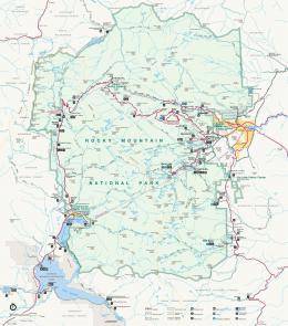


![[pdF] Download Kaufman Field Guide to Insects of ... - Google Sites](https://m.moam.info/img/260x300/pdf-download-kaufman-field-guide-to-insects-of-goo_647852ef097c474b228d116d.jpg)