SLCO4A1. PRDM16. ADRB2. ELOVL3. DIO2. PGC1ACPT1B. 0. 5. 10. 15. Re la tive m. RNA. E xp re ssio n vehicle. Rosi. GQ-16 average ± SEM; *p
Md Shakir Hussain Haider, Wajihul Hasan Khan, Farah Deeba, Sher Ali, Anwar Ahmed, Irshad H Naqvi, Ravins Dohare,. Hytham A. Alsenaidy, Abdulrahman M.
K3,8,10,14R, the four lysines at K3, 8, 10, and 14 of endogenous Htz1 were mutated to ... Data are the average of at least three independent experiments ± SD.
SUPPLEMENTAL FIGURE LEGENDS. Figure S1. Mmp2 and Mmp9 are expressed in discrete populations within hemogenic regions. Related to Figure 1.
progressive allograft vasculopathy; (b) representative photomicrographs of elastin van Gieson stained paraffin sections depicting typical fibroproliferative arterial ...
Chryssa Kanellopoulou, Timothy Gilpatrick, Gokhul Kilaru, Patrick Burr, Cuong K. Nguyen, Aaron Morawski, Michael J. Lenardo, and Stefan A. Muljo ...
Quantibody arrays using Genepix Pro software. .... PBS and serum-free MesenCult® XF media (Stem Cell Technologies) was added, stimulated with 10 ng/ml TNF-α ..... Lee, R.H., Pulin, A.A., Seo, M.J., Kota, D.J., Ylostalo, J., Larson, B.L., ...
... involving the 3'Eκ and MiEκ bait across the Igk locus in pre-B, Immature B, DN and DP cells and their replicates. This matrix is denoted as M3 in the main text.
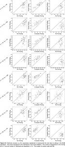
May 29, 2015 - Complete Pooling. No Pooling. Figure S1 Prediction accuracy rı (for populations represented in training set) for trait days to silking in the NAM.
ascidians suggests the presence of multiple paralogues (Wnt5a and Wnt5b) which originated in the. Stolidobranchia ... Despite the fact that the number of Wnt5 genes is the same in species of the Molgula ..... S12.g13523â¡. -. Wnt9 ..... KR, Eppig JT
The samples for this study were obtained from a patient with a primary diffuse gastric cancer tumor and metastases located in each ovary (Krukenberg tumors).
Poignard, Elise Landais, James C. Paulson, Ian A. Wilson, and Andrew B. Ward ... (A) C-terminally truncated (âCT) and full-length (FL) Env constructs ...
lilah-Seyfried, Thomas Scheper, Henning Kempf, Robert Zweigerdt, and .... Cultures were dissociated using Collagenase II (Worthington, Lakewood, USA) and ...
negative inputs of Nkx2.2, Olig2 and Nkx6.1. Related to Figures 5 and 6. Supplemental Tables (available as separate files). Table S1. Genes, their expression ...
Transcriptome alignment (%). 0e+00. 1e+06. 2e+06. 3e+06. 4e+06. 5e+06. GBM. DMSO. GBM. RG7388. Number of reads d. L1210. Number of genes detected.
Expression of murine microglial signature genes in human cells. ..... microscope, equipped with Olympus DP30BW black and white digital .... values in excel.
Sep 2, 2015 - Figure S1 Amino acid sequences of RAE1/An-1 in O. glabeerima and O. sativa. Amino acid ... sequence are shown below the mutation sites.
**The structure is an extruded hexagon and cannot be approximated by a single ... Ïss not reported in paper, but calculated from the estimate Ïss â¼ r2/D where r is the typical length ..... the concentration c of the signal due to a single cell th
MDA-HGSC-1 cells grew as spheroids rather than individual attached cells in the absence of PTX and grew as spheroids with multiple giant cells (black arrows) ...
Images were cropped in Adobe Photoshop 6.0, and then sized and placed in figures using Adobe Illustrator CS2 (Adobe Systems). Live-Cell Imaging. Live cell ...
Erik van der Wal, Pablo Herrero-Hernandez, Raymond Wan, Mike Broeders, Stijn L.M. in. 't Groen, Tom J.M. van Gestel, Wilfred F.J. van IJcken, Tom H. Cheung, ...
19. TAG-11 36.5 M July 12. No. July 28. -. Sept. 21. 55. TAG-12 18.0 M July 11. Yes. Aug. 22. -. Sept. 21. 30. TAG-13 22.0 M July 18. Yes. Aug. 30. -. Sept. 21. 22.
X-CGD mice that were secondary transplant recipients of .... found to be represented significantly higher (pt < 0.05, â) in either the 950-1100 cGy or 300 cGy.
Figure S1. in vivo 30S and 50S ribosome assembly maps adapted from (16). (A) 30S ... Ribosomes of the indicated strains were isolated by sucrose cushion ...
A
5'
3' 16S RNA
S17
S4
S8
S16
S12
21S
S15
S20
S6:S18
S5
S11
S7 S9
S19
S10
S14
S3
S2
S21
B
S13
5'
3' 23S RNA
L24
32S
L20
L21
L22
43S
L23
L17
L3 L13
L34
L29
L19
L32
L5
L2
L14
L6 L33
L28
L30
L10
L18
5S
L11
L9
L16
L7/12 L35
L1
L15
L4
L27
L25 L31
L36
Figure S1. in vivo 30S and 50S ribosome assembly maps adapted from (16). (A) 30S assembly map reflecting order and interdependency of r-protein interaction with the nascent small ribosomal subunit. Early interacting r-proteins are shaded in dark gray, late interacting ones in light gray. Boxed proteins are contained in the 21S precursor of the 30S subunit. S2 is circled in red. (B) 50S assembly map reflecting order and interdependency of r-protein interaction with the nascent large ribosomal subunit. Early interacting r-proteins are shaded in dark gray, late interacting ones in light gray. Proteins contained in the 32S or in the 43S precursor of the large ribosomal subunit are boxed or cycled in black, respectively. L19 is circled in green.
.
S2-mCherry L19-EGFP
*
S2
28 17 10 *
MC rg
MC 41 0 MC 0 r MC g 75 63 48 35
*
L19
Figure S2. Immunoblot analysis of the supernatant after sucrose cushion centrifugation. Ribosomes of the indicated strains were isolated by sucrose cushion centrifugation. The protein content of the supernatant was TCA precipitated and analyzed by SDS-PAGE and subsequent immunoblotting using S2 and L19 specific antisera. Asterisks denote unspecific protein bands.
+IPTG
-IPTG
MC4100 MCΔsQ MCΔlC MCrg MCrgΔsQ MCrgΔlC Figure S3. Viability test of rpsQ and rplC conditional gene knock-out. Serial dilutions of cultures of the indicated strains were spotted on LB agar plates containing IPTG (left panel), or not (right panel). Cells were incubated at 37°C until visible colonies had formed.
A
debries
none
de
br ie
s
70S
2
B
10
20
30S
30
50S
40
50
60
70
23S rRNA 16S rRNA (fraction)
50
60
70
23S rRNA 16S rRNA (fraction)
70
23S rRNA 16S rRNA (fraction)
debries
de
br ie
s
Cam
2
C
10
20
30
40
debries
de br ie
s
Ery
2
10
20
30
40
50
60
Figure S4: A254 detection and fluorescence determination of scrose density fractions Sucrose density gradient (10-25%) centrifugation profiles from (A) control cells with no antibiotic (none), (B) chloramphenicol (Cam) and (C) erythromycin (Ery) treated cells. A254 profiles and fluorescence bar charts were superimposed and sucrose fractions were analyzed for presence of 16S and 23S rRNA by agorse gelelectrophoresis. Gray circles: low-intermediate ammounts of rRNA, black circles: high amounts of rRNA