BR. F2-1 shR. N. A. 0.0. 0.5. 1.0. U. 6. /5. S. E x p re s s io n. (R e la tiv e to. C o n tro l). 5S rRNA Control. U6 snRNA. C on trol. BRF2-1 shRNA. H520. Figure S3 a.
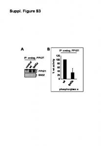
IP: endog. PPH21 controlw t tpd3. â. PPH21. RRD2. 0. 20. 100. 60. 80. 40. 120 tpd3. â w t phosphorylase a. IP: endog. PPH21. % w. t-a c tiv ity. Suppl. Figure S3.
>Soybean aphid c002 homolog c cg crc Trc cGA TCT Arc ccc GrG TAG CAc AGG ATA Gcg ATT ATA 47. 48 AAC AGT ATG GGA CGT TAC CAA TTA TAT GTG ...
No antibiotic. High antibiotic. Interference competition. Exploitation competition. Non cross. â feeding. Cross. â feeding. 0. 15. 30. 0. 15. 30. 0.0. 0.5. 1.0. 0.0. 0.5.
Platypus. Opossum. Cow. Dog. Mouse. Rat. Macaque. Human. E. F. G. D. C. B. A e1 e2 f1 f2 g1 g2 d1 d2 c1 c2 b1 b2 a1 a2. A e2 e1. G g2 g1. F f1 f2. D d1 d2. C.
0. -2 -1. TTT. |. -2 -1. R = 0.32 m = 0.67. R = 0.5 m = 0.51. R = 0.18 m = 0.09. TT. TTT. 2. 2. -2 -1 0 1 2. Exiqon. -2 -1 0 1. Exiqon. -2 -1 0 1. Illumina. **. *. 5. 5. 5. 0.
2 X 10⁻². 2 X 10⁻³. 2 X 10⁻⁴. 2 X 10⁻⁵. 2 X 10⁻⁶. 2 X 10⁻⁷. 2 X 10⁻⁸ time(hr). RF. U. S2 Figure mule deer CWD+ brain homogenate. 2 hr NaPTa incubation. 0. 10.
Figure S3. Neutralization titres against H1N1pdm. The location of each pie chart indicates neutralization tire at baseline (x-axis) and at follow-up (y-axis).
Methylation sensitive enzymes cleave the amplicon at 11 sites: 60°C. BstUI cleaves at CGCG. 5 cuts at bases 43, 81, 93, 95, 105. 37°C. HhaI cleaves at GCGC.
Page 1. 0. 10. 20. 30. 40. 50. 60. 70. HIF004Bay x. HIF004Sha. R e la tive. P rim a ry R o o t le n g th. (%. ) HIF004Bay. HIF004Sha a a b. Figure S3.
Figure S3. 4.%. TEM. HIC PTC HIC. | p=0.002. TIM. РТС. РТС ÐÐС. TCM. 1 E + F T T + + P T T T + E + F = F = | ? 1 : 0 I T = T T T I C 1 C 1 T T T Ft T + T + 1.
Figure S3. (A) rs12150660. (B) rs6258. (C) rs5934505. GOOD. HH. FHS. H. H. H. SHIP. HH. Discovery. ÐÐÐС. HH. InCHIANTI. HH. RS1. KORA. MrOS Sweden.
Mobility of TTDA with and without actD. 0. 10. 20. 30. 40. 50. 0.4. 0.6. 0.8. 1.0. Time (sec.) R e la tiv e flu o rescenc e. Figure S3.
FIGURE S3. Homer la Knockout. Oh. 48 h. 200%. |. UH. NREM Delta %. Days. 20 IL. 09%. 1 2 3 4 5 6 7 8 9 10 11 12 13 14 15. Interval. --WT HE HET O-KO.
Figure S3: Detection of transcripts displaying circadian accumulation profiles in mice fed with normal chow (-Dox). A) Phase map of the 131 hepatic feature sets ...
DQ138152 VIET NAMA/CHICKEN/VIET NAM/TG-023/2004 GALLIFORMES. DQ138153 VIET NAM A/CHICKEN/VIETNAM/LA-024/2004 GALLIFORMES.
Supplementary Figure 3. Conserved domains among class III heme-peroxidases. Amino acid sequence alignment of POX12 with selected members of secreted ...
Shapiro-Wilk P= 0.597. Shapiro-Wilk P=0.685. Acetate.obs. Ethanol.obs. 0.04. 0.05. 0.00. O. O. O. Oo oo. O. -0.05. -0.04. TITI. -1.5 -0.5 0.5 1.5. Shapiro-Wilk P= ...
221. HAALSS. --CDP S. SVNV SEL HHTASPS-S. hTRNâSR1. 223 K A. DK-- N ... A HL. S D QR к ÑÑн. V. T E YD-. QYI R. SY P NEKQS VLPR-. â YQES KA SDI.
C. D. 1. 4. /1. 9. A le x a. -7. 0. 0. 0. 0. 50K. 100K. 150K. 200K. 250K. SSC-A. 85.5. 0. 10 3. 10 4. 10 5. C. D. 3. A. P. C. -H. 7. 0. 50K. 100K. 150K. 200K. 250K.
FIGURE S3 perinuclear region lamellar region. (μ m. 2. ) 10-3. 10-2. 10-1. 100. 10-3. 10-2. 10-1. 100. Time Lag, Ï (s). 10-1. 100. 101.