Am J Physiol Cell Physiol 283: C1133–C1143, 2002. First published June 20, 2002; 10.1152/ajpcell.00104.2002.
Fluid shear- and time-dependent modulation of molecular interactions between PMNs and colon carcinomas SAMEER JADHAV AND KONSTANTINOS KONSTANTOPOULOS Department of Chemical Engineering, The Johns Hopkins University, Baltimore, Maryland 21218 Received 6 March 2002; accepted in final form 4 June 2002
Jadhav, Sameer, and Konstantinos Konstantopoulos. Fluid shear- and time-dependent modulation of molecular interactions between PMNs and colon carcinomas. Am J Physiol Cell Physiol 283: C1133–C1143, 2002. First published June 20, 2002; 10.1152/ajpcell.00104.2002.—This study compares the effects of fluid shear on the kinetics, adhesion efficiency, stability, and molecular requirements of polymorphonuclear leukocyte (PMN) binding to two colon adenocarcinoma cell-lines, the CD54-negative/sLex-bearing LS174T cells and the CD54-expressing/sLex-low HCT-8 cells. The efficiency of PMN-colon carcinoma heteroaggregation decreases with increasing shear, with PMNs binding HCT-8 more efficiently than LS174T cells at low shear (50–200 s⫺1). In the low shear regime, CD11b is sufficient to mediate PMN binding to LS174T cells. In contrast, both CD11a and CD11b contribute to PMN-HCT-8 heteroaggregation, with CD54 on HCT-8 cells acting as a CD11a ligand at early time points. At high shear, only PMN-LS174T heteroaggregation occurs, which is initiated by PMN L-selectin binding to a sialylated, O-linked, protease-sensitive ligand on LS174T cells. PMNLS174T heteroaggregation is primarily dependent on the intercellular contact duration (or shear rate), whereas PMNHCT-8 binding is a function of both the intercellular contact duration and the applied force (or shear stress). Cumulatively, these studies suggest that fluid shear modulates the kinetics and molecular mechanisms of PMN-colon carcinoma cell aggregation.
cancerous cells undergo extensive interactions with various host cells, including polymorphonuclear leukocytes (PMNs), in the circulatory system before they eventually colonize a distant tissue. Several lines of evidence suggest that these adhesive interactions may affect the ability of tumor cells to metastasize. Some studies indicate that PMNs may exert a direct cytotoxic effect on tumor cells (7), and their tumoricidal ability relies on intimate contact between PMNs and tumor cells (6, 19). However, several independent studies have shown that PMNs may enhance tumor metastasis. This has been demonstrated by experiments showing that PMNs facilitate tumor cell extravasation in vitro (35, 38) and promote the arrest and deposition of tumors in the microvasculature of target organs in animal models (3, 11, 35).
Light and electron microscopic studies also reveal close association of PMNs with metastatic cancer cells in vivo (5). Hence, whether PMNs abet hematogenous metastasis or are cytotoxic to tumor cells remains controversial. Nevertheless, PMN-tumor cell binding appears to be critical to both these processes, and the molecular mechanisms mediating these cell-cell interactions require further investigation. Published data indicate that PMNs can bind certain melanoma, neuroblastoma, and colon adenocarcinoma cells through the CD18 (2) integrin receptor on PMN surface in the absence of any selectin contribution under static conditions (16, 22, 26). Alternatively, PMNs have been reported to interact with certain colon carcinomas via PMN CD62L (L-selectin) in a CD18independent manner at 4°C, a temperature that renders integrins inactive (20). However, static binding assays fail to replicate the hydrodynamic shear environment of the vasculature in which PMN-tumor cell binding typically occurs and, thus, may not be sufficient to determine the precise molecular requirements of this adhesion process. It is now well established that fluid shear affects the binding kinetics and receptor specificity of PMN homotypic aggregation (37) as well as PMN heterotypic interactions with platelets (15) and endothelial cells (13, 34). Furthermore, we recently showed that CD11b (Mac-1) is sufficient to mediate binding of chemotactically stimulated PMNs to CD54 (ICAM-1)-negative/sLex-bearing LS174T colon carcinoma cells at low shear (100 s⫺1) (12). In marked contrast, L-selectin, CD11a (lymphocyte function-associated antigen LFA-1), and CD11b are all requisite for optimal PMN-LS174T binding under high shear conditions (800 s⫺1) (12). Along these lines, the CD54-expressing/sLex-low HCT-8 colon carcinoma cells fail to aggregate with PMNs at 800 s⫺1. However, a detailed quantitative analysis of PMN binding to LS174T vs. HCT-8 colon carcinoma cells as well as the relative contributions of L-selectin, CD11a, CD11b, and their respective ligands in mediating these heterotypic adhesive interactions as a function of fluid shear and time have yet to be investigated. In the present study, we utilized a rheometric-flow cytometric methodology to compare the aggregation kinetics, stability, and molecular requirements of PMN
Address for reprint requests and other correspondence: K. Konstantopoulos, Dept. of Chemical Engineering, The Johns Hopkins Univ., 3400 N. Charles St., Baltimore, MD 21218-2694 (E-mail:
[email protected]).
The costs of publication of this article were defrayed in part by the payment of page charges. The article must therefore be hereby marked ‘‘advertisement’’ in accordance with 18 U.S.C. Section 1734 solely to indicate this fact.
CD11a/CD18; CD11b/CD18; L-selectin; polymorphonuclear leukocytes; LS174T cells; HCT-8 cells
DURING BLOOD-BORNE METASTASIS
http://www.ajpcell.org
0363-6143/02 $5.00 Copyright © 2002 the American Physiological Society
C1133
C1134
PMN-COLON CARCINOMA AGGREGATION UNDER SHEAR
binding to LS174T cells with that to HCT-8 cells under defined shear conditions in the presence of the chemotactic agent N-formyl-methionyl-leucyl-phenylalanine (fMLP). To quantify cell-cell interactions independent of physical parameters such as shear rate, cell size, and initial cell concentration, we calculated the efficiency of PMN homotypic and PMN-colon carcinoma heterotypic aggregation by using a mathematical model based on Smoluchowski’s two-body collision theory (33, 36, 37). Moreover, the contributions of intercellular contact duration (dictated by shear rate) and tensile forces (exerted by shear stress) on receptor-ligand bonds mediating PMN-colon carcinoma heteroaggregation were differentiated by varying the viscosity of the cell suspension (10, 29, 37). MATERIALS AND METHODS
Reagents. The IgG murine monoclonal antibodies (MAbs) 6.7(blocking anti-CD18), HI111 (blocking anti-CD11a), and ICRF44(44) (blocking anti-CD11b) were purchased from BD Pharmingen (San Diego, CA). The blocking F(ab⬘)2 antiCD54 MAb MHCD54F was from Caltag (Burlingame, CA). Red-out and PMN isolation media were obtained from Robbins Scientific (Sunnyvale, CA). Citrate-phosphate-dextrose (CPD) solution, fMLP, chymotrypsin, trypsin, benzyl-2-acetamido-2-deoxy-␣-D-galactopyranoside (Bzl GalNAc), tunicamycin, dextran sulfate, and fucoidan were from Sigma-Aldrich (St. Louis, MO). Heparin was from Elkins-Sinn (Cherry Hill, NJ). Dulbecco’s phosphate-buffered saline (D-PBS) and trypsin/EDTA were acquired from Life Technologies (Gaithersburg, MD). Phosphatidylinositol-specific phospholipase C (PI-PLC) was purchased from Glyko (Novato, CA), and Vibrio cholerae neuraminidase was from Roche Molecular Biochemicals (Indianopolis, IN). d,l-Threo-1-phenyl-2-amino3morpholino-1-propanol hydrochloride (threo-PPPP) was procured from Matreya (State College, PA). CellTracker orange dye CMTMR [5(6)-(4-chlormethylbenzoylamino)tetramethylrhodamine] and green dye CFDA-SE [5(6)-carboxyfluorescein diacetate-succinimidyl ester] were purchased from Molecular Probes (Eugene, OR). CMTMR and CFDA-SE are excited at 488 nm by the argon laser of a flow cytometer, and their emission spectra are well separated (570 nm for CMTMR and 515 nm for CFDA-SE), thereby allowing simultaneous twocolor immunofluorescence measurements. Colon carcinoma cell culture and staining. The LS174T and HCT-8 human colon adenocarcinoma cell lines were obtained from American Type Culture Collection (Manassas,VA) and cultured in the recommended media. Before each experiment, LS174T or HCT-8 cells were detached from the tissue culture substrate by mild trypsinization (0.25% trypsin/EDTA for 2 min at 37°C). The colon carcinoma cells were resuspended at a concentration of 1 ⫻ 107 cells/ml in the recommended media and incubated for 2 h at 37°C to regenerate surface glycoproteins (12, 20, 21). During this period, the cells were incubated with 1 M CMTMR (orange dye) at 37°C for 1 h. The cells were washed once to remove excessive dye and resuspended at a concentration of 2.5 ⫻ 106 cells/ml in D-PBS containing Ca2⫹/Mg2⫹/0.1% BSA (Sigma-Aldrich) and maintained at 4°C for no longer than 3 h before being used in binding assays or flow cytometry. PMN isolation and labeling. Human blood was drawn by venipuncture from healthy volunteers into a sterile syringe containing CPD solution (2.8 ml CPD/20 ml blood). A red blood cell agglutinating agent (Red-out; 1/100 volume) was AJP-Cell Physiol • VOL
added to the blood to minimize erythrocyte contamination in PMNs (12, 14). The blood was gently mixed, layered over the PMN isolation medium, and centrifuged to allow for density separation of cell populations. The PMN layer was carefully aspirated and washed once, and the PMNs were resuspended in Ca2⫹/Mg2⫹-free D-PBS at a concentration of 1 ⫻ 107 cells/ml. The PMNs were then incubated with 0.1 M CFDA-SE at 4°C for 1 h, washed once to remove excess dye, resuspended in Ca2⫹/Mg2⫹-free D-PBS containing 0.1% BSA at a concentration of 0.5 ⫻ 107 cells/ml, and maintained at 4°C for no longer than 3 h before being used in aggregation assays. L-selectin was preserved during PMN isolation and staining with CFDA-SE as assessed by flow cytometry (12). PMN-colon carcinoma cell aggregation assay. CFDA-SElabeled PMNs and CMTMR-stained colon carcinoma cells were mixed at final cell concentrations of 1 ⫻ 106 and 2 ⫻ 106 cells/ml, respectively, and allowed to equilibrate at 37°C for 2 min. This mixed suspension (500 l) was then placed on the plate of a cone-and-plate rheometer (RS150, 60-mm-diameter plate, 0.5°Cone angle; Haake, Paramus, NJ) and stimulated with 1 M fMLP 1 s before initiation of shear at 37°C. The suspension was subjected to well-defined hydrodynamic conditions with shear rates ranging from 50 to 1,200 s⫺1 for durations of 10, 30, 60, 120, 180, and 300 s. Upon termination of shear, 100-l aliquots were immediately fixed with 1% formaldehyde (final concentration) at room temperature (RT) and subsequently analyzed in a FACSCalibur flow cytometer (Becton Dickinson, San Jose, CA), as previously described (12). The fixative was kept in the specimens during the flow cytometric analysis to preserve aggregate integrity (12). In selected experiments, 6% Ficoll was used to increase the viscosity of the cell suspension (10, 37). Cell treatment with MAbs and enzymes. For some blocking experiments, PMNs (1 ⫻ 107 cells/ml) were pretreated for 10 min at 37°C with anti-CD11a and/or anti-CD11b MAbs (20 g/ml), which were kept present during the aggregation assays. In some studies, the HCT-8 cells were incubated with F(ab⬘)2 anti-CD54 MAb (10 g/ml) for 10 min at 37°C before being mixed with PMNs in the cone-and-plate rheometer. For selected inhibition studies, PMNs were incubated with chymotrypsin (1 U/ml) for 20 min at RT to cleave L-selectin [and P-selectin-glycoprotein ligand-1 (PSGL-1)] from the PMN surface and washed once before being used in aggregation assays (12). For others, heparin (100 U/ml) (25), dextran sulfate (100 g/ml) (32), or fucoidan (10 g/ml) (8) were added to the suspension just before the application of shear. For experiments with biosynthetic inhibitors, LS174T cells were cultured for 48 h in the presence of either Bzl GalNAc (2 mM) to inhibit O-linked glycosylation or tunicamycin (200 ng/ml) to inhibit N-linked glycosylation of surface glycoproteins before being used in aggregation assays (30). In other studies, LS174T cells were cultured with 5 M threo-PPPP for 96 h, which blocks the transfer of UDP-glucose to ceramide, thereby blocking the biosynthesis of glycosphingolipids having a glucosylceramide core (1). Colon carcinoma cells treated with the aforementioned biosynthetic inhibitors were washed twice before being used in aggregation assays (22). In selected experiments, LS174T cells were incubated at 37°C for 30 min with either 20 g/ml trypsin to cleave cell surface glycoproteins or 0.1 U/ml Vibrio cholerae neuraminidase to cleave terminal cell surface sialic acid residues, or for 1 h with 1 U/ml PI-PLC, which cleaves glycosyl phosphatidylinositol (GPI)-linked surface glycoproteins (21), and washed once (twice in the case of trypsin) before being mixed with PMNs. Cell viability was consistently ⬎97% after treatment with each of the aforementioned enzymes and inhibitors, as detected by the trypan blue exclusion assay.
283 • OCTOBER 2002 •
www.ajpcell.org
C1135
PMN-COLON CARCINOMA AGGREGATION UNDER SHEAR
Quantitation of receptor expression. For indirect immunofluorescence, colon carcinoma cells were incubated with saturating concentrations of primary MAbs directed against either sLex or CD54 for 30 min at 4°C and then washed once with D-PBS/0.1%BSA. After an additional 30-min incubation with 15 g/ml FITC-labeled IgM or phycoerythrin-labeled IgG (Vector Laboratories, Burlingame, CA), the specimens were washed again, fixed with 1% formaldahyde, and analyzed by flow cytometry (21). Appropriate isotype-matched MAbs were also included for background fluorescence determination. Flow cytometric analysis of PMN-colon carcinoma aggregation. A dual-color flow cytometric methodology (FACSCalibur flow cytometer) was employed to analyze the sheared samples for size distribution and cellular composition of formed aggregates. The PMN and colon carcinoma cell populations were identified on the basis of their forward-scatter, side-scatter, and fluorescence profiles. Single PMN (P) and single tumor cell (T) populations were identified on the green (FL1) vs. orange (FL2) fluorescence dot plots, and the aggregates were resolved as integral multiples of the mean fluorescence values of single P and T populations (12). The homotypic PMN aggregates were resolved into doublets (P2) and higher order aggregates (P3⫹), whereas heterotypic aggregates consisted of a single colon carcinoma cell adhering to one (TP), two (TP2), and three or more (TP3⫹) PMNs. The aggregation was quantified as the fraction of total PMN in homotypic or heterotypic aggregates as described previously (12). %PMNs in homotypic aggregates ⫽ 共2P2 ⫹ 3P3 ⫹ 兲 ⫼ 共P ⫹ 2P2 ⫹ 3P3 ⫹ ⫹ TP ⫹ 2TP2 ⫹ 3TP3 ⫹ 兲 %PMNs in heterotypic aggregates ⫽ 共TP ⫹ 2TP2 ⫹ 3TP3 ⫹ 兲 ⫼ 共P ⫹ 2P2 ⫹ 3P3 ⫹ ⫹ TP ⫹ 2TP2 ⫹ 3TP3 ⫹ 兲 Aggregates with more than three PMN were ⬍10% of the total neutrophil population and resulted in ⬍3% error in estimation of percent aggregation. Estimation of PMN homotypic and PMN-colon carcinoma cell heterotypic adhesion efficiencies. PMN homotypic and PMN-colon carcinoma heterotypic aggregation observed in the cone-and-plate rheometer result from intercellular collisions induced by subjecting cell suspensions to fluid shear. The aggregation process may be expressed as a system of chemical reactions, where single PMN and colon carcinoma cells represent two monomer species and (k,l) denotes a general aggregate comprised of k PMN and l colon carcinoma cells. Each interaction between (k,l) and another aggregate (i,j) represents an elementary reaction step, and K[(i,j),(k,l)] is the aggegation rate coefficient for this reaction. The twospecies aggregation kinetics may then be expressed in the form of a population balance equation (PBE) (17) k
aggregate (k,l), resulting in the formation of a larger aggregate. kmax and lmax denote the maximum number of PMNs and colon carcinoma cells, respectively, that can be found in an aggregate. For our system, lmax ⫽ 1 because colon carcinoma cells do not aggregate homotypically under the experimental conditions of this study, and kmax ⫽ 3 because aggregates containing more than three PMNs are rare events. The rate of aggregate formation depends on the intercellular collision frequency as well as the adhesion efficiency of these collisions. The collision frequency per unit volume f[(i, j),(k,l)] for particles of radii r(i, j) and r(k, l) with concentrations C(i, j) and C(k,l), respectively, at a shear rate of G (s⫺1) is given by (33) f 关共i, j兲,共k,l兲兴 ⫽ 4 ⁄ 3 G关r共i, j兲 ⫹ r共k, l兲兴 3 C共i, j兲C共k, l兲 The radius of PMNs was taken to be 3.75 m (36). The radii of LS174T and HCT-8 cells were measured by light microscopy and found to be 6.0 and 6.65 m, respectively. The adhesion efficiency is defined as the fraction of collisions resulting in the formation of stable aggregates and is given by E 关共i, j兲,共k,l兲兴 ⫽
l max kmax
兺 兺 关C共k, l兲
exp
F⫽
l
兺兺
⫺ C共k, l兲cal兴2
l⫽0 k⫽0 lmax kmax
兺 兺 关C共k, l兲
2 exp
兴
l⫽0 k⫽0
kmax lmax
兺 兺 关共1 ⫹ ␦
f关共i, j兲,共k,l兲兴
If j ⫽ l ⫽ 0, then E[(i, j),(k,l)] is the homotypic adhesion efficiency for PMNs, EPP. If i ⫽ k ⫽ 0, then E[(i, j),(k,l)] is the homotypic adhesion efficiency for colon carcinoma cells, ETT (ETT ⫽ 0, because tumor cells do not aggregate homotypically in this study); otherwise, E[(i, j),(k,l)] is the heterotypic adhesion efficiency, EPT. Here, we assume that heterotypic aggregation results from the adhesion of PMN or PMN homotypic aggregates to colon carcinoma cells and not to PMN already bound to colon carcinoma cells. This assumption is a good approximation when the concentration of TP1 is much larger than that of heterotypic aggregates containing more than one PMN (TP2 and TP3⫹), as observed at early time points in this study. Specifically, the fraction of colon carcinoma cells with more than one adherent PMNs was ⬍3% after 30 s of shear. The PBE represents a set of ordinary differential equations with two unknown parameters, EPP and EPT, and is integrated by using the fourth-order Runge-Kutta-Gill method (9). The PMN homotypic and PMN-colon carcinoma cell heterotypic adhesion efficiencies (EPP and EPT) are obtained by minimizing the objective function given below by using the Nelder and Mead simplex method (2). C(k,l)exp and C(k,l)cal are the experimentally observed concentrations and those calculated by the integration of the PBE, respectively.
dC共k, l兲 关0.5共1 ⫹ ␦i,k ⫺ i␦j,l ⫺ j兲K关共i, j兲,共k ⫺ i,l ⫺ j兲兴 ⫽ dt i⫽0 j⫽0 ⫻ C共i, j兲C共k ⫺ i, l ⫺ j兲兴 ⫺
K 关共i, j兲,共k,l兲兴 C共i, j兲C共k, l兲
␦
兲
k,i ⫺ k l,j ⫺ l
i⫽k j⫽l
⫻ K关共k,l兲,共i ⫺ k, j ⫺ l兲兴C共k, l兲C共i ⫺ k, j ⫺ l兲兴 where C(k,l) is the concentration of the aggregate and ␦i, j is the Kronecker delta function. The first term on the righthand side of the PBE represents the formation of the aggregate (k,l) by the combination of two smaller aggregates, whereas the second term represents the consumption of the AJP-Cell Physiol • VOL
For pure PMN suspensions, a single-species PBE was numerically integrated by using the Runge-Kutta-Gill method, and the homotypic adhesion efficiency was estimated by fitting the experimental data using the Golden section search method (2). Statistics. Data are expressed as means ⫾ SE. Statistical significance of differences between means was determined by one-way ANOVA. Posttests were performed by using the Tukey method. Values of P ⬍ 0.05 were selected to be statistically significant.
283 • OCTOBER 2002 •
www.ajpcell.org
C1136
PMN-COLON CARCINOMA AGGREGATION UNDER SHEAR
RESULTS
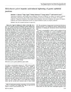
Comparative efficiency of heteroaggregation of LS174T and HCT-8 colon carcinoma cells with fMLP-stimulated PMNs under hydrodynamic shear. At low shear rates (50–200 s⫺1), PMN-LS174T heteroaggregation increased with time until 120 s, when as many as 60% PMNs were in heterotypic aggregates (Fig. 1A). Partial disaggregation of these heterotypic aggregates was observed at longer periods (180–300 s) of shear exposure for all shear rates examined. Similarly, HCT-8 cells aggregated extensively with PMNs under these low shear conditions, with maximal values ⱖ75% after 120 s of shear exposure (Fig. 1B). Partial disaggregation of heterotypic aggregates followed at later times for shear rates of 100–200 s⫺1. In marked contrast to LS174T cells, PMN-HCT-8 cell heteroaggregates did not undergo disaggregation at 50 s⫺1 (Figs. 1, A and B). In the low-shear regime (50–200 s⫺1), PMN homotypic aggregation in the presence of colon carcinoma cells was ⬃15% at early times (10–30 s) and either remained unchanged (PMN-LS174T cell suspensions, Fig. 1C) or showed a slight decrease (PMN-HCT-8 cell suspensions, Fig. 1D) at longer shear exposure times. The low percentage of PMNs in homotypic aggregates is attributed to the extensive recruitment of PMNs by colon carcinoma cells at low shear. PMN homotypic aggregation was observed to increase with increasing shear in PMN-HCT-8 cell sus-
pensions, reaching maximal values of ⬃40 and 65% at 400 and 800 s⫺1, respectively, after 60 s of shear (Fig. 1D). The percent PMNs in homotypic aggregates was observed to decrease subsequently on exposure to longer duration (ⱖ120 s) of shear. The decrease in the extent of PMN homotypic aggregation at 800 s⫺1 is entirely a disaggregation process, because the incorporation of PMNs into PMN-HCT-8 cell heterotypic aggregates at this level of shear is negligible (Fig. 1D). However, at 400 s⫺1, the heterotypic aggregation was observed to increase with time to ⬃40% after 300 s of shearing (Fig. 1D) and could be partially responsible for the low PMN homotypic aggregation observed at later time points. This is also corroborated by previous observations showing the absence of any appreciable disaggregation of PMN homotypic aggregates when pure PMN suspensions were subjected to a shear rate of 400 s⫺1 (37). PMN homotypic aggregation was also found to increase with increasing shear in PMN-LS174T cell suspensions with ⬃35–40% PMNs in homotypic aggregates at high shear rates (800–1,200 s⫺1, Fig. 1C). In distinct contrast, PMN-LS174T heteroaggregation decreased with increasing shear, and only ⬃25% PMNs were recruited by LS174T cells in the high-shear regime (Fig. 1A). Partial disaggregation of the PMNLS174T aggregates was observed at later times (120– 300 s) for all shear rates.
Fig. 1. Kinetics of polymorphonuclear leukocyte (PMN)-colon carcinoma cell and PMN-PMN aggregation. CMTMR (orange dye)-labeled colon carcinoma cells (LS174T or HCT-8, 2 ⫻ 106 cells/ ml) were incubated with CFDA-SE (green dye)-labeled PMNs (1 ⫻ 106 cells/ml) at 37°C for 2 min, stimulated with 1 M fMLP, and sheared at prescribed shear rates and times. Sheared specimens were immediately fixed with 1% formaldehyde and analyzed with a FACSCalibur flow cytometer. A and C: percentage of total PMNs in heterotypic and homotypic aggregates in the presence of LS174T cells, respectively. B and D: percentage of total PMNs in heterotypic and homotypic aggregates in the presence of HCT-8 cells, respectively. Values are means ⫾ SE (n ⫽ 3–13).
AJP-Cell Physiol • VOL
283 • OCTOBER 2002 •
www.ajpcell.org
PMN-COLON CARCINOMA AGGREGATION UNDER SHEAR
To further investigate how PMN homotypic aggregation is affected by the presence of colon carcinoma cells, we compared homotypic aggregation in pure PMN suspensions with that observed in PMN-colon carcinoma cell suspensions. Because disaggregation is insignificant at early times (37), the extents of aggregation occurring over the first 30 s were determined. Moreover, to quantify cell-cell interactions independent of physical parameters such as aggregate size, cell concentration, and intercellular collision frequency, we estimated PMN homotypic and PMN-colon carcinoma heterotypic adhesion efficiencies by fitting the aggregation data over the first 30 s to a mathematical model based on Smoluchowski’s two-body collision theory. After 30 s of shear, the extent of PMN homotypic aggregation in the presence LS174T cells was significantly less than that observed when PMNs were sheared alone over the entire range of shear rates examined (Fig. 2A). However, PMN homotypic aggregation in the presence of HCT-8 cells was significantly lower than that observed for pure PMN suspensions only at low shear rates (50–200 s⫺1), whereas no appreciable change was detected in the high-shear regime (ⱖ400 s⫺1). Nevertheless, our data indicate that PMN homotypic adhesion efficiency in the presence of either LS174T or HCT-8 cells is similar to that observed for pure PMN suspensions over the entire range of shear
C1137
rates examined (Fig. 2B). Cumulatively, these data suggest that PMN homotypic aggregation is reduced in the presence of tumor cells because of PMN recruitment by tumor cells and not because of the release of any inhibitory enzymes by tumor cells. In all three cases, the homotypic adhesion efficiency decreased sharply from 0.12 to 0.04 with increasing shear rate between 50 and 200 s⫺1, after which it was almost constant till 1,600 s⫺1 (Fig. 2B). At low shear rates, initial PMN recruitment by HCT-8 cells was significantly higher than that by LS174T cells (Fig. 2C). Consequently, the PMN-HCT-8 cell heterotypic adhesion efficiency was higher than the PMN-LS174T cell adhesion efficiency (0.09 for HCT-8 and 0.06 for LS174T at 50 s⫺1). However, in the high-shear regime, LS174T cells displayed significant PMN recruitment, whereas PMN-HCT-8 cell heteroaggregation was close to background levels. Along these lines, at shear rates ⱖ400 s⫺1, heterotypic adhesion efficiency for HCT-8 cells fell below that for LS174T cells. At 800 s⫺1, the heterotypic adhesion efficiencies for LS174T and HCT-8 cells were 0.0033 and 0.0008, respectively. Relative contribution of CD11a, CD11b, and CD54 to PMN-colon carcinoma cell adhesion. Function-blocking MAbs were used against PMN CD11a and CD11b to investigate their relative roles in PMN-LS174T heteroaggregation in the shear range of 100–800 s⫺1. Block-
Fig. 2. PMN homotypic and PMN-colon carcinoma heterotypic adhesion efficiencies as a function of shear rate. The percentages of PMNs in homotypic (A) and heterotypic (C) aggregates after 30 s of shear exposure of either pure PMN or PMN-colon carcinoma cell suspensions were determined as a function of hydrodynamic shear by a dual-color flow cytometric methodology. The adhesion efficiencies for homotypic (B) and heterotypic (D) aggregation were calculated by using a mathematical model based on Smoluchowski’s theory. The experimental protocol was similar to that mentioned in the legend of Fig. 1. Values are means ⫾ SE (n ⫽ 3–6).
AJP-Cell Physiol • VOL
283 • OCTOBER 2002 •
www.ajpcell.org
C1138
PMN-COLON CARCINOMA AGGREGATION UNDER SHEAR
Fig. 3. The relative contributions of CD11a, CD11b, CD54, and L-selectin to PMN-colon carcinoma cell heteroaggregation. CFDA-SE-labeled PMNs (1 ⫻ 106 cells/ml) were incubated with anti-CD11a (20 g/ml) and/or antiCD11b (20 g/ml) function-blocking MAbs at 37°C for 10 min before being mixed with CMTMR-labeled colon carcinoma cells (2 ⫻ 106 cells/ml) at 37°C for 2 min. The cell mixture was then stimulated with 1 M fMLP 1 s before application of shear. Sheared samples were immediately fixed with 1% formaldehyde and analyzed by flow cytometry. A: the extent of PMN-LS174T heteroaggregation after shearing for 30 s at prescribed shear rates. B: the extent of PMN-HCT-8 cell heteroaggregation after 30 s of shearing at 100 s⫺1. C: the extent of PMN-HCT-8 cell heterotypic aggregation after 300 s of shear exposure at 100 or 400 s⫺1. In selected experiments, PMNs were treated with 1 U/106 cells chymotrypsin for 20 min at room temperature before being mixed with LS174T cells and subjected to shear. In some experiments, HCT-8 cells were incubated with an anti-CD54 function-blocking MAb at 37°C for 10 min before being mixed with PMNs. Values are means ⫾ SE (n ⫽ 4–10). *P ⬍ 0.05 with respect to control (untreated) samples. #P ⬍ 0.05 with respect to CD54-treated samples.
ing CD11b function abrogated PMN binding to LS174T cells under all shearing conditions (Fig. 3A). In distinct contrast, use of an anti-CD11a MAb did not appreciably affect PMN-LS174T heteroaggregation in the lowshear regime (100–200 s⫺1). However, PMN binding to LS174T cells was reduced by 40 and ⬎60% in the presence of anti-CD11a MAb relative to control values at shear rates of 400 and 800 s⫺1, respectively. Together, our data illustrate that CD11b is requisite for PMN-LS174T binding over the entire range of shear rates examined, whereas CD11a contribution becomes evident only in the high-shear regime. The relative contributions of CD11a and CD11b in mediating PMN binding to HCT-8 cells were assessed at 100 s⫺1 after 30 s of shear exposure. In distinct contrast to PMN-LS174T heteroaggregation, PMN attachment to HCT-8 cells was partially inhibited by either anti-CD11a (30%) or anti-CD11b (50%) (Fig. 3B). However, simultaneous blockade of CD11a and CD11b completely abrogated PMN-HCT-8 cell binding after 30 s of shear at 100 s⫺1 (Fig. 3B). Subsequent studies aimed to identify the counterreceptor(s) for CD11a and CD11b. Our flow cytometric data indicate that CD54, a potential ligand to CD11a and CD11b, is expressed on HCT-8 cells (Table 1). To assess its potential involvement in this process, HCT-8 cells were treated with anti-CD54 F(ab⬘)2 MAb before the cells were sheared with PMNs. This treatment significantly AJP-Cell Physiol • VOL
reduced PMN binding to HCT-8 cells by ⬃35% after 30 s of shear at 100 s⫺1 (Fig. 3B). The extent of heteroaggregation of anti-CD54-treated HCT-8 cells with anti-CD11a-treated PMNs was similar to the binding of anti-CD54-treated HCT-8 cells to untreated PMNs (Fig. 3B). However, simultaneous treatment of HCT-8 cells with anti-CD54 and PMNs with antiCD11b was more effective than anti-CD11b treatment alone, although it did not completely abrogate adhesion (Fig. 3B). Cumulatively, these results indicate that, under the above conditions (100 s⫺1, 30 s), CD54 preferentially binds CD11a, whereas CD11b appears to interact with a yet unidentified counterreceptor on the HCT-8 cell surface. Table 1. Flow cytometric analysis of adhesion receptor expression on LS174T and HCT-8 colon adenocarcinoma cells Geometric Mean Fluorescence Receptor
Control IgG (IgM) sLex CD54
LS174T cells
5.4 ⫾ 0.8 (10.6 ⫾ 2.1) (224.1 ⫾ 64.9) 6.0 ⫾ 0.9
HCT-8 cells
9.5 ⫾ 2.3 (4.4 ⫾ 0.1)* (10.2 ⫾ 0.1)* 48.1 ⫾ 0.8*
Values are geometric mean fluorescence intensities ⫾ SE of 3–5 experiments, with different batches of cells used each time. * n ⫽ 2; mean ⫾ range. Values in parentheses represent those experiments carried out with an anti-mouse secondary antibody.
283 • OCTOBER 2002 •
www.ajpcell.org
PMN-COLON CARCINOMA AGGREGATION UNDER SHEAR
We next examined the contribution of CD18 integrins in mediating PMN-HCT-8 cell heteroaggregation at longer shear exposure (300 s) at both 100 and 400 s⫺1. Use of either anti-CD11a or anti-CD11b partially inhibited PMN-HCT-8 cell heteroaggregation by 40 and 60%, respectively, at 100 s⫺1 (Fig. 3C). It is noteworthy that under high-shear conditions (400 s⫺1, 300 s), PMN binding to HCT-8 cells was completely abrogated by anti-CD11b, whereas it was also dramatically inhibited by anti-CD11a. Interestingly, use of an anti-CD54 MAb failed to significantly reduce PMN adhesion to HCT-8 cells on longer shear exposure at both 100 and 400 s⫺1. Together, our data suggest that at longer exposure to hydrodynamic shear, CD54 does not play a significant role in PMN-HCT-8 adhesion, suggesting that both CD11a and CD11b bind other yet unidentified ligands on the HCT-8 cells. CD54 is minimally expressed on LS174T cells (Table 1) and, therefore, does not appear to be involved in mediating PMNLS174T cell adhesion (12). The ligand on LS174T binding PMN CD18 integrins has yet to be characterized. Role of L-selectin in PMN-LS174T adhesion and characterization of L-selectin ligand on colon carcinoma cells. We next wanted to systematically investigate the relative contribution of L-selectin to the PMNLS174T adhesion process over a wide range of shear rates (100–800 s⫺1) and to characterize the L-selectin ligand on LS174T cells. Chymotrypsin treatment (1 U/ml) has been shown to cleave L-selectin from PMN surface (12). In the low-shear regime (100–200 s⫺1; Fig. 3A), the extent of LS174T cell binding to chymotrypsin-treated PMNs was similar to that to untreated PMNs. In marked contrast, chymotrypsin treatment reduced PMN-LS174T heteroaggregation by 50% at 400 s⫺1 and by ⱖ85% at 800 s⫺1. These data suggest that the L-selectin contribution in mediating adhesion becomes progressively larger with increasing shear. Fucoidan, which has been shown to block PMN binding to L-selectin (8), abrogated PMN-LS174T heteroaggregation at 800 s⫺1 (Fig. 4), providing further evidence for L-selectin involvement in this process under highshear conditions. We have recently shown that the L-selectin ligand on LS174T cells is a sialylated molecule, as evidenced by abrogation of PMN binding to neuraminidase-treated LS174T cells (12). We wanted to further characterize the L-selectin ligand and determine whether it is a glycoprotein or a glycosphingolipid. Enzymatic treatment of LS174T cells with trypsin abrogated PMNLS174T cell aggregation at high (Fig. 4) but not low shear (data not shown). In distinct contrast, LS174T cells cultured in the presence of an inhibitor of ceramide:UDP glucose transferase, threo-PPPP (1), retained their ability to aggregate effectively with PMNs under high shear (Fig. 4). Cumulatively, these data suggest that the L-selectin ligand on LS174T colon carcinoma cells is a protease-sensitive glycoprotein rather than a glycosphingolipid. We next investigated whether the L-selectin ligand is a GPI-linked molecule by treating LS174T cells with PI-PLC, an enzyme that AJP-Cell Physiol • VOL
C1139
Fig. 4. Characterization of L-selectin ligand on LS174T cells. CFDASE-labeled PMNs (1 ⫻ 106 cells/ml) were incubated with CMTMRlabeled LS174T cells (2 ⫻ 106 cells/ml) for 2 min at 37°C, stimulated with 1 M fMLP, and sheared at 800 s⫺1 for 30 s. Sheared samples were immediately fixed with 1% formaldehyde and analyzed with a FACSCalibur flow cytometer. In selected experiments, LS174T cells were cultured in the presence of either benzyl-2-acetamido-2-deoxy␣-D-galactopyranoside (Bzl GalNAc; 2 mM) or tunicamycin (200 ng/ml) for 48 h or with d,l-threo-1-phenyl-2-amino3-morpholino-1propanol hydrochloride (threo-PPPP; 5 M) for 96 h. Some experiments were conducted with LS174T cells treated with either trypsin (20 g/ml for 30 min) or phosphatidylinositol-specific phospholipase C (PI-PLC; 1 U/ml for 1 h). Others were conducted in the presence of dextran sulfate (100 g/ml), heparin (100 U/ml), or fucoidan (10 g/ml). Values are means ⫾ SE (n ⫽ 3–5). *P ⬍ 0.05 with respect to control (untreated) samples.
cleaves GPI-anchored glycoproteins (21). However, this treatment failed to affect PMN-LS174T heteroaggregation at 800 s⫺1 (Fig. 4). Further studies aimed at testing whether critical L-selectin binding determinants on LS174T colon carcinoma cells are presented on O-linked and/or N-linked glycans. To this end, we cultured LS174T cells in the presence of either Bzl GalNAc or tunicamycin, which are known to inhibit O- and N-linked glycosylation, respectively (30). PMN binding to LS174T treated with these inhibitors of glycosylation was not significantly different from that to untreated LS174T cells at low shear rates (data not shown). However, PMN-LS174T heteroaggregation was drastically inhibited by Bzl GalNAc, but not by tunicamycin, at 800 s⫺1 (Fig. 4). Together, these data illustrate that the L-selectin ligand is an O-linked, sialylated, protease-sensitive structure and does not require N-glycans for binding. Sulfated polysaccharides such as heparin and dextran sulfate have been shown to inhibit L-selectin binding to sLex (25). Our data indicate that both heparin and dextran sulfate significantly inhibited PMN binding to LS174T cells at 800 s⫺1 (Fig. 4), but not in the low-shear regime (data not shown), presumably by
283 • OCTOBER 2002 •
www.ajpcell.org
C1140
PMN-COLON CARCINOMA AGGREGATION UNDER SHEAR
interfering with L-selectin-sLex binding. The inhibitory effects of heparin and dextran sulfate may be suggestive of the potential presence of sulfated groups on the L-selectin ligand (18). On the other hand, HCT-8 cells express near background levels of sLex (Table 1). The lack of PMN recruitment by HCT-8 cells at high shear reveals the absence of functional PMN L-selectin ligands on these colon carcinoma cells. Effect of shear stress on PMN-colon carcinoma cell aggregate formation. Exposure of cell suspensions to increasing levels of shear rate increases the frequency of cell-cell collisions but decreases the intercellular contact duration. Concomitantly, there is an increase in the magnitude of tensile forces acting on the adhesive contact region to dissociate intercellular bonds. To differentiate the contributions of contact duration and tensile forces on cell aggregation, we used 6% Ficoll to double the viscosity of the suspending medium, thereby increasing the shear stress at a constant shear rate or contact time (10, 29, 37). PMN homotypic aggregation was unaffected by the increase in viscosity in the presence of LS174T (Fig. 5A) as well as HCT-8 cells (data not shown). Similarly, increasing the viscosity did not appreciably affect PMN-LS174T binding over the entire range of shear rates examined (Fig. 5B), suggesting that these interactions are dependent on contact duration and that the aggregates are sufficiently stable to resist the increased shear forces. How-
ever, in the case of HCT-8 cells, heterotypic aggregation at 30 s remained ⬃45% at low shear rates (50–200 s⫺1) for 0.8-cP suspensions, whereas heteroaggregation decreased sharply for 1.6-cP suspensions from ⬃55% at 50 s⫺1 to 22% at 200 s⫺1 (Fig. 5C). The significant reduction in PMN-HCT-8 heterotypic adhesion observed at 1.6 cP compared with that at 0.8 cP at a shear rate of 200 s⫺1 suggests that the adhesive bonds are not strong enough to resist the increased shear force acting on them. For shear rates ⬎400 s⫺1, PMN-HCT-8 cell heteroaggregation was close to background levels in 0.8-cP as well as 1.6-cP suspensions (Fig. 5C). We next wanted to investigate whether the presence of L-selectin ligands on LS174T cells is responsible for the differential effects detected with LS174T and HCT-8 cells when the high viscosity (1.6 cP) cell suspensions are subjected to shear. Treating LS174T cells with neuraminidase cleaved cell surface sialic acid residues (12) and significantly impaired PMN-LS174T heteroaggregation at ⱖ400 s⫺1 (Fig. 5, B and D). We also observed that PMN binding to neuraminidasetreated LS174T cells in 0.8-cP suspensions was similar to that detected in 1.6-cP suspensions at shear rates of 50–100 s⫺1 (Fig. 5D). However, PMN-LS174T heterotypic aggregation was significantly lower in 1.6-cP suspension than in 0.8-cP buffer at 200 s⫺1 (Fig. 5D), which is in accord with the results obtained with sLexlow HCT-8 cells.
Fig. 5. Effect of shear stress on PMNcolon carcinoma cell aggregate formation. Neuraminidase (0.1 U/ml)treated or untreated CMTMR-labeled colon carcinoma cells (LS174T or HCT-8, 2 ⫻ 106 cells/ml) were incubated with CFDA-SE-labeled PMNs (1 ⫻ 106 cells/ml) at 37°C for 2 min. In selected experiments, the viscosity of the suspending medium was increased from 0.8 cP to 1.6 cP at 37°C with 6% Ficoll. The cell mixture was stimulated with 1 M fMLP and sheared for 30 s at prescribed shear rates. Sheared specimens were immediately fixed with 1% formaldehyde and analyzed with a FACSCalibur flow cytometer. A and B: percentage of PMNs in homotypic and heterotypic aggregates, respectively, in the presence of untreated LS174T cells. C and D: percentage of PMNs in heterotypic aggregates in the presence of HCT-8 cells and neuraminidase-treated LS174T cells, respectively. Values are means ⫾ SE (n ⫽ 3–4). *P ⬍ 0.05 with respect to the aggregation in 0.8-cP suspensions.
AJP-Cell Physiol • VOL
283 • OCTOBER 2002 •
www.ajpcell.org
C1141
PMN-COLON CARCINOMA AGGREGATION UNDER SHEAR DISCUSSION
The main findings of this work are as follows: 1) the adhesion efficiency of PMN homotypic aggregation is not affected by the presence of colon carcinoma cells over the entire range of shear rates examined in this work; 2) the efficiency of PMN-colon carcinoma cell heterotypic aggregation decreases with increasing shear, with PMN binding to CD54-bearing HCT-8 cells being more efficient than that to CD54-negative LS174T cells at low shear; 3) under these low-shear conditions, both CD11a and CD11b contribute to PMNHCT-8 cell aggegation, with CD54 on HCT-8 cells acting as a CD11a ligand only at early time points; 4) CD11a involvement becomes progressively larger with increasing shear by binding to a yet unidentified ligand distinct from CD54; and 5) in the high-shear regime, only PMN-LS174T cell aggregation occurs, which is initiated by PMN L-selectin binding to a sialylated, O-linked, protease-sensitive ligand on LS174T cells. Thus we provide evidence that both fluid shear and shear exposure time modulate the molecular interactions between PMNs and colon carcinoma cells. PMN homotypic adhesion efficiency is unaffected by the presence of colon carcinoma cells. PMN homotypic aggregation in PMN-colon carcinoma cell suspensions was significantly lower than that in pure PMN suspensions whenever appreciable PMN-colon carcinoma heteroaggregation occurred. We argue that the reduction in PMN homotypic aggregation occurred because of recruitment of PMNs by colon carcinoma cells and not because of the release of any inhibitory enzyme. This concept is corroborated by the fact that PMN homotypic adhesion efficiency estimated in the presence of colon carcinoma cells did not differ significantly from that calculated for pure PMN suspensions. PMN homotypic adhesion efficiency decreased with increasing shear in the low-shear regime. This decrease is attributed to the lower probability of CD18 integrin receptors to mediate stable adhesion at increased shear rates and correspondingly shorter intercellular contact durations in the absence of any selectin contribution. Nevertheless, the efficiency is nearly unchanged at higher shear rates (ⱖ200 s⫺1) because of the involvement of L-selectin, which binds to its counterreceptor, PSGL-1, and mediates transient tethering between PMNs, thereby increasing the collisional contact duration and allowing the CD18 integrins to mediate stable adhesion (37). The PMN homotypic adhesion efficiency in our studies with pure suspensions was consistently lower than that previously reported (37). This discrepancy may be ascribed to three reasons. First, the coefficient for collision frequency used by Taylor et al. (37) was in error by a factor of 2, as acknowledged by the authors in a later publication (10). Second, omission of (1 ⫹ ␦i,j␦k,l) from the stoichiometric coefficients in the PBE (17) results in a significant error, as shown in Table 2. Last, the extent of PMN homotypic aggregation, which varies from donor to donor, was observed to be lower in our study than that reported by Taylor et al. (37). AJP-Cell Physiol • VOL
The molecular mechanisms of PMN-binding to LS174T and HCT-8 colon carcinoma cells are shear and time dependent. The extent of PMN binding to HCT-8 cells was consistently higher than that to LS174T cells at low shear (50–200 s⫺1) and could be at least partially attributed to the presence of CD54 on the HCT-8 carcinoma cell surface. Under these low shear conditions, CD11a and CD11b are involved in PMN-HCT-8 binding at both short and long shear exposure times. This finding is in contrast to PMN homotypic aggregation (24) as well as PMN binding to CD54-transfected mouse cells (23), where adhesion was almost entirely CD11b dependent at long shear exposure times. However, this discrepancy may be attributed to the presence of distinct CD11a ligands with markedly different binding kinetics in each of these cell types. The lack of any additive inhibitory effect upon simultaneous use of CD11a (on PMNs) and CD54 (on HCT-8 cells) suggests that PMN CD11a binds to CD54 expressed on HCT-8 colon carcinoma cells at short shear exposure times. However, at longer shear exposure, the inability of anti-CD54 mAb alone to substantially reduce PMN-HCT-8 cell adhesion suggests the presence of another ligand for CD11a with a higher affinity than CD54. Furthermore, CD11b appears to interact with a yet unidentified ligand on HCT-8 cells, rather than CD54 at all shear rates and shear exposure times examined here. Along these lines, PMN CD11b binds to CD54-negative LS174T colon carcinoma cells and mediates heteroaggregation under shear (12). It is noteworthy that at low shear (100 s⫺1), CD11a does not seem to be involved in PMN binding to LS174T cells. This is in accord with previous studies performed under static conditions showing that transmigration of fMLP-activated PMNs across CD54negative T84 human colon adenocarcinoma cell layers is mediated by CD11b and is independent of CD11a (26–28). However, the contribution of CD11a to PMNcolon carcinoma heteroaggregation becomes evident (LS174T cells) or even more pronounced (CD54-expressing HCT-8 cells) in the high-shear regime (ⱖ400 s⫺1). In distinct contrast, a previous study shows that PMN homotypic aggregation via CD11a alone is barely detectable at 400 s⫺1 and becomes entirely CD11aTable 2. PMN homotypic adhesion efficiency PMN Homotypic Adhesion Efficiency
Shear Rate, s⫺1
(1 ⫹ ␦i, j␦k,l) K[(i, j),(k,l )]
K[(i, j),(k,l )]
Percent Error
50 100 200 400 800 1600
0.129 ⫾ 0.017 0.073 ⫾ 0.012 0.056 ⫾ 0.005 0.039 ⫾ 0.002 0.050 ⫾ 0.007 0.026 ⫾ 0.006
0.254 ⫾ 0.033 0.144 ⫾ 0.022 0.109 ⫾ 0.009 0.075 ⫾ 0.004 0.089 ⫾ 0.011 0.046 ⫾ 0.011
96.9 97.3 94.6 92.3 78.0 76.9
Polymorphonuclear leukocyte (PMN) homotypic adhesion efficiency was calculated with different stoichiometric coefficients from aggregation data of pure PMN suspensions subjected to prescribed shear rates for 30 s in the presence of fMLP. See text for explanation of adhesion rate coefficient (K[(i, j),(k,l )]. Values are means ⫾ SE of 3–6 experiments.
283 • OCTOBER 2002 •
www.ajpcell.org
C1142
PMN-COLON CARCINOMA AGGREGATION UNDER SHEAR
independent/CD11b-dependent at even higher shear rates (24). The partial disaggregation of PMN-colon carcinoma cell aggregates observed at long shear exposure times may reflect a decay in the avidity of PMN CD11a and CD11b (24) as well as of their respective counterreceptors on the carcinoma cell surface. L-selectin-mediated PMN-colon carcinoma cell binding depends on intercellular contact duration and is shear stress resistant. PMN homotypic and PMNLS174T heterotypic aggregation were unaffected when the shear stress was doubled independently of the shear rate by increasing the viscosity of the medium from 0.8 to 1.6 cP. However, the twofold increase in shear stress caused a significant reduction of PMN binding to sLex-low HCT-8 cells and neuraminidasetreated (sLex-low) LS174T cells at 200 s⫺1. These data suggest that L-selectin-ligand interactions increase the contact duration between PMNs and sLex-expressing LS174T cells, thereby allowing a sufficient number of CD18 integrin bonds to form that can withstand the increase in shear stress by elevating the viscosity of the suspending medium. PMN L-selectin is not involved in PMN-LS174T heteroaggregation at low shear rates, but its contribution increases progressively with increasing shear rate, which is in agreement with previously published data on PMN homotypic aggregation (37). However, unlike PMN homotypic adhesion efficiency, the PMN-LS174T cell heterotypic efficiency falls with increasing shear (200–800 s⫺1), even though L-selectin is involved. This decrease may occur because the L-selectin ligand on LS174T cells may not be expressed in large numbers or may have a lower binding affinity for L-selectin. On the other hand, PMNs expressing L-selectin as well as its counterreceptor, PSGL-1, may preferentially bind other PMNs, rather than LS174T cells, which express only the L-selectin ligand and are devoid of L-selectin. The L-selectin ligand on LS174T colon carcinoma cells appears to be a sialylated, O-glycosylated, protease-sensitive molecule distinct from PSGL-1 (12). Moreover, the lack of any inhibitory effects on PMN binding to LS174T cells at high shear with the use of certain specific enzymes and biosynthetic inhibitors suggests that the L-selectin ligand is neither a glycosphingolipid nor a GPI-anchored molecule and does not require N-glycans for binding to L-selectin. Recent findings illustrate the presence of activated PMNs in the circulatory system of patients with metastatic adenocarcinomas of the colon, pancreas, and breast (31). The activation of PMNs could be induced by cytokines or chemokines produced by the tumor or an inflammatory response to bacterial or viral infection (4, 31). Hence, the interaction of PMNs activated by bacterial products with tumor cells could be physiologically important. Although this study does not address the effect of PMNs on blood-borne metastasis, which is currently controversial (4, 7), it provides a quantitative analysis of the dynamics and molecular mechanisms mediating PMN-neoplastic emboli formation that could potentially promote the hematogenous dissemination of tumor cells (3, 5, 35, 38). A recent study AJP-Cell Physiol • VOL
through the use of L-selectin-deficient mice suggested that L-selectin facilitates the metastatic spreading of tumor cells, thereby implicating leukocytes as enhancers of blood-borne metastasis (3). Consequently, this study provides a mechanistic interpretation of potential adhesion events between PMNs and tumor cells occurring in vivo. It is noteworthy that at the shear rate of 100 s⫺1, ⬃4 of 100 collisions between PMNs and LS174T cells result in stable binding compared with ⬃2 of 1,000 collisions between thrombin-treated platelets and LS174T cells (21a). Taking into account the physiological concentrations of platelets (2 ⫻ 108 per ml) and PMNs (6 ⫻ 106 per ml), the probability of PMN-LS74T colon carcinoma cell aggregate formation is of the same order of magnitude as that of plateletLS174T binding. Together, our data clearly show that the hydrodynamic shear environment and shear exposure time influence the kinetics, stability, and the molecular constituents of PMN-colon carcinoma cell adhesion. We thank Dr. Ronald L. Schnaar (Johns Hopkins University) for helpful discussions. This work was supported by National Science Foundation Grants BES 9978160 and BES 0093524. REFERENCES 1. Abe A, Radin NS, Shayman JA, Wotring LL, Zipkin RE, Sivakumar R, Ruggieri JM, Carson KG, and Ganem B. Structural and stereochemical studies of potent inhibitors of glucosylceramide synthase and tumor cell growth. J Lipid Res 36: 611–621, 1995. 2. Belegundu AD and Chandrupatla TR. Optimization Concepts and Applications in Engineering. Upper Saddle River, NJ: Prentice-Hall, 1999. 3. Borsig L, Wong R, Hynes RO, Varki NM, and Varki A. Synergistic effects of L- and P-selectin in facilitating tumor metastasis can involve non-mucin ligands and implicate leukocytes as enhancers of metastasis. Proc Natl Acad Sci USA 99: 2193–2198, 2002. 4. Coussens LM and Werb Z. Inflammatory cells and cancer: think different! J Exp Med 193: F23–F26, 2001. 5. Crissman JD, Hatfield J, Schaldenbrand M, Sloane BF, and Honn KV. Arrest and extravasation of B16 amelanotic melanoma in murine lungs. A light and electron microscopic study. Lab Invest 53: 470–478, 1985. 6. Dallegri F, Ottonello L, Ballestrero A, Dapino P, Ferrando F, Patrone F, and Sacchetti C. Tumor cell lysis by activated human neutrophils: analysis of neutrophil-delivered oxidative attack and role of leukocyte function-associated antigen 1. Inflammation 15: 15–30, 1991. 7. Di Carlo E, Forni G, Lollini P, Colombo MP, Modesti A, and Musiani P. The intriguing role of polymorphonuclear neutrophils in antitumor reactions. Blood 97: 339–345, 2001. 8. Fuhlbrigge RC, Alon R, Puri KD, Lowe JB, and Springer TA. Sialylated, fucosylated ligands for L-selectin expressed on leukocytes mediate tethering and rolling adhesions in physiologic flow conditions. J Cell Biol 135: 837–848, 1996. 9. Gupta SK. Numerical Methods for Engineers. New Delhi, India: Wiley Eastern, 1995. 10. Hentzen ER, Neelamegham S, Kansas GS, Benanti JA, McIntire LV, Smith CW, and Simon SI. Sequential binding of CD11a/CD18 and CD11b/CD18 defines neutrophil capture and stable adhesion to intercellular adhesion molecule-1. Blood 95: 911–920, 2000. 11. Ishikawa M, Koga Y, Hosokawa M, and Kobayashi H. Augmentation of B16 melanoma lung colony formation in C57BL/6 mice having marked granulocytosis. Int J Cancer 37: 919–924, 1986.
283 • OCTOBER 2002 •
www.ajpcell.org
PMN-COLON CARCINOMA AGGREGATION UNDER SHEAR 12. Jadhav S, Bochner BS, and Konstantopoulos K. Hydrodynamic shear regulates the kinetics and receptor specificity of polymorphonuclear leukocyte-colon carcinoma cell adhesive interactions. J Immunol 167: 5986–5993, 2001. 13. Konstantopoulos K, Kukreti S, and McIntire LV. Biomechanics of cell interactions in shear fields. Adv Drug Deliv Rev 33: 141–164, 1998. 14. Konstantopoulos K, Kukreti S, Smith CW, and McIntire LV. Endothelial P-selectin and VCAM-1 each can function as primary adhesive mechanisms for T cells under conditions of flow. J Leukoc Biol 61: 179–187, 1997. 15. Konstantopoulos K, Neelamegham S, Burns AR, Hentzen E, Kansas GS, Snapp KR, Berg EL, Hellums JD, Smith CW, McIntire LV, and Simon SI. Venous levels of shear support neutrophil-platelet adhesion and neutrophil aggregation in blood via P-selectin and 2-integrin. Circulation 98: 873–882, 1998. 16. Kushner BH and Cheung NKV. Absolute requirement of CD11/CD18 adhesion molecules, FcRII, and the Phosphatidylinositol-Linked FcRIII for monoclonal antibody-mediated neutrophil antihuman tumor cytotoxicity. Blood 79: 1484–1490, 1992. 17. Laurenzi IJ and Diamond SL. Monte Carlo simulation of the heterotypic aggregation kinetics of platelets and neutrophils. Biophys J 77: 1733–1746, 1999. 18. Ley K, Cerrito M, and Arfors KE. Sulfated polysaccharides inhibit leukocyte rolling in rabbit mesentery venules. Am J Physiol Heart Circ Physiol 260: H1667–H1673, 1991. 19. Lichtenstein A. Stimulation of the respiratory burst of murine peritoneal inflammatory neutrophils by conjugation with tumor cells. Cancer Res 47: 2211–2217, 1987. 20. Mannori G, Crottet P, Cecconi O, Hanasaki K, Aruffo A, Nelson RM, Varki A, and Bevilacqua MP. Differential colon cancer cell adhesion to E-, P-, and L-selectin: role of mucin-type glycoproteins. Cancer Res 55: 4425–4431, 1995. 21. McCarty OJ, Mousa SA, Bray PF, and Konstantopoulos K. Immobilized platelets support human colon carcinoma cell tethering, rolling, and firm adhesion under dynamic flow conditions. Blood 96: 1789–1797, 2000. 21a.McCarty OJ, Jadhav S, Burdick MB, Bell WR, and Konstantopoulos K. Fluid shear regulates the kinetics and molecular mechanisms of activation-dependent platelet binding to colon carcinoma cells. Biophys J 83: 836–848, 2002. 22. Miyata R, Iwabuchi K, Watanabe S, Sato N, and Nagaoka I. Short exposure of intestinal epithelial cells to TNF-␣ and histamine induces Mac-1-mediated neutrophil adhesion independent of protein synthesis. J Leukoc Biol 66: 437–446, 1999. 23. Neelamegham S, Taylor AD, Burns AR, Smith CW, and Simon SI. Hydrodynamic shear shows distinct roles for LFA-1 and Mac-1 in neutrophil adhesion to intercellular adhesion molecule-1. Blood 92: 1626–1638, 1998. 24. Neelamegham S, Taylor AD, Shankaran H, Smith CW, and Simon SI. Shear and time-dependent changes in Mac-1, LFA-1,
AJP-Cell Physiol • VOL
25.
26.
27.
28.
29. 30. 31.
32. 33. 34. 35. 36. 37.
38.
C1143
and ICAM-3 binding regulate neutrophil homotypic adhesion. J Immunol 164: 3798–3805, 2000. Nelson RM, Cecconi O, Roberts WG, Aruffo A, Linhardt RJ, and Bevilacqua MP. Heparin oligosaccharides bind L- and P-selectin and inhibit acute inflammation. Blood 82: 3253–3258, 1993. Parkos CA, Colgan SP, Bacarra AE, Nusrat A, Delp-Archer C, Carlson S, Su DHC, and Madara JL. Intestinal epithelia (T84) possess basolateral ligands for CD11b/CD18-mediated neutrophil adherence. Am J Physiol Cell Physiol 268: C472– C479, 1995. Parkos CA, Colgan SP, Diamond SL, Nusrat A, Liang TW, Springer TA, and Madara JL. Expression and polarization of intercellular adhesion molecule-1 on human intestinal epithelia: consequences for CD11b/CD18-mediated interactions with neutrophils. Mol Med 2: 489–505, 1996. Parkos CA, Delp C, Arnaout MA, and Madara JL. Neutrophil migration across a cultured intestinal epithelium. Dependence on a CD11b/CD18-mediated event and enhanced efficiency in physiological direction. J Clin Invest 88: 1605–1612, 1991. Rinker KD, Prabhakar V, and Truskey GA. Effect of contact time and force on monocyte adhesion to vascular endothelium. Biophys J 80: 1722–1732, 2001. Sawada T, Ho JJ, Chung YS, Sowa M, and Kim YS. Eselectin binding by pancreatic tumor cells is inhibited by cancer sera. Int J Cancer 57: 901–907, 1994. Schmielau J and Finn OJ. Activated granulocytes and granulocyte-derived hydrogen peroxide are the underlying mechanism of suppression of t-cell function in advanced cancer patients. Cancer Res 61: 4756–4760, 2001. Skinner MP, Lucas CM, Burns GF, Chesterman CN, and Berndt MC. GMP-140 binding to neutrophils is inhibited by sulfated glycans. J Biol Chem 266: 5371–5374, 1991. Smoluchowski MV. Versuch einer mathematichen Theorie der koagulationskinetik Kolloider losungen. Z Phys Chem 1992: 129–168, 1917. Springer TA. Traffic signals on endothelium for lymphocyte recirculation and leukocyte emigration. Annu Rev Physiol 57: 827–872, 1995. Starkey JR, Liggitt HD, Jones W, and Hosick HL. Influence of migratory blood cells on the attachment of tumor cells to vascular endothelium. Int J Cancer 34: 535–543, 1984. Tandon P and Diamond SL. Kinetics of 2-integrin and Lselectin bonding during neutrophil aggregation in shear flow. Biophys J 75: 3163–3175, 1998. Taylor AD, Neelamegham S, Hellums JD, Smith CW, and Simon SI. Molecular dynamics of the transition from L-selectinto 2-integrin-dependent neutrophil adhesion under defined hydrodynamic shear. Biophys J 71: 3488–3500, 1996. Wu QD, Wang JH, Condron C, Bouchier-Hayes D, and Redmond HP. Human neutrophils facilitate tumor cell transendothelial migration. Am J Physiol Cell Physiol 280: C814– C822, 2001.
283 • OCTOBER 2002 •
www.ajpcell.org