Fluid Shear Stress Regulates the Invasive Potential of Glioma Cells via Modulation of Migratory Activity and Matrix Metalloproteinase Expression Henry Qazi, Zhong-Dong Shi, John M. Tarbell* Department of Biomedical Engineering, City College of New York, City University of New York, New York, New York, United States of America
Abstract Background: Glioma cells are exposed to elevated interstitial fluid flow during the onset of angiogenesis, at the tumor periphery while invading normal parenchyma, within white matter tracts, and during vascular normalization therapy. Glioma cell lines that have been exposed to fluid flow forces in vivo have much lower invasive potentials than in vitro cell motility assays without flow would indicate. Methodology/Principal Findings: A 3D Modified Boyden chamber (Darcy flow through collagen/cell suspension) model was designed to mimic the fluid dynamic microenvironment to study the effects of fluid shear stress on the migratory activity of glioma cells. Novel methods for gel compaction and isolation of chemotactic migration from flow stimulation were utilized for three glioma cell lines: U87, CNS-1, and U251. All physiologic levels of fluid shear stress suppressed the migratory activity of U87 and CNS-1 cell lines. U251 motility remained unaltered within the 3D interstitial flow model. Matrix Metalloproteinase (MMP) inhibition experiments and assays demonstrated that the glioma cells depended on MMP activity to invade, and suppression in motility correlated with downregulation of MMP-1 and MMP-2 levels. This was confirmed by RT-PCR and with the aid of MMP-1 and MMP-2 shRNA constructs. Conclusions/Significance: Fluid shear stress in the tumor microenvironment may explain reduced glioma invasion through modulation of cell motility and MMP levels. The flow-induced migration trends were consistent with reported invasive potentials of implanted gliomas. The models developed for this study imply that flow-modulated motility involves mechanotransduction of fluid shear stress affecting MMP activation and expression. These models should be useful for the continued study of interstitial flow effects on processes that affect tumor progression. Citation: Qazi H, Shi Z-D, Tarbell JM (2011) Fluid Shear Stress Regulates the Invasive Potential of Glioma Cells via Modulation of Migratory Activity and Matrix Metalloproteinase Expression. PLoS ONE 6(5): e20348. doi:10.1371/journal.pone.0020348 Editor: Sumitra Deb, Virginia Commonwealth University, United States of America Received May 11, 2010; Accepted April 30, 2011; Published May 26, 2011 Copyright: ß 2011 Qazi et al. This is an open-access article distributed under the terms of the Creative Commons Attribution License, which permits unrestricted use, distribution, and reproduction in any medium, provided the original author and source are credited. Funding: This study was supported by National Heart, Lung, and Blood Institute Grants RO1-HL-35549 and RO1-HL-57093. This study was also supported in part by the CCNY/MSKCC Partnership for Cancer Research NIH/NCI U54CA137788/U54CA132378. Competing Interests: The authors have declared that no competing interests exist. * E-mail:
[email protected]
[1,2,13,14]. Moreover normalization of tumor vasculature alters the intratumor interstitial flow rates thereby modifying shearing forces on cells throughout the tumor [13]. In spite of the aforementioned characteristics, the contributions of the fluid dynamic microenvironment and the effect of normalization on the migratory activity of tumor cells have been largely overlooked. There have been no assessments of the effect of fluid shear stress on the migratory activity of glioma cells. It has, however, been theorized that spatial and temporal heterogeneities in flow, elevated fluid flow at the periphery, and fluid shear stress may modulate metastasis, growth, and invasion [1,15,16]. The defining step of cell invasion into normal tissue is the degradation of the extracellular matrix (ECM), within and around the tumor, by the activity of matrix metalloproteinases (MMPs) [17–21]. The enhanced expression of proteases by gliomas indicates that MMPs play a major role in tissue invasion and degradation of the extracellular matrix [22]. Many MMP genes are susceptible to modulation by extracellular stimuli and fluid shear stress might be one such stimulus [23]. Since MMP
Introduction Developing glioma vasculature is convoluted with temporally and spatially heterogeneous flow and enhanced neovascularization [1–5]. Angiogenesis-induced breakdown of normal vasculature leads to hyperpermeable vessels that are associated with elevated interstitial convection into the parenchyma and consequently elevated fluid shear stress on tumor cell surfaces [6–13]. Solid brain tumors are also characterized by elevated fluid flux into the parenchyma at the tumor boundary [13]. Interstitial fluid in the brain eventually drains through white matter tracts into cerebrospinal fluid or into the subarachnoid space [6,10]. It should be noted that since the central nervous system does not have ‘true’ lymphatic vessels, enlarged tumors in the brain lead to edema and flow velocities come to a near halt unless antiangiogenic therapy is applied [6]. Normalization of the tumor vasculature via antiangiogenic interventions decreases the fluid flow heterogeneity to improve fluid drainage through the parenchyma and white matter tracts PLoS ONE | www.plosone.org
1
May 2011 | Volume 6 | Issue 5 | e20348
Flow Regulates Glioma Invasion
expression and activity are modulated by fluid shear stress in various (non-tumor) cell types [24–26], shearing forces could regulate the migratory behavior of glioma cells. Therefore any observed modulations of MMP expression in this study may be reflective of migratory activities and invasive potentials. Modified Boyden chamber models have proven to be an effective way to analyze the migration response of glioma cells to a variety of stimuli [20,23,27]. One study utilized a modified Boyden chamber to demonstrate that flow-induced chemokine gradients lead to directional migration of cells [28]. The present study attempts to show that in addition to the previously recognized extrinsic roles of fluid flow, shear stress can modulate intrinsic characteristics of cells thus altering their motility and invasive potential. This study utilizes a three-dimensional modified Boyden chamber to model the effects of fluid shear stress on the motility of tumor cells. Another motivation for this study was to identify shear stress as a key regulator of motility that may explain discrepancies between in vitro and in vivo invasiveness of glioma cell line models. Several in vitro studies claimed that U87 cells exhibited one of the highest migratory activities among glioma cell line models and displayed biological properties and characteristics similar to human glioblastomas obtained through surgical interventions [4,20,21]. Contrary to these findings, in vivo studies have shown that the U87 cell line is minimally to non-invasive and lacks pseudopalisading unlike the CNS-1 and U251 cell lines [29]. Therefore we investigated the influence of shear stress on the motility of these cells and demonstrated that the invasive potentials of these three cell lines were altered differently by shearing forces.
Materials and Methods Cell culture and chemoattractant
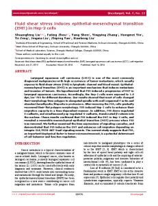
Figure 1. Darcy Flow Experimental Apparatus for the application of shear stress in 3D and Modified Boyden chamber invasion assay. (A) The flow apparatus applied a constant hydrostatic pressure via a double reservoir system composed of a larger reservoir [A] feeding flow media into the smaller syringe reservoir [B] and a pressure release tube [C], both of which were fastened within a rubber seal [D] to the cell culture insert [E]. The pressure release tube [C] ensured that there were no fluctuations in pressure applied across the collagen/cell suspension [F]. Hydrostatic pressure [DP] drove media throughout the thickness of the gel [L] and exerted shear stress on the cell membranes. Flow media filtrate was collected in another reservoir [G]. (B) At the end of the flow period, the inserts containing the cell suspensions [E] were decoupled from the apparatus and the invasion assay ensued. During the migration period, 10 nM TGF-a directed cells to migrate through 8 mm pores towards the underside of the filter. At the end of the migration period, cells on the underside of the inserts were stained and migration rates were quantified. doi:10.1371/journal.pone.0020348.g001
U87 human glioma (HTB-14; ATCC), rat CNS-1 glioma (Dr. William F. Hickey and Dr. David J. Graber, Dartmouth Medical School), and U251 human glioma (Dr. Eric C. Holland, Memorial Sloan-Kettering Cancer Center) cell lines were cultured in DMEM (Sigma) supplemented with 10% FBS (HyClone) and 1% Penicillin/Streptomycin (Sigma). Cells were grown to a minimum of 70% confluence, and then all experiments were conducted in humidified incubators. TGF-a (Sigma) was chosen as the chemoattractant since it is one of the most potent stimulators of migration [27]. The optimal concentration of TGF-a was determined via Boyden chamber experiments (data not shown); ultimately, 10 nM TGF-a in DMEM without serum was added to the companion wells for all invasion assays unless otherwise specified.
Three-dimensional (3D) cell suspensions culture inserts for proper gelation to reduce settling of cells at the bottom due to gravity. The cell/collagen suspensions were incubated for 12 hours with 800 ml of serum-containing media in each well to allow sufficient time for cell spreading.
The boundaries between brain tumors and parenchyma have been shown to contain interstitial collagen type I among other matrix components, and some implanted glioma cell lines form interstitial ECM that consists primarily of interstitial collagen [20,23,30]. A model was developed in which glioma cells suspended in type I collagen were exposed to 3D fluid shear stress via the Darcy Flow Experimental Apparatus (Fig. 1A). High concentration Rat Tail Collagen Type I (BD Biosciences) was utilized as the stock and all dilutions were carried out using serumcontaining media (DMEM containing 10% FBS; culture media). The walls of 12-well cell culture inserts (containing 8.0 mm pore filters [BD Falcon]) were pre-coated with 50 ml of 1 mg/ml collagen to prevent gel detachment or contraction by the suspended cells. 50,000 cells were suspended in 400 ml of 2 mg/ ml collagen and immediately incubated within the pre-coated PLoS ONE | www.plosone.org
Three-dimensional (3D) shear stress model The three-dimensional model simulated the interstitial flow forces that cells would encounter within the interstitium in order to study the effects of shear stress on chemotactic migration (Fig. 1A). DMEM with 10% serum was used as the flow media, and both time of exposure to flow (up to four hours) and shear stress levels were varied. The shear stress (t) on the cell surface was calculated knowing the Darcy permeability coefficient (Kp) of the 3D collagen/cell suspension and assuming cylindrical or spherical cell geometries based on theory by Wang and Tarbell [31], and 2
May 2011 | Volume 6 | Issue 5 | e20348
Flow Regulates Glioma Invasion
Kp ~mðJv =AÞ=ðDP=LÞ
ð1Þ
(L), to quantify the effect of compaction on cell distribution (from initiation of flow), and to ensure proper attachment of the gels to the filters. Confocal imaging was repeated after a 48 hour migration period on other cell suspensions in order to observe cell morphology.
pffiffiffiffiffiffi t&m(Jv =A)= Kp
ð2Þ
Three-dimensional (3D) shear stress invasion assays
Brinkman [32]:
where m is the fluid viscosity, Jv is the volumetric flow rate, A is the area of the filter, L is the thickness of the gel, and DP is the pressure drop [17,31,32]. The shear stress equation is an approximation; for spherical cells it should be multiplied by p/3, and for cylindrical cells by a factor of p/4 [31]. These model equations were also utilized to determine the physiologic range of shear stress based on established permeability and interstitial flow velocity measurements in developing tumors and in brain tumors. During the flow period, the fluid filtrate was quantified every 10 minutes after initiation of flow through the gel to determine the volumetric flow rate, Jv. After it was determined that Jv was stable after the first 10 minutes of flow, when the collagen compacts to a fixed point (discussed below), then the filtrate was measured on an hourly basis. The flow properties of the collagen/cell suspension along with the parameters characterizing the 3D model are presented in Table 1.
Following completion of the flow period, all cells on the underside of the inserts were mechanically removed to ‘‘zero’’ the initial migration count [26]. The inserts containing the cell suspensions were then incubated with 700 ml of 10 nM TGF-a in the well for 48 hours of migration (without flow; Fig. 1B). Thus the flow (shear) and migration periods were separated so that flow effects could not be interpreted as resulting from the convection of chemoattractant or signaling molecules produced by the suspended cells. At the end of the migration period, cells that had migrated to the underside of the inserts were fixed, stained with DiffQuik (Dade Behring), and counted in 5 fields to quantify migration rates; method established by Garanich et al [24]. It should be noted that cells that migrated through the pores adhered to the underside of the filter and there was no evidence of cells detaching from the filter and floating in or attaching to the bottom of the well.
Compaction of cell suspensions
Broad-spectrum MMP inhibitor invasion assays GM6001 (Calbiochem), a broad-spectrum MMP Inhibitor, and GM6001-NC (Calbiochem), as a negative control, were utilized to assess the extent to which the glioma cell lines within the collagen/ cell suspensions depended on active MMP expression for invasion. Standard 48 hour invasion assays were conducted with the glioma cells migrating from the suspensions towards 10 nM TGF-a solutions containing 10 mM GM6001 or 10 mM GM6001-NC.
It became evident that the initial application of fluid flow was permanently changing the thickness of the gels by compaction of the collagen and thereby altering the cell distribution, collagen density and permeability. Therefore, following the initial 12-hour incubation period, 8 cmH2O of differential pressure was applied across all gels inducing flow for 10 minutes to pre-compact the gels before the flow experiment. The gels that were allowed to compact with no additional flow were designated as compacted controls. Assuming minimal collagen degradation by the cells during the initial incubation and flow periods, a mass of collagen/volume analysis was performed by collecting and measuring the volume of the gels (from both compacted controls and flow cases) to determine the collagen density of the compacted gels. During all incubation periods, to ensure that the gels would not detach from the filter, the media in the well was maintained at a level below the upper surface of the gel, a normal pressure of 0.05 cm, to prevent back flow of media and to maintain compaction of the gel under its own weight.
Isolation of chemotactic motility To confirm that autologous chemotaxis flow effects had been isolated and removed for all cell lines being used, the Darcy flow experiment and invasion assay were carried out without the use of TGF-a as a chemoattractant. Additionally, the potency of TGF-a (without flow) was determined for each cell line in the modified 3D Boyden chamber to ensure that 10 nM TGF-a was able to effectively provide a directional cue for cells to migrate so that motility effects could be established.
Cell apoptosis and viability assay Cell distribution and viability
To determine if apoptosis played a role in the invasion response brought on by flow, after the 48 hour migration period, both control gels and gels exposed to a differential pressure drop of 7 cmH2O were stained with the Vybrant Apoptosis Assay Kit no. 2 (Invitrogen) following the manufacturer’s instructions. Gels
Following the flow period, some of the 12-well inserts containing the 3D cell suspension were stained with Calcein AM (Invitrogen) and imaged using a Leica Confocal microscope. Fluorescence at different depths was obtained to determine the thickness of the gels Table 1. Flow Properties of Collagen Gel and Cell Suspensions.*
Pressure, DP (cm H20)
Flow Rate, Jv (ml/min)
Permeability, Kp (10215 m2)
Velocity (mm/sec)
Shear Stress, t (dynes/cm2)#
1.0
4.3260.42
0.8360.08
4.1660.41
0.1160.01
5.0
9.5861.18
1.8560.23
1.8460.23
0.3660.02
7.0
15.9769.89
3.0862.01
2.0561.27
0.5560.18
*Darcy permeability and shear stresses obtained by utilizing equations (1) and (2). *Viscosity (m) 20.84 cP; Area (A) 20.865 cm2; Length (L) 2600 mm. #Each shear stress level was different from the other levels (p,0.005). Data presented as mean6standard deviation. doi:10.1371/journal.pone.0020348.t001
PLoS ONE | www.plosone.org
3
May 2011 | Volume 6 | Issue 5 | e20348
Flow Regulates Glioma Invasion
were also stained with Calcein AM to further confirm viability after the migration period. Cell apoptosis and viability images were acquired utilizing a Nikon inverted fluorescent microscope.
further confirm the findings (data not shown). Specificity of the amplified products was verified by both dissociation curve analysis and by gel electrophoresis.
MMP activity assays
RNA interference
Following the migration period, the conditioned media from the wells of sheared and control (non-sheared) glioma cells were collected and stored. MMP-1 and MMP-2 are two of the most important MMPs for the degradation of the ECM in gliomas [18]. Triple helical collagen is cleaved by interstitial collagenases (mainly active MMP-1) and subsequent degradation of denatured collagen fibrils and basement membrane ECM by gelatinases (predominantly active MMP-2) [22,33]. Changes in total (active and pro-) and active levels of MMP-1 and MMP-2 were determined utilizing the AnaSpec SensoLyte Plus 520 MMP-1 Assay Kit and the AnaSpec SensoLyte 520 MMP-2 Assay Kit (AnaSpec, San Jose, CA). Both kits used the fluorogenic substrate 5-FAM/QXL520 and upon cleavage by their respective MMP, the fluorescence intensity could be measured at 490/520-nm wavelength [26]. Manufacturer’s instructions were followed to determine relative concentrations of the MMPs in the conditioned media.
To determine if shear-dependent modulation of MMPs could affect migration, specific MMP mRNAs were silenced by short hairpin RNA (shRNA). MMP-1 shRNA oligonucleotide corresponding to bases 305 to 323 of the MMP-1 mRNA, previously cloned into the pSuper-retro-puro expression vector (OligoEngine), was utilized to silence MMP-1 gene expression in U87 cells (shRNA designated as ‘305’-a kind gift from Dr. Constance E. Brinckerhoff, Dartmouth Medical School) [35]. As a control vector, a scrambled sequence cloned into the pSuper-retro-puro plasmid was also provided (control shRNA shMAMMX designated as ‘MMX’; another kind gift from Dr. Brinckerhoff) [35]. Both vectors were amplified utilizing XL1-Blue Competent Cells (Stratagene) and purified utilizing the QIAprep Spin Miniprep Kit (Qiagen) following the suppliers’ instructions. MMP-2 shRNA Plasmid (r) and its corresponding Control shRNA Plasmid-A (Santa Cruz Biotechnology) were utilized to silence MMP-2 gene expression in CNS-1 cells. Puromycin resistance was designed into all vectors utilized in this study so that cells stably expressing the shRNA could be selected.
RNA extraction and isolation Cell lysis and RNA extraction from the collagen suspension was carried out with the use of TRIzol Reagent (Invitrogen) following the manufacturer’s instructions. After homogenization (decoupling of proteins from nucleic acids) insoluble matrix was removed via centrifugation as recommended by the supplier. For total mRNA isolation and purification the RNeasy Mini Kit (Qiagen) and the associated QIAvac 24 vacuum manifold (Qiagen) were utilized following the supplier’s instructions.
shRNA transfection and puromycin selection The shRNA plasmids were transfected into their respective cell lines using Lipofectamine LTX and PLUS reagents (Invitrogen) and cells were grown to confluence. Following transfection, cells stably expressing their respective shRNA plasmids were isolated by puromycin selection. The optimal puromycin concentration of 1.5 mg/ml was chosen such that non-transfected cells would die within 2 days of culture (data not shown). For the duration of the MMP gene knock-down experiments, tranfected cells were grown in culture media supplemented with 1.5 mg/ml puromycin.
Reverse transcription-polymerase chain reaction RT-PCR was performed to validate that interstitial flow was modulating MMP levels. Reverse transcription to cDNA was performed following the Cells-to-cDNA II Kit (Ambion) procedures. Quantitative real-time PCR (RT-qPCR) was performed on the ABI PRISM 7000 sequence detection system (Applied Biosystems) with the reactions containing SYBR Green PCR Master Mix (Applied Biosystems). In addition to the RT-qPCR, representative samples of the amplified mRNA products were isolated through gel electrophoresis and visualized under excitation by ultraviolet light in the presence of 0.1 mg/ml ethidium bromide in 2.75% agarose gels. It was determined that modulation of MMP-1 levels was the primary effect of shearing the U87 cells. Therefore a 274-bp MMP-1 product was amplified using the sense primer as 59-TGA GGG GAA CCC TCG CTG GG -39 and its antisense primer as 59-TCC CCT CCA ATA CCT GGG CCT G-39 (Genebank accession no. NM_002421.3), and a 267-bp GAPDH product was amplified using the sense primer as 59-CCT GAC CTG CCG TCT AGA AA-39 and its antisense primer as 59TTA CTC CTT GGA GGC CAT GT-39 (Genebank accession no. NM_002046) [34]. It was further established that modulation of MMP-2 levels was the primary effect of shearing the CNS-1 cells. Therefore a 200-bp rat MMP-2 product was amplified using the sense primer as 59-GAT GGA TAC CCA TTT GAC GG-39 and its antisense primer as 59-CTG CTG TAT TCC CGA CCA TT-39 (Genebank accession no. NM_031054) [26], and a 232-bp GAPDH product was amplified using the sense primer as 59-TCT TCA CCA CCA TGG AGA A-39 and its antisense primer as 59ACT GTG GTC ATG AGC CCT T-39 (Genebank accession no. NM_017008) [26]. GAPDH served as an internal control for each sample. b-actin was also used as a secondary housekeeping gene to PLoS ONE | www.plosone.org
RNA interference invasion assay The invasive potential of glioma cells stably expressing their respective shRNA vectors was determined. Standard invasion assays were conducted and the suspended glioma cells were allowed to migrate towards 10 nM TGF-a for 48 hours. After the migration period, cells suspended inside the gels were lysed, RNA was extracted and purified, and reverse transcription was performed as previously described. RT-qPCR was performed and representative samples for the amplified mRNA products were isolated through gel electrophoresis and visualized in the presence of ethidium bromide. Cells that had migrated to the underside of the inserts were fixed and stained with DiffQuik, and migration rates were quantified.
Collagen and gelatin zymography Following the migration period, the conditioned media from the wells of glioma cells transfected by the control vectors and knockdown vectors were collected and stored. Collagen and gelatin zymography were performed as detailed by Shi et al [26] to confirm knockdown of MMP expression by shRNA vectors. Collagen zymography was used to confirm knockdown of pro- and active levels of MMP-1 expression in U87 cells and gelatin zymography was used to confirm knockdown of pro- and active levels of MMP-2 expression in CNS-1 cells brought on by their respective shRNA gene silencing. MMP expressions were quantified using the Quantity One software (Bio-Rad) and were presented as percentage of their respective controls [26]. 4
May 2011 | Volume 6 | Issue 5 | e20348
Flow Regulates Glioma Invasion
a two-factor analysis of variance (ANOVA) was utilized followed by a post-hoc analysis for intensity and distance.
Specific MMP inhibitor invasion assay To further verify invasion effects by shRNA knockdown, specific MMP inhibitors were used in invasion assays with non-transfected cells. Standard 48 hour invasion assays were conducted with the glioma cells migrating from the suspensions towards 10 nM TGFa solutions containing 10 mM of either MMP Inhibitor I or MMP2 Inhibitor I (Calbiochem). MMP Inhibitor I was utilized at a concentration that would inhibit MMP-1 activity in U87 cells and MMP-2 Inhibitor I was used to inhibit MMP-2 activity in CNS-1 cells.
Results Effect of fluid compaction on cell distribution The collagen concentration of gels exposed to flow (sheared) and compacted (non-sheared) controls was 8.0960.26 mg/ml-4 times more concentrated than the original gels before compaction. Compaction only affected cell distribution perpendicular to the filter (vertical direction); cells remained viable, morphologically normal, and well distributed in every horizontal plane throughout the thickness of the gels (Supplementary Fig. S1). The cumulative effect of compaction by flow through the gels was most apparent closer to the filter (Supplementary Fig. S2). The cell distribution of the compacted controls and the suspensions exposed to four hours of shear were similar, whereas the distribution of non-compacted controls was different from the compacted control and sheared
Statistical analysis All data were normalized by their respective controls and are presented as mean6standard error of the mean. The two-tailed Student’s t-test (type 3) was utilized to determine statistical significance and Bonferroni corrections were applied for multiple comparisons (#). For cell distribution and fluorescence in the gels
Figure 2. Fluorescence intensity of gels containing cells stained with Calcein to quantify cell distribution within the collagen suspension. (A) Fluorescence of horizontal slices (10 mm each) up to 130 microns above the filter for gels that were compacted, not compacted, and suspensions that were exposed to four hours of flow. The cell distribution in non-compacted gels was distinctive when compared to the compacted control and four-hour flow gels (# p,0.015). (B) Intensity of the 600 micron thick collagen suspensions (50 mm slices) to verify similar cell distribution of the compacted control and the experimental flow cases at 0.36 dynes/cm2 (low) and 0.55 dynes/cm2 (high) shear stress. Compacted control and flow cell suspensions utilized for the migration study had similar cell distributions (p