cyclic monophosphate-dependent protein kinase (ATP:protein ... state provide a highly sensitive assay of cyclic AMP-dependent protein kinase and ...
Proc. Natl Acad. Sci. USA Vol. 78, No. 10, pp. .6048-6050, October 1981 Biochemistry
Fluorometric assay for adenosine 3',5'-cyclic monophosphatedependent protein kinase and phosphoprotein phosphatase activities (peptide substrate/phosphorylation/enzyme kinetics/fluorescence)
DAVID E. WRIGHT*, EMILY SHACTER NOIMANt, P. BOON CHOCKt, AND VINCENT CHAUtt *Laboratory of Nutrition and Endocrinology, National Institute of Arthritis, Diabetes, and Digestive and Kidney Diseases; and tLaboratory of Biochemistry, National Heart, Lung, and' Blood Institute, National Institutes of Health, Bethesda, Maryland 20205
Communicated by E. R. Stadtman, June 22, 1981
ABSTRACT A novel peptide substrate for adenosine 3',5'cyclic monophosphate-dependent protein kinase (ATP:protein phosphotransferase, EC 2.7.1.37), Leu-Arg-Arg-Trp-Ser-Leu-Gly, was synthesized. Phosphorylation of the peptide causes a 20% increase in the peptide fluorescence intensity at 358 nm. Values of K. and keat for the phosphorylation reaction at pH 7.0 (25"C), were determined to be 2.7 ± 0.5 pM and 5.5 ± 0.4 sec', respectively. The phosphorylated peptide was shown to be an effective substrate for phosphoprotein phosphatase (phosphoprotein phosphohydrolase, EC 3.1.3.16) with a Km of 113 ± 10 FM and a kcat of 2.4 ± 0.2 sec- in the presence of 2.5 mM MnCI2. Changes in the peptide fluorescence intensity as a function of its phosphorylation state provide a highly sensitive assay of cyclic AMP-dependent protein kinase and phosphoprotein phosphatase activities.
the serine phosphorylation site. It is shown that phosphorylation of the peptide leads to a significant change in the tryptophan fluorescence ofthe peptide. This fluorescence change can be used as a highly sensitive measure of cAMP-dependent protein kinase and phosphoprotein phosphatase (phosphoprotein phosphohydrolase, EC 3.1.3.16) activities. MATERIALS AND METHODS Protein Purifications. Bovine cardiac cAMP-dependent protein kinase was purified as described by Rubin et aL (9) with Sigma histone ILA as substrate. The purified enzymewas judged to be approximately 90% pure when analyzed on sodium dodecyl sulfate polyacrylamide gels. Phosphoprotein phosphatase (Mr 35,000) was purified 4500-fold to near homogeneity from bovine cardiac muscle by the procedure of Chou et aL (10) with phosphorylated histone IIA as substrate. Protein concentrations were determined by the method of Bradford (11). Mrs of174,000 and 35,000 for cAMP-dependent protein kinase and phosphoprotein phosphatase, respectively, were used to calculate the enzyme concentrations. Peptide Synthesis. The peptide, Leu-Arg-Arg-Trp-Ser-LeuGly, was synthesized by the Merrifield solid-phase procedure (12). The Boc amino acids used had the following side-chain protecting groups: Arg(NO2), Trp(CHO), and Ser(Bzl). The peptide was cleaved from the resin with liquid HF. The formyl group was removed by treatment with aqueous piperidine (13). The peptide was purified by gel filtration on Sephadex G-10 and by a SEP-PAK-C-18 cartridge on high-pressure liquid chromatography. Enzymatic hydrolysis of the peptide gave the following amino acid values: Arg, 2.1; Trp, 1.0; Ser, 1.0; Gly, 1.0; and Leu, 1.9. Phosphopeptide and Phosphoamino Acid Analysis. Two-dimensional peptide mapping was carried out with Eastman thinlayer cellulose sheets as described by Gracy (14). The phosphorylated peptide was subjected to electrophoresis at pH 3.7 (pyridine/glacial acetic acid/water, 1:10:89, vol/vol) in the first dimension and to chromatography (1-butanol/glacial acetic acid/ water/pyridine, 15:3:10:12, vol/vol) in the second dimension. Autoradiography of the cellulose sheet showed that greater than 97% of the phosphate incorporation was associated with a single peptide spot. Phosphoamino acids were analyzed as described by Noiman (15). Enzyme Assays. All assays were carried out at 25°C. cAMPdependent protein kinase activity was assayed in a reaction
Adenosine 3',5'-cyclic monophosphate (cAMP)-dependent protein kinase (ATP:protein phosphotransferase, EC 2.7.1.37) catalyzes the transfer of the terminal phosphoryl group of adenosine 5'-triphosphate to specific serine or threonine residues of substrate proteins (1). In addition, the enzyme also catalyzes the phosphorylation of serine residues in a number of synthetic peptides containing amino acid sequences similar to the local phosphorylation site sequences of substrate proteins (2, 3). Studies using these synthetic peptide substrates have provided valuable information concerning the substrate specificity of cAMP-dependent protein kinase (2-4) and the molecular basis of enzyme-substrate recognition (5, 6). Although the enzymatic activity of cAMP-dependent protein kdnase can be readily assayed by quantitating the transfer of labeled phosphoryl groups from adenosine 5'-[y-32P]triphosphate to the substrate protein or peptide, continuous and rapid assay of the enzymatic activity with this method is not possible. The use of adenosine 5'-[y-32P]triphosphate also places severe constraints on the methodology that can be used for detailed physicochemical study of the enzymatic reaction. Therefore, an alternate method of assay is highly desirable. Recently, Kemp (7) reported that the phosphorylation of an a-N-dansyl derivative of a substrate peptide can be quantitated spectros opically after separation of the phosphorylated peptide from the unmodified substrate by high-pressure liquid chromatography. Bramson et al. (8) found that phosphorylation of a substrate peptide containing a nitrotyrosine residue adjacent to the serine phosphorylation site causes a measurable spectral change at 430 nm and can be used for the continuous assay of protein kinase activity. In this communication, we report the synthesis of a substrate peptide containing a tryptophan residue adjacent to
Abbreviation: cAMP, cyclic AMP.
* To whom reprint requests should be addressed at the present address:
The publication costs ofthis article were defrayed in part by page charge payment. This article must therefore be hereby marked "advertisement" in accordance with 18 U. S. C. §1734 solely to indicate this fact.
Dept. of Biochemistry and Molecular Biology, J. Hillis Miller Health Center, Box J-245, Univ. of Florida, Gainesville, FL 32610. 6048
Biochemistry: Wright et aL
Proc. Nad Acad. Sci. USA 78 (1981)
6049
Phosphoprotein phosphatase was assayed in a reaction mix4-morpholinepropanesulfonic acid (pH 7.0), 2 mM dithiothreitol, 2.5 mM MnCl2, 0.3 mg of bovine serum albumin per ml, and [32P]phosphorylated peptide. Release of inorganic phosphate was determined as described by Titanji (17). The [3 P]phosphorylated peptide was prepared by incubation of the peptide with [y-32P]ATP and cAMP-dependent protein kinase. Excess [y-32P]ATP was removed by passing the reaction mixture through a small AGL-X8 anion-exchange column equilibrated with 30% acetic acid. The eluate, containing the phosphorylated peptide, was lyophilized and washed ture containing 25 mM
z z 0
LLI
twice with water to remove residual acetic acid. Fluorescence spectra and time courses of fluorescence
300
325
350
375
400
425
change for the phosphorylation and dephosphorylation reactions were obtained in an SLM spectrofluorometer fitted with a thermostated microcell (0.1 ml) equilibrated at 250C.
450
WAVELENGTH, nm
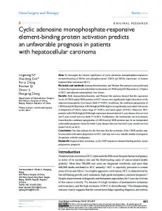
FIG. 1. Fluorescence emission spectra of the phosphorylated and unmodified peptide. Spectra were obtained by using a peptide concentration of 20 /AM in 25 mM 4-morpholinepropanesulfonic acid (pH 7.0) at 2500.
mixture containing 25 mM 4-morpholinepropanesulfonic acid (Mops) (pH 7.0), 10 mM MgCl2, 2 ,uM cAMP, and 0.2-1.0 mM [y-3 P]ATP (100-250 cpm/pmol) or unlabeled ATP. The concentration of the peptide substrate Leu-Arg-Arg-Trp-Ser-LeuGly was determined by using a molar extinction coefficient of £280 nm = 5550, and those of Leu-Arg-Arg-Ala-Ser-Leu-Gly and histone IIA were determined by using Mrs of 772 and 12,000, respectively. Incorporation of labeled phosphate into peptide was measured as described by Glass et aL (16) with Whatman P81 paper circles (2.3 cm). The reaction was stopped by spotting an aliquot of the reaction mixture onto the paper disk, which previously had been moistened with 40 /,u of 30% (vol/vol) acetic acid.
RESULTS The heptapeptide Leu-Arg-Arg-Trp-Ser-Leu-Gly was synthesized, characterized, and found to be readily phosphorylated in the presence of cAMP-dependent protein kinase and ATP. A stoichiometry of0.95-1.0 mol of PJmol of peptide was found when the peptide was maximally phosphorylated. The isolated phosphorylated peptide can be completely dephosphorylated by phosphoprotein phosphatase purified from bovine cardiac muscle according to the. procedure of Chou et aL (10). Phosphoamino acid analysis of the phosphorylated peptide showed that phosphate was incorporated into the single serine residue. Fig. 1 shows the fluorescence emission spectra of the phosphorylated and dephosphorylated forms of the peptide. Although both spectra showed the characteristics of tryptophancontaining peptides, the relative peptide fluorescence intensity at the emission maximum (358 nm) was found to be 20% greater for the phosphorylated form. The increase in the fluorescence intensity that occurred upon phosphorylation of the peptide was
8
100 -
0
0
80
6
801 -0
O
IO0
a)
C
cO 01
C).
401-
Q) .C
C/)0~~
oL
s~~~o
9) >
0 °~~0
0 CL
60 F
o 0~~o
0
C =
It
_
0)
0
60
CIo U a)
0
0
o2
SO.. -
20
_o
a
o 0
20 -
o--
~~~~~~
' ~
300
Time,
sec
2. Time course of phosphorylation of the peptide. The reaction was monitored either by quantitating the incorporation of [32P]phosphate into the peptide or by the changes in the peptide fluorescence intensity at 358 nm. The reaction mixture contained 25 mM 4-morpholinepropanesulfonic acid (pH 7.0), 10 mM MgCI2, 21AM cAMP, 0.2 mM [_32P]ATP (150 cpm/pmol) or unlabeled ATP, 36 pM peptide, and 30 nM cAMP-dependent protein kinase. FIG.
600
900
1200
1500
Time, sec FIG. 3. Time course of dephosphorylation of the phosphorylated peptide. The reaction was monitored either by quantitating the release of [32P~phosphate from the peptide or by changes in the peptide fluorescence intensity at 358 nm. The reaction mixture contained 25 mM 4-morpholinepropanesulfonic acid (pH 7.0), 2 mM dithiothreitol, 2.5 mM MnCl2, 0.3 mg of bovine serum. albumin per ml, 50 AM peptide, and 84 nM phosphoprotein phosphatase.
6050
Biochemistry: Wright et al.
Proc. Natl. Acad. Sci. USA 78 (1981)
Table 1. Comparison of kinetic constants of cAMP-dependent protein kinase and phosphoprotein phosphatase* Phosphoprotein cAMP-dependent protein phosphatase kinase Substrate Km, IM kcat, sec-' Kmi, kMt, sec1 2.4 ± 0.2 113 ± 10 1.4 ± 0.1 2.7 ± 0.5 Leu-Arg-Arg-Trp-Ser-Leu-Gly 2.3 114 5.8 17 Leu-Arg-Arg-Ala-Ser-Leu-Gly 0.11 87 3.2 Histone IHA * Km and kat were determined from linear least-squares analysis of double reciprocal plots. Data for the pyruvate kinase analog peptide and histone ILA were obtained at 370C. The ranges of substrate concentrations used for these studies are from 0.25 Km to 3 Km.
completely reversed when the peptide was dephosphorylated by phosphoprotein phosphatase. Fig. 2 shows the time-dependent increase in the fluorescence intensity at 358 nm when the peptide was undergoing phosphorylation in the presence of cAMP-dependent protein kinase and ATP. Excellent agreement was obtained when [32P]phosphate incorporation into the peptide was used to monitor the phosphorylation reaction under identical conditions (Fig. 2). The same agreement was obtained when the dephosphorylation reaction was monitored either by fluorescence change or by the release of [32P]phosphate from the peptide (Fig. 3). To determine the Kmn and kcat, initial rates of the phosphorylation reaction were obtained at various concentrations of the peptide (Table 1). For comparison, values obtained using histone IIA and the pyruvate kinase analog peptide Leu-Arg-ArgAla-Ser-Leu-Gly are also included. Values of K. and kst for the dephosphorylation reaction, derived from analysis of initial rate data obtained at various concentrations of the phosphorylated peptide, are also in Table 1. DISCUSSION In this communication, we report the synthesis of a novel heptapeptide, Leu-Arg-Arg-Trp-Ser-Leu-Gly. It was found to be an excellent substrate for cAMP-dependent protein kinase, and the phosphorylated peptide can be readily dephosphorylated by phosphoprotein phosphatase. The peptide is unique in that phosphorylation of the serine residue of the peptide leads to a significant change in the peptide fluorescence intensity. It has been shown that this change in fluorescence can be used as a continuous assay to monitor the enzymatic activities of cAMPdependent protein kinase and phosphoprotein phosphatase. The use of a small peptide as substrate for cAMP-dependent protein kinase was first reported by Daile and Carnegie (18), who used a 17-residue peptide derived from myelin basic protein. Subsequently, Kemp et al. (3) reported that the synthetic peptide Leu-Arg-Arg-Ala-Ser-Leu-Gly, corresponding to the local phosphorylation site sequence of porcine pyruvate kinase, is an excellent substrate of cAMP-dependent protein kinase. The heptapeptide reported here represents a slight modification of the pyruvate kinase analog in that the alanine residue was replaced by a tryptophan. Judging from the Km and kcat reported in Table 1, we find that the single amino acid substi-. tution does not have a significant effect on the ability of the peptide and its phosphorylated form to serve as effective substrates for cAMP-dependent protein kinase and phosphoprotein phosphatase, respectively. Substrate peptides of cAMP-dependent protein kinase have been used extensively to study the substrate specificity of the enzyme (2-4). Because variations of the amino acid sequence can be obtained readily, studies using the substrate peptides have yielded important information concerning the nature of the enzyme-substrate interactions and recognition. We have
used a substrate peptide for a different purpose by introducing a tryptophan residue adjacent to the serine phosphorylation site. Our results show that the tryptophan fluorescence can be used to monitor the extent of peptide phosphorylation. The results reported here are similar to those obtained by Bramson et al. (8) in which a nitrotyrosine was introduced to form the substrate peptide, Leu-Arg-Arg-(o-NO2)Tyr-Ser-Leu-Gly. Both spectral probes can be used to monitor phosphorylation of the peptide. However, the changes in fluorescence intensity of the heptapeptide reported here give a far more sensitive measure of the enzymatic activities. -Phosphorylation of the nitrotyrosine-containing peptide results in a decrease in extinction coefficient at 430 nm by 210 M-1 cm'- (8). Thus, the detection limit of this peptide is roughly 0.1 ,umol. In contrast, the fluorescent peptide can detect phosphorylation changes in the picomole range, comparable to the sensitivity of the common assays that use radioactive phosphate. In addition, the fluorescent changes resulting from phosphorylation and dephosphorylation of this peptide can be observed and continuously recorded within milliseconds. Thus, the fluorescent peptide opens up another dimension for studying the mechanism of action of cAMP-dependent protein kinase and phosphoprotein phosphatase. The authors thank Dr. A. Komoriya for the use of the HF apparatus and members of the Laboratory of Biochemistry for helpful discussions. 1. Krebs, E. B. & Beavo, J. A. (1979) Annu. Rev. Biochem. 48, 923-959. 2. Zetterquist, O., Ragnarsson, U., Humble, E., Berglund, L. & Engstrom, L. (1976) Biochem. Biophys. Res. Commun. 70, 696-703. 3. Kemp, B. E., Graves, D. J., Benjamini, E. & Krebs, E. G. (1977) J. Biol. Chem. 252, 4888-4894. 4. Kemp, B. E., Benjamini, E. & Krebs, E. G. (1976) Proc. Nat. Acad. Sci. USA 73, 1038-1042. 5. Small, D., Chou, P. Y. & Fasman, G. D. (1977) Biochem. Biophys. Res. Commun. 79, 341-346. 6. Matsuo, M., Huang, C. H. & Huang, L. C. (1978) Biochem. J. 173, 441-447. 7. Kemp, B. E. (1980) J. Biol. Chem. 255, 2914-2918. 8. Bramson, H. N., Thomas, N., DeGrado, W. F. & Kaiser, E. T. (1980) J. Am. Chem. Soc. 102, 7156-7157. 9. Rubin, C. S., Erlichman, J. & Rosen, 0. M. (1972)J. Biol. Chem. 247, 36-44. 10. Chou, C. K., Alfano, J. & Rosen, 0. M. (1977)J. Biol. Chem. 252, 2855-2859. 11. Bradford, M. (1976) Anal. Biochem. 72, 248-254. 12. Merrifield, R. B. (1964) Biochemistry 3, 1385-1390. 13. Ohno, M., Tsukamoto, S. & Izumiya, N. (1972) J.C.S. Chem. Comm. 663-664. 14. Gracy, R. W. (1977) Methods Enzymol. 47, 195-204. 15. Noiman, E. S. (1980)J. Biol. Chem. 255, 11067-11070. 16. Glass, D. B., Masaracchia, R. A., Feramisco, J. R. & Kemp, B. E. (1978) Anal. Biochem. 87, 566-575. 17. Titanji, P. V. K. (1977) Biochim. Biophys. Acta 481, 140-151. 18. Daile, P. & Carnegie, P. R. (1974) Biochem. Biophys. Res. Commun. 61, 852-858.