are strictly periodic and always oriented in parallel, interrow bridges are not strictly periodic ... microtubules in adjacent rows (the so-called inter- row bridges) have a ... is a promising system in which to study correla- ..... The bracket indicates a presumptive microtubule profile and the horizontal arrowheads indicate material ...
FREEZE-FRACTURE
OF
BRIDGES
AXOSTYLES
IN M O T I L E
MICROTUBULES
R O B E R T A. B L O O D G O O D and K E N N E T H
AND
R. M I L L E R
From the Department of Molecular, Cellular, and Developmental Biology, University of Colorado, Boulder, Colorado 80302
ABSTRACT A freeze-fracture study of the motile axostyles of the flagellate protozoa
Saccinobaculus and Pyrsonympha has been undertaken in order to obtain a view of the relationships of microtubules and their cross bridges not dependent on conventional preparative procedures. Reactivation studies using isolated axostyles prepared for freeze-fracture and then thawed demonstrate that we are observing the structure of a potentially functional axostyle. Cross fractures through the axostyle demonstrate more extensive interrow bridging than expected on the basis of observations of thin-sectioned material. Each microtubule has approximately sixfold bridge-binding sites with connections to as many as four interrow bridges. Measurements of microtubule diameter and spacing are significantly larger than those made from sectioned material and may indicate that conventional processing for electron microscopy results in the loss of structurally important water within the microtubule in addition to loss of intertubule material. Longitudinal fractures through the axostyle at various orientations demonstrate a minimum longitudinal periodicity of 160 A for both the spacing of the globular subunits within the microtubule wall and the spacing of the intrarow bridges. While intrarow bridges are strictly periodic and always oriented in parallel, interrow bridges are not strictly periodic and can be oriented at varying angles to the microtubule axis. INTRODUCTION In many cases of microtubule-associated motility, the microtubules are observed to possess arms or to be connected by bridges (for reviews, see Tilney, 1971; Mclntosh, 1974). Motile axostyles are composed only of microtubules and bridges connecting these microtubules (Grass6, 1956; Grimstone and Cleveland, 1965; Brugerolle, 1970; Hollande and Carruette-Valentin, 1970; Mclntosh et al., 1973). These axostyles can be isolated and reactivated using an ATP-containing medium. The axostyle contains a protein with enzymatic activity and electrophoretic mobility similar to that of flagellar and ciliary dynein (Mooseker and Tilney, 1973).
660
Bend formation is accompanied by the sliding of rows of microtubules relative to one another (McIntosh, 1973). It is presumed that the periodic connections between microtubules within a row (the so-called intrarow bridges) are structural in nature while the less regular connections between microtubules in adjacent rows (the so-called interrow bridges) have a mechanochemical role in axostyle motility. Most observations on bridges between microtubules in various systems have been limited to material fixed, sectioned, and stained with heavy metals, and some difficulty exists in identifying
T H E JOURNAL OF CELL BIOLOGY . VOLUME 62, 1974 . p a g e s 6 6 0 - 6 7 1
bridges, particularly nonperiodic ones, prepared in this m a n n e r . We have u n d e r t a k e n the e x a m i n a t i o n of bridges and microtubules in motile axostyles using the technique of freeze-fracture which permits a view of the axostyle based more on physical t h a n chemical properties. The techniques of freeze-fracture and freezeetch have been applied to the study of yeast spindle microtubules (Moor, 1966 a, b; Moor, 1967; G u t h et al., 1972), plant cortical microtubules (Northcote and Lewis, 1968), a presumed microtubule paracrystal within an alga (Brown and Franke, 1971), and cytoplasmic microtubules in an insect ovary (Stebbings and Willison, 1973). N o n e of these systems possesses both a highly ordered array of cross-bridged microtubules and a proven association with motility. On the other hand, the axostyle possesses both of these qualities and hence is a promising system in which to study correlations of structure with function. MATERIALS
AND METHODS
to give a higher percentage of reactivated axostyles and more normal axostyle motility than that of Mooseker and Tilney (1973). Addition of 5 mM dithiothreitol (DDT) to the isolation and reactivation media prolonged the motility. Optical transforms of electron micrographs were prepared by the procedure of Mclntosh (1973). Optical transforms of electron micrographs of shadowed material (freeze-fracture replicas) have a characteristic artifact which involves the absence of some of the pattern in the first and third, or second and fourth quadrants; sometimes causing the layer lines to appear nonsymmetrical around the meridian. All measurements were made on negatives using a Nikon Shadowgraph 6C (Nikon, Inc., D]v. of EPOI, Garden City, N.Y.). Measurements of microtubule diameters were made perpendicular to the direction of shadow. Measurements of microtubule spacing and longitudinal periodicity were made from optical transforms. The electron microscope was calibrated with a 28,800 line/inch diffraction grating replica (Ernest F. Fullam, Inc., Schenectady, N.Y.) and the diffractometer was calibrated with a copper wire mesh screen with a spacing of 0.212 4- 0.004 mm,
Axostyle-bearing flagellate protozoa used in this study fall into the genera Pyrsonympha and Saccinobaculus. O B S E R V A T I O N S Species of Pyrsonympha were obtained from the hindgut of individuals of the worker caste of Reticulitermes Functional Competence of the Axostyles flavipes, and species of Saccinobaculus were obtained Prepared for Freeze-Fracture from the hindgut of Cryptocercuspunctulatus. Two observations lead us to believe that the For freeze-fracture studies, hindgut contents were image of the axostyle obtained through the techisolated into Trager's salt solution U (Trager, 1934) and infiltrated with glycerol to a final concentration of nique of freeze-fracture is representative of the 25-30% over the course of several hours, with or without structure under physiological conditions. (a) Reacprior fixation in a phosphate-buffered mixture of glutar- tivation of isolated axostyles is not affected by the aldehyde and acrolein at pH 7.0 (Mcintosh et al., 1973). presence of 25% glycerol. Virtually 100% of the The cells were concentrated by centrifugation and frozen axostyles are reactivated and the pattern of motilin liquid Freon 12 before transfer and storage in liquid ity is representative of that observed in vivo. (b) nitrogen. The samples were fractured in a Balzer's Isolated axostyles infiltrated with glycerol for 5 freeze-etch apparatus (Balzer's High Vacuum Corp., min to a concentration of 25%, frozen in Freon 12, Santa Ana, Calif.) at - 100°C according to the method of Moor and Mfihlethaler (1963) and immediately transferred to liquid nitrogen, and thawed after 1 h replicated with platinum and carbon. The replicas were can be reactivated. In this case, the efficiency of cleaned in bleach and 40% chromic acid and photo- reactivation is low (10 15%) but m a n y normally graphed in a Philips EM300 or JEM 100B electron beating axostyles have been observed. microscope. For reactivation studies, Cryptocercus were fed only General Considerations of the Freezeon filter paper for several days before sacrifice and all glassware was soaked in 0.5 mM ethylenediamine tetraa- Fracture Replicas cetic acid (EDTA) before use. Axostyles were isolated by No differences have been observed between lysing the protozoa in a medium containing 1.0% Triton fixed and unfixed, freeze-cleaved material. All X-100, 3 mM MgClz, and 30 mM Tris-HCl, pH 7.5 observations have been m a d e through u n b e n t (Stephens and Linck, 1969). The axostyles were reacregions of the axostyle. N o preferential fracture tivated using a medium containing 0.1 M piperazineN-N'-bis(2-thane)sulfonic acid (PIPES), 1.0 mM [eth- planes have been detected; all degrees of obliquity thylenebis(oxyethylene-nitrilo)] tetraacetic acid (EGTA), of the fracture planes through the axostyle have been seen. The fracture images shown in this paper 2.0 mM adenosine triphosphate (ATP), and 1.0 mM MgCI~, pH 6.9. This reactivation medium was observed have been selected to show interpretable and
R. A. BLOODGOOD AND K. R. MILLER
Freeze-Fractureof the Axostyle
661
structurally significant planes through the axostyle. No major differences have been observed between the freeze-fracture images of the Pyrsonyrnpha and the Saccinobaculus axostyles. Interrow bridges are less easily observed in the Pyrsonympha axostyle. The majority of our observations have been made on the axostyle of Saccinobaculus because these protozoa can be obtained in larger numbers and the axostyle has a larger volume than that of Pyrsonympha. Further, more detailed correlative observations on sectioned and stained material are available.
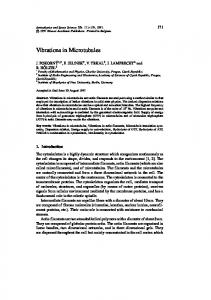
Cross Fractures through the Axostyle Fig. 1 shows the image of the Saccinobaculus axostyle seen in thin sections of material prepared by conventional procedures (Mclntosh et al., 1973). The microtubules are ordered into rows and each microtubule is always linked to its neighbors within a row by densely staining intrarow bridges. The angle of the intrarow bridge with respect to the tubule-tubule axis was measured for 150 of the intrarow bridges in Fig. 1. The average angle is 11 ° with a range of 0-30 °, and 40% of the bridges are oriented at an angle greater than 15 ° . Occasionally, more weakly staining bridges are seen to connect microtubules in adjacent rows of the axostyle (arrowheads in Fig. 1). A low-magnification view of a cross fracture through the axostyle of Saccinobaculus is shown in Fig. 2. This axostyle has the characteristic crescent shape and consists of 30 32 curving rows of microtubules, and the packing of the microtubules varies from square at the top to hexagonal at the bottom of the micrograph. Even at this magnifica-
tion, the extensive cross bridging of the microtubules is visible. A higher magnification view of the outlined region of the axostyle in Fig. 2 is shown in Fig. 3. The rows of the axostyle are oriented horizontally and most of the microtubules can be seen to be bridged to their neighbors within a row. All of the microtubules within a row are probably cross-bridged, as is seen in sectioned material, but the plane of the fracture may sometimes pass from one microtubule to the next in a row at a level above or below that of the bridge. The intrarow bridges can sometimes be seen to be oriented at an angle from the line joining the centers of neighboring microtubules. The extent of bridging of microtubules in adjacent rows as seen in freeze-fractured images appears to be substantially greater than that in axostyles prepared by conventional methods. Each microtubule has up to four sites of attachment to interrow bridges (see arrows in Fig. 3), although it is seldom that all four interrow bridge-binding sites are occupied. Occasionally, what appear to be two interrow bridges are observed between the same pair of microtubules (circle in Fig. 3). The intrarow bridges differ from the interrow bridges in being shorter and thicker (160 A x 90 A versus 210 A x 60 A) and being positioned at an angle to the tubuletubule axis. Table I presents data on the diameter and spacing of the axostyle microtubules in freeze-fracture replicas compared to those from sectioned m a t e r i a l prepared by c o n v e n t i o n a l m e t h o d s (Mclntosh et al., 1973). The diameters of the microtubules are increased 20-40% while both the intrarow and interrow spacings are increased approximately 50%.
FIGURE 1 Cross section of the axostyle of Saccinobaculus. The rows of the axostyle extend horizontally and every pair of microtubules within a row is connected by a densely staining bridge. Certain pairs of microtubules in adjacent rows are connected by slender, more weakly staining bridges (arrowheads). x 110,000 FIGURE 2 Cross fracture of the axostyle of Saccinobaculus. The curving microtubule rows extend vertically. The microtubules are hexagonally packed at the lower portion and square packed at the upper portion of the micrograph. The area within the box is shown at higher magnification in Fig. 3. The arrowhead in the lower left corner indicates the direction of shadow. × 34,000. FIGURE 3 Cross fracture of the Saccinobaculus axostyle at high magnification, with the microtubule rows extending horizontally. In addition to the connections between microtubules within a row, there is extensive cross-bridging of microtubules in adjacent rows. The arrows indicate some of the interrow bridges. The circle encloses a double interrow bridge extending between two microtubules. The arrowhead in the lower left corner indicates the direction of shadow, x 165,000.
662
THE JOURNAL OF CELL BIOLOGY . VOLUME
62,
1974
R. A. BLOODGOOD AND K. R. MILLER Freeze-Fracture o f the Axostyle
663
TABLE I
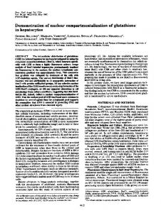
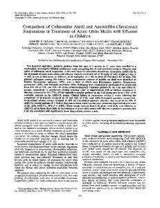
bridges oriented obliquely or projecting from the plane of the section. The optical transform shown Longitudinal as an inset to Fig. 5 has prominent layer lines at Microtubule perioMicrotuble 160 A representing the center-to-center spacing of space* dicity diameter the subunits comprising the microtubule wall. ,4 ,4 ,4 Fig. 6 shows a micrograph of a Saccinobaculus Saccinobaculus axostyle where the fracture plane has passed Freeze-fracture replica 525 x 560 160 300 400 through the axostyle almost normal to the miFixed and unfixed crotubule rows so that the long axis of the interrow Saccinobaculus bridges should be in the plane of the fracture while Thin sections 350 x 425 150 250 the long axis of the intrarow bridges should be Pyrsonympha normal to the plane of the fracture (see Fig. 4 for Freeze-fracture replica 570 x 670 160 300 350 orientation). The single rows of regularly spaced, Fixed large (80 A) particles (see vertical arrowhead in Pyronympha Fig. 6) are interpreted as strings of intrarow Thin sections 350 x 400 155 250 bridges viewed on end. The spacing of the particles * The first value is the row-to-row spacing; the second value is the in these rows is 160 A. A microtubule profile is center-to-center spacing within a row. These measurements were marked by the lower bracket in Fig. 6. If this made from optical diffraction patterns of electron micrographs. profile is followed upward, it changes to a single row of large particles (interpreted again as intraLongitudinal Fractures through row bridges on end), and further on again assumes the Axostyle a double row appearance. We interpret this image In the absence of preferential fracture planes, as a microtubule leaving and entering the plane of many different views of the axostyle can be the fracture. Nonperiodically spaced material can obtained with longitudinal fractures. The stippled be seen in the space to the right of this microtubule lines in Fig. 4 indicate the approximate fracture profile (horizontal arrowheads in Fig. 6). If Fig. 6 planes of the three views of the axostyle chosen for is tilted approximately 75 ° about the horizontal presentation. Fig. 5 shows a longitudinal view of axis and viewed in this manner, the particulate the Pyrsonympha axostyle. Each pair of parallel matter seemingly bridging the gap between this rows of globular subunits (see arrows in Fig. 5) microtubule and its neighbor to the right can now separated by a space containing little material is be seen more clearly and is interpreted as repreinterpreted as the image of a microtubule. Each senting nonperiodic, interrow bridges lying with row of globular subunits represents the view seen their long axis in the plane of the fracture. when the fracture plane passes through the wall of Fig. 7 shows a fracture whose plane follows a the microtubule. The space within each pair of row of microtubules and hence is almost normal to closely spaced rows of globular subunits represents the plane represented by the micrograph in Fig. 6. the lumen of the microtubule. The wide spacing of The periodic, intrarow bridges now have their long the microtubules is interpreted as resulting from a axes in the plane of the fracture (see Fig. 4 for fracture plane oriented as shown in Fig. 4. Based orientation). Numerous profiles of intrarow on this interpretation of the fracture plane, only bridges are visible in Fig. 7 and although many of interrow bridge material would show up between the bridges are tilted as much as 30 ° from the the microtubules and hence the absence of periodic normal to the microtubule axis (which is vertical), material in this region is not surprising. The all of the bridges within a vertical row are strictly interrow bridges expected to be present between oriented in parallel. The strictly periodic nature of these microtubules would be oriented end-on, these presumed intrarow bridges is shown by the making recognition difficult. Comparison of mea- optical transform inset to Fig. 7 which has promisurements of microtubule diameter and spacing nent layer lines at 160 A. made on cross fractures of the Pyrsonympha Fig. 8 is a micrograph of a rather irregular axostyle with measurements made on the micro- fracture which gives two very different views of the graph shown in Fig. 5 exactly fit the above Saccinobaculus axostyle. Most of the micrograph interpretation, and render unlikely the possibility shows row on row of periodic intrarow bridges that we are looking at rows of periodic intrarow viewed both superimposed on microtubule profiles
664
THE JOURNAL OF CELL BIOLOGY - VOLUME 62, 1974
FIGURE 4 Diagrammatic representation of a transverse view of the axostyle based on information from cross sections and cross fractures. The stippled lines represent the approximate fracture planes of the images in Figs. 5 7. FIGURE 5 Longitudinal fracture through the axostyle of Pyrsonympha. The orientation of the fracture is shown in Fig. 4. Pairs of lines of globular subunits are interpreted as microtubule profiles. The wall of the microtubule is composed of globular subunits (arrows) with a center-to-center spacing of 160 ,~ as indicated by the prominent layer lines in the optical transform shown in the inset. The area enclosed in the box represents the area of the photograph used to obtain the optical transform. The arrowhead in the lower left corner indicates the direction of shadow. × 200,000.
and viewed between microtubule profiles (in particular, see the area within the box, which corresponds to the optical transform inset). This image is similar to that seen in Fig. 7. The only periodicity observed in the entire micrograph is at 160 A (see prominent layer lines in the optical transform), which is interpreted as being due to both the spacing of the intrarow bridges and the spacing within the microtubule walls. In the far right portion of Fig. 8, profiles of microtubule walls can be seen to be connected by rather widely spaced, narrow, nonperiodic bridges (see arrowheads in Fig. 8). These are interpreted as interrow bridges with their long axes in the plane of the fracture and can be seen oriented at various angles to the microtubule axis. The orientation of this portion of the fracture is similar to that in Fig. 6. DISCUSSION By means of reactivation experiments, we have demonstrated that the preparative procedures used in this study leave the axostyle in a potentially functional state. Without the need for chemical fixation, dehydration in organic solvents, or staining with heavy metals, we have confirmed the previously described globular subunit nature of the microtubule wall in both transverse and longitudinal view and have demonstrated the presence of two morphologically distinguishable kinds of microtubule-associated bridges. The nonperiodic bridges are more readily visualized by the freezefracture technique than by conventional preparative procedures. We observe that interrow bridges are longer and more slender than the intrarow bridges and are arranged in a less periodic manner. In longitudinal fractures, neighboring interrow bridges can assume different angles to the microtubule axis while the intrarow bridges are always arranged strictly in parallel. Tilney et al. (1973) have recently demonstrated,
using the tannic acid-glutaraldehyde technique of Mizuhira and Futaesaku (1971), that the wall of microtubules from virtually any source, including the axostyle, as viewed in transverse section is composed of 13 symmetrically arranged globular subunits. The present study demonstrates the presence of nearly symmetric sixfold bridge-binding sites on the axostyle microtubules. In addition, Tucker (1968) has demonstrated hexagonal packing and sixfold symmetric cross bridging of microtubules in the cytopharyngeai rods of Nassula. The problem arises as to how a microtubule with 13 equally spaced subunits and hence 13-fold symmetry can have sixfold symmetric bridge-binding sites. The measurements given in this paper and observations on tannic acid-glutaraldehydetreated axostyles suggest that the intrarow bridges are associated with more than one wall subunit. If each bridge need not be associated with a single microtubule subunit, various models for the relationship of the sixfold bridge-binding sites to the 13 subunits can be made which give approximately sixfold symmetry. Occasionally, what appear to be double interrow bridges can be seen to connect the same pair of microtubules. These images are not, at this time, understood. There are several possible interpretations: (a) the double bridge image is an anomalous one resulting from some combination of fracturing and shadowing and actually represents a single bridge; (b) the fracture plane transiently traveled along the length of the microtubule revealing bridges at different levels. This is extremely unlikely since interrow bridges are spaced at minimum intervals of 150-160 A along the microtubule and the shadowing would probably reveal the change in position of the fracture plane; (c) a microtubule can be bridged to another tubule in an adjacent row by two bridges and hence there is the possibility that each microtubule can have more
FIGURE 6 Longitudinal fracture through the Saccinobaculus axostyle with an orientation as indicated in Fig. 4. The bracket indicates a presumptive microtubule profile and the horizontal arrowheads indicate material located between microtubule profiles and interpreted as being interrow bridges. The vertical arrow indicates a row of large globular subunits with a center-to-center spacing of 160 A and interpreted as end-on images of intrarow bridges. The arrowhead in the lower left corner indicates the direction of shadow, x 168,000. FIGURE 7 Longitudinal fracture through the Saccinobaculus axostyle with an orientation as indicated in Fig. 4. The periodic structures are interpreted as intrarow bridges. The optical transform in the inset shows the outlined area of the micrograph and has prominent layer lines at 160 A. The arrowhead in the lower left corner indicates the direction of shadow, x 109,000. 666
THE JOURNALOF CELL BIOLOGY - VOLUME62, 1974
R. A. BLOODGOOD AND K. R. MILLER
Freeze-Fracture of the Axostyle
667
FIGURE 8 Longitudinal fracture through the Saccinobaculus axostyle. The arrowheads indicate presumptive interrow bridges connecting microtubule profiles. The optical transform inset shows the outlined area of the micrograph and has layer lines at 160 fit. The arrowhead in the lower left corner indicates the direction of shadow. × 120,000. t h a n four interrow bridge-binding sites. In any case, each microtubule can be connected by interrow bridges to a m a x i m u m of four other microtubules, two in each adjacent row. The d i a m e t e r of the microtubules observed in the freeze-fracture replicas is significantly larger
668
t h a n the 2 4 0 - 2 6 0 - A d i a m e t e r observed for axostyle microtubules and most other microtubules in thin sections. The latter procedure involves dehydration t h a t m a y remove water molecules import a n t to the spacing of the protofilaments within the microtubule. R e m o v a l of this water m a y allow the
THE JOURNAL OF CELL BIOLOGY VOLUME 62, 1974 •
protofilaments to pack together more tightly, reducing the diameter of the microtubule without necessarily affecting its longitudinal dimension. Stebbings and Willison (1973) noted a 30% increase in microtubule diameter as measured from freeze-etch replicas versus sectioned material. Cohen et al. (1971) have shown that the distribution of diffracted intensity from the surface lattice of wet microtubules observed by X-ray analysis changes when the preparation is dried. Support for the hypothesis that the larger diameter of microtubules observed by freeze-fracture more accurately reflects the true diameter comes from the work of Fern~ndez-Mor~n and Finean (1957). These authors showed that both osmium tetroxide fixation and dehydration in ethanol resulted in a reduction of the spacing of myelin over fresh material as measured by X-ray diffraction. On the other hand, myelin infiltrated with 25% glycerol and examined by freeze-etching has the same spacings as fresh myelin examined by X-ray diffraction (Bischoff and Moor, 1967 a, b). The spacings between microtubules in the axostyle are also significantly larger in freeze-fracture images than in sectioned material. It is conceivable that dehydration with organic solvents during processing by conventional methods may remove intertubule material necessary for maintaining the proper spacing of the microtubules. Indeed, it is possible that each microtubule has a layer of material associated with its surface analogous with the surface coat or "glycocalyx" found on many cell surfaces, and that dehydration may remove this material. A 40-80-A clear zone or exclusion zone devoid of recognizable structure has been observed around microtubules in thin sections (Ledbetter and Porter, 1963; Porter, 1966; Silver and McKinstry, 1967; Lane and Treherne, 1970; Macgregor and Stebbings, 1970) and freeze-etch replicas (Stebbings and Willison, 1973). It may be normally unstained surface material that is being visualized when microtubules are observed in negative contrast after treatment with lanthanum (Lane and Treherne, 1970; Bannister, 1972; Burton and Fernandez, 1973), ruthenium red (Tani and Ametani, 1970), thiourea (Warner and Satir, 1973), or tannic acid (Mizuhira and Futaesaku, 1971; Tilney et al., 1973), or even without specific treatment (Ledbetter and Porter, 1964). Both lanthanum and ruthenium red bind to polyanionic substances, and Lane and Treherne (1970) have suggested that the "microtubule glycocalyx" occupying the clear zone may be polysaccharide in nature.
It can be argued that the increase in diameter and spacing of axostyle microtubules observed in freeze-fracture replicas may be due to artifacts of freezing, glycerination, or shadowing. The 20-40% increase in diameter and the 50% increase in the spacing of the microtubules in the freeze-fracture replicas cannot be due entirely to the increase in volume of water upon freezing, since this accounts for only a 9% increase in volume and is even less for glycerol-water mixtures (Newman, 1968). Leonard et al. (1972) observed the lattice spacing of Xenopus yolk crystals to be 92-105/~ in thin-sectioned material, and 80-100 A in freeze-etched material using no cryoprotective agent. Olive et al. (1973) examined cytochrome b~ crystals by negative staining and freeze-etching and found the spacing values to be 165 ± 5 /~ and 164 ± 5 A, respectively, for trigonal crystals, and to be 95 ± 5 A and 94 ± 6 A, respectively, for tetragonal crystals. It appears that for protein crystals, at least, the freezing process induces little or no change in dimensions. The possibility that the diameter of the microtubules could be increased by the shadowing process has been eliminated by making measurements perpendicular to the direction of shadow. Few data are available concerning the effects of glycerol on the dimensions of structures in biological systems. Artificial lecithin bilayers are increased in diameter by 0-17% in the presence of 20% glycerol, depending upon the concentration of lecithin (Buckingham and Staehelin, 1969). This is, of course, a purely lipid system, while the axostyle is composed almost solely of protein. As mentioned previously, myelin infiltrated with 25% glycerol and examined by freeze-etching has the same spacings as untreated myelin examined by X-ray diffraction (Bischoff and Moor, 1967 a, b). Although little is known about glycerol-protein interactions, the present work has shown that the axostyle is perfectly functional in the presence of 25% glycerol. The minimum periodicity observed in the freezefracture replicas of the axostyle is 160 ,~ and corresponds to both the periodicity of the intrarow bridges and a longitudinal periodicity of the microtubule wall. This correlates well with the minimum observed longitudinal periodicity of thin-sectioned axostyles (Mclntosh et al., 1973) even after application of the tannic acid-glutaraldehyde technique (Bloodgood, unpublished observations). Moor (1966 a) observed 40-A and 80-A periodicities in freeze-etched yeast spindles, and Stebbings and Willison (1973) observed a 40-A periodicity in
R. A. BLOODGOOD AND K. R. MILLER Freeze-Fractureof the Axostyle
669
freeze-etched cytoplasmic microtubules from an insect ovary, but neither of these is a highly cross-bridged system of microtubules. Virtually all microtubules observed by negative staining have a basic 40 × 50-,~, subunit with the 4 0 - ~ dimension as the longitudinal periodicity of the microtubule. Based on the observed subunit structure in the wall of axostyle microtubules (Tilney et al., 1973) and the chemical similarity of axostyle microtubules to other microtubules ( M o o s e k e r and Tilney, 1973), it is likely that axostyle microtubules are also composed of 40 x 50-,~ globular subunits. The 160-A periodicity observed in the microtubule wall by freeze fracture m a y reflect a slightly aperiodic a r r a n g e m e n t of the typically observed 40-A microtubule subunits, thereby providing bridge-binding sites with a periodicity greater than that of the basic subunit. The authors thank Dr. L. A. Staehelin for the use of his freeze-etch facilities, and Drs. Staehelin and Mclntosh for advice during the course of this work and for a critical reading of the manuscript. Cryptocercus punctulatus were provided by Mr. Keith H. Griffith and Dr. Hope T. M. Ritter, and Reticulitermesflavipes were provided by Dr. Richard V. Smythe. Fig. I was provided by J. R. Mclntosh. A preliminary report of this work was presented at the 13th Annual Meeting, in November 1973, of the American Society for Cell Biology, Miami Beach, Florida. This work was supported by National Science Foundation grant GB 25876 to J. R. Mclntosh and National Institute of General Medical Science grant GM 18639 to L. A. Staehelin. Received for publication 27 December 1973, and in revised form 12 April 1974.
REFERENCES
BANNISTER, L. H. 1972. Lanthanum as an intracellular stain. J. Microsc. 95:413-419. BISCHOFF, A., and H. MOOR. 1967 a. Ultrastructural differences between the myelin sheaths of peripheral nerve fibers and CNS white matter. Z. Zellforsch. Mikrosk, Anat. 81:303-310. BtSCHOFF, A., and H. MOOR. 1967 b. The ultrastructure of the "Difference Factor" in the myelin. Z. Zellforsch. Mikrosk. Anat. 81:571-580. BROWN, R. M., and W. W. FRANKE. 1971. A microtubule crystal associated with the golgi field of Pleurochrysis scherfelii. Planta Med. 96:354-363. BRUGEROLLE, G. 1970. Sur l'ultrastructure et la position syst6matique de Pyrsonympha vertens (Zooflagellata Pyrsonymphina). C. R. Hebd. Seances Acad. Sci. Ser. Sci. Nat. D. 270:966-969.
670
BUCKINGHAM,J. H., and L. A. STAEHEL1N. 1969. The effect of glycerol on the structure of lecithin membranes; a study by freeze-etching and X-ray diffraction. J. Microsc. 90:83 106. BURTON, P., R., and H. L. FERNANDEZ. 1973. Delineation by lanthanum staining of filamentous elements associated with the surfaces of axonal microtubules. J. Cell Sci. 12:567-583. COHEN, C., S. C. HARRISON, and R. E. STEPHENS. 1971. X-ray diffraction from microtubules. J. Mol. Biol. 59:375-380. FERIqANDEz-MoRAN, H., and J. B. FINEAN. 1957. Electron microscope and low-angle X-ray diffraction studies of the nerve myelin sheath. J. Cell Biol. 3:725-748. GRASSI~ P.-P. 1956. L'ultrastructure de Pyrsonympha vertens (Zooflagellata Pyrsonymphina): les flagelles et leur coaptation avec le corps, l'axostyle contractile, le paraxosty|e, le cytoplasme. Arch. Biol. 67:595 609. GRIMSTONE, A. V., and L. R. CLEVELAND. 1965. The fine structure and function of the contractile axostyles of certain flagellates. J. Cell Biol. 24:387-400. GUTH, E., T. HASHIMOTO, and S. F. CONTI. 1972. Morphogenesis of ascospores in Saccharomyces cerevisiae. J. Bacteriol. 109:869 880. HOLLANDE, A., and J. CARRUETTE-VALENTIN. 1970. La lign6e des Pyrsonymphines et les caract[res infrastructuraux communs aux genres Opisthomitus, Oxymonas, Saccinobaculus, Pyrsonympha et Streblomastix. C. R. Hebd. Seances Acad. Sci. Ser. D. Sci. Nat. 270:966 969. LANE, N. J., and J. B. TREHFRNE. 1970. Lanthanum staining of neurotubules in axons from cockroach ganglia. J. Cell Sci. 7:217-23 I. LEDBETTER, M. C., and K. R. PORTER. 1963. A microtubule in plant cell fine structure. J. Cell Biol. 19:239-250. LFDBETTER, M. C., and K. R. PORTER. 1964. The morphology of microtubules of plant cells. Science ( Wash. D.C.)144:872-874. LEONARD, R., D. W. DEAMER, and P. ARMSTRONG. 1972. Amphibian yolk platelet ultrastructure wisualized by freeze-etching. J. Ultrastruct. Res. 40:1-24. MACGREGOR, H. C., and H. STEBBINGS. 1970. A massive system of microtubules associated with cytoplasmic movement in teletrophic ovarioles. J. Cell Sci. 6:431-449. MClNTOSH, J. R. 1973. The axostyle of Saccinobaculus. II. Motion of the microtubule bundle and a structural comparison of straight and bent axostyles. J. Cell Biol. 56:324-339. MCINTOSrt, J. R. 1974. Bridges between microtubules. J. Cell Biol. 62:000. MCINTOSH, J. R., E. S. OGATA, and S. C. LANDIS. 1973. The axostyle of Saccinobaculus. 1. Structure of the organism and its microtubule bundle. J. Cell Biol. 56:304-323. MIZUH1RA, V., and Y. FUTAESAKU. 1971. On the new approach of tannic acid and digitonine to the biologi-
THE JOURNAL OF CELL BIOLOGY . VOLUME 62, 1974
cal fixatives. 29th Annual Proceedings of the Electron Microscope Society of America. 494 495. MooR, H. 1966 a. The performance of freeze-etching and the interpretation of results concerning the surface structure of membranes and the fine structure of microtubules and spindle fibers. Balzers High Vacuum Report. Balzers High Vacuum Corp., Santa Ana, Calif. 9:1-12. MOOR, H. 1966 b. Ultrastrukturen im Zellkern der Backerhefe. J. Cell Biol. 29:153-155. MOOR, H. 1967. Der Feinbau der Mikrotubuli in Here nach Gefrieratzung. Protoplasma. 64:89 103. MOOR, H., and K. MI~HLETHALER. 1963. Fine structure in frozen-etched yeast cells. J. Cell Biol. 17:609-628. MOOSEKER, M. S., and L. G. TILNEY. 1973. Isolation and reactivation of the axostyle. Evidence for a dynein-like ATPase in the axostyle. J. Cell Biol. 56:13-26. NEWMAN, A. A. 1968. Glycerol. Chemical Rubber Company, Cleveland, Ohio. NORTHCOTE, D. H., and D. R. LEWIS. 1968. Freeze etched surfaces of membranes and organelles in the cells of pea root tips. J. Cell Sci. 3:199 209. OLIVE, J., J. N. BARBOTIN, and J. L. RISLER. 1973. The crystal and molecular structure of yeast L-lactate dehydrogenase (cytochrome b2). An electron microscope study by negative staining and freeze-etching technique. Int. J. Protein Peptide Res. 5:219-228. PORTER, K. R. 1966. Cytoplasmic microtubules and their function. In Principles of biomolecular organization. G.E.W. Wolstenholme, and M. O'Connor, editors.
J. & A. Churchill Ltd., London, England. 308-345. SILVER, M. D., and J. E. McKINSTRY. 1967. Morphology of microtubules in rabbit platelets. Z. Zellforsch. Mikrosk. Anat. 81:12 17. STEBBINGS,H., and J. H. M. WILLISON. 1973. Structure of microtubules: a study of freeze-etched and negatively stained microtubules from the ovaries of Notonecta. Z. Zellforsch. Mikrosk. Anat. 138:387-396. STEPHENS, R. E., and R. W. LINCK. 1969. A comparison of muscle actin and ciliary microtubule protein in the mollusk Pecten irradians. J. Mol. Biol. 40:497 501. TAM, E., and T. AMETANI. 1970. Substructure of microtubules in brain nerve cells as revealed by ruthenium red. J. Cell Biol. 46:159-165. TILNEY, L. G. 1971. Origin and continuity of microtubules. In Origin and continuity of cell organelles. J. Reinert and H. Ursprung, editors. Springer-Verlag New York Inc., New York. TILNEY, L. G., J. BRYAN, D. J. BUSH, K. FUJIWARA, M. S. MOOSEKER, D. B. MURPHY, and D. H. SNYDER. 1973. Microtubules: evidence for 13 protofilaments. J. Cell Biol. 59:267-275. TRAGER, W. 1934. The cultivation of a cellulose-digesting flagellate, Trichomonas termopsidis, and of certain other termite protozoa. Biol. Bull. (Woods Hole). 66:182 190. TUCKER, J. B. 1968. Fine structure and function of the cytopharyngeal basket in the ciliate Nassula. J. Cell Biol. 3:493-514. WARNER,F. D., and P. SATIR. 1973. The substructure of ciliary microtubules. J. Cell Sci. 12:313-326.
R. A. BLOODGOOD AND K. R. MILLER Freeze-Fracture of the Axostyle
671