THE ORIGIN AND KINETICS OF MONONUCLEAR PHAGOCYTES. BY RALPH VAN FURTH,* M.D., ~o ZANVIL A. COLIN, M.D.. (From The Rockefeller University ...
THE ORIGIN AND KINETICS OF MONONUCLEAR PHAGOCYTES BY RALPH VANFURTH,* M.D., ~ o ZANVIL A. COLIN, M.D.
(From The Rockefeller University, New York 10021) (Received for publication 8 May 1968) Mononuciear phagocytes can be separated into two groups of cells: the circulating mononuclear phagocytes or monocytes in the peripheral blood and tissue macrophages in organs such as the spleen, lymph nodes, liver (Kupffer's cells), the lung (alveolar macrophages), the peritoneal cavity, and the subcutaneous tissues. Tissue macrophages are mainly involved in the clearance and destruction of bacteria, nonmicrobial foreign materials and damaged tissue cells from the bloodstream, lymph, or outer world (1). The function and fate of the monocytes in the circulation is not clearly understood. I n vitro these cells can mature into a cell with characteristics of tissue macrophages (2) a n d a similar transformation was shown to occur in vivo at extravascular sites (3). Recently it has been reported that macrophages in inflammatory exudates originate from monocytes
(4-7). The aim of this s t u d y was to investigate the relationship between the free and fixed mononuclear phagocytes in different parts of the body under normal, steady state conditions. This communication will concern the origin and turnover of the blood monocyte and peritoneal macrophage of the mouse with observations on the properties of the bone marrow progenitor.
Materials and Methods Animals.--In all studies Swiss mice of the pathogen-free NCS strain, maintained at the Rockefeller University, were employed. Only male mice with a bodywdght between 25 and 30 g were used. Peritoneal Mc~rophages.--Mice were killed with chloroform. The skin over the abdomen was reflected and 1 ml of phosphate-buffered saline (pH 7.2) (Dffco Laboratories, Detroit, Mich.) with 50 uuits/ml heparin, without preservative, was injected into the peritoneal cavity. The abdomen was kneaded gently and after about 1 min the cell-rich fluid was removed with a capillary pipette and placed in a plastic tube (Falcon Plastics, Los Angeles, Calif.). These exudates, which contain about 2-5 X 100 cells, consist of about 40% macrophages, 60% lymphocytes, and a few mast cells. The cells were centrifuged for 8 rain, at 150 g, at room temperature. 2-ml culture medium was then added to the pellet. The cells were thoroughly mixed by repeated pipetting and two cultures of each sample were prepared which contained 1 ml of the cell suspension. Blood Monocytes.--Monocytes of the peripheral blood were studied mainly from smears, * Present address: Department of Microbiological Diseases, University Hospital, Leiden, The Netherlands. 415
416
ORIGIN AND KINETICS 0]~ ~rONONUCLEAR PHAGOCYTES
prepared from tail vein blood. The slides were fixed in absolute methyl alcohd, for 30 min at 4°C. Blood was aspirated from the axillary pouch with heparinized pipettes and employed for the preparation of monocyte cultures. Two parts of blood were mixed with one part of 3 % gelatine solution (Difco Laboratories) in buffered saline and the tubes were incubated for 30-50 rain at 37°C. During that period the red cells sedimented, leaving a leukocyte-rich supernatant. This was removed, centrifuged at 180 g, and washed once with medium 199. The cells were then suspended in the culture medium, and 1 ml of the suspension containing about 10 X 106 leukocytes per mm*, of which 1.5-5% were monocytes, were then incubated in a Leighton tube. Bone Marrow.--Both femurs of the mouse were isolated and removed unimpaired from the hind limb. After cleaning the bone from the adherent muscles, the femur was cut at both ends in the region of the metaphysis. A needle (26 gauge) on a 2.5 ml syringe was inserted into one end of the femur, and bone marrow tissue was expressed by flushing the femur shaft with 2-ml culture medium. The bone marrow tissue was collected in a sterile Petri dish and teased with needles. The cell suspension was then transferred to a plastic tube and the cells were further dispersed by repeated, gentle aspiration in a pipette. The cell suspension, divided into equal parts of about 1 nil, which contained about 8 X I0 e nucleated cells, was pipetted into two Leighton tubes. Tissue Culture Te~hniclues.--The tissue culture methods have been outlined previously (8), and will be described briefly in respect to the different tissues used in this study. The culture medium consisted of medium 199 (Microbiologieal Associates Inc. Bethesda, Md.), 20 or 50% newborn calf (NBC) serum (Grand Island Biological Co., Grand Island, N. Y.), and 200 anits/ml penicillin G. The medium for the bone marrow cultures also contained 50 ~g/ml streptomycin. All cultures were prepared in Leighton tubes, containing 10 X 35 nun flying cover slips, which were marked by cutting off one comer. The tubes were gassed with a 5% COrair mixture, closed with rubber stoppers, and incubated at 37°C. Peritoneal cell cultures were incubated in medium 199 and 50% NBC serum for 2 hr, during which time the macrophages attached to the cover slips. At this time point the medium was removed and the cell layer on the cover slip was washed three times with 1.5 ml of medium 199 by vigorous shaking. After the fluid was removed for the last time, the slides were fixed with 2 % giutaraldchyde (pH 7.4), for 30 rain at 4°C. The cover slips were then removed, washed three times in distilled water, and dried. The slides were stored in capped plastic tubes until they were prepared for radioautography. Peripheral blood leukocytes were incubated in medium 199 and 50% newborn calf serum for 2 hr, allowing the cells to attach to the cover slip. The supernatant was then removed, the cell layer on the cover slip was washed as described for the peritoneal cells, and again incubated for 24 or 48 hr. The slides were then prepared for phase-contrast microscopy or for radioautography. Bone marrow cells were incubated in medium 199 and 20% newborn calf serum for 2 hr. After the supernatant was removed, the cover slip was thoroughly washed three times with medium 199 and then incubated again with 2 ml of culture medium for 24 hr. After that period the cover slips were washed again with medium 199, and incubated once more for 24 hr. The additional washes were required to remove degenerating cells of the granulocyte series. The cover slips were finally washed again, fixed in 2% giutaraldchyde (pH 7.4) and prepared for phase-contrast microscopy, light microscopy, or radioautography. Labd/ng.--The cells were labeled with methyl thymidine-SH (spec. act. 6.7 c/mmole; New England Nuclear Corporation, Boston, Mass.). The in vivo labeling was done according to two different schedules. The animals were labeled by one intravenous injection of thymidineSH (1 tae/g body weight) or by four intramuscular injections of thymidine-SH (1 ~c/g body
RALPH VAN FURTH AND ZANVIL A. COHIq
417
weight) each 3 hr. According to the second schedule the last injection, which was given 12 hr after the first, represents the zero point on the graphs. The cells were then harvested for the first time period 12 hr after the last injection. The in vitro labeling of peritoneal macrophages, monocytes, and bone marrow cells was performed by incubating the cells for a given time period, in a medium which contained 0.1 ~c/ml thymidine-SH. All procedures prior to the incubation of the ceils in a radioactive culture medium were also performed with solutions which contained 0.1 ~c/ml thymidine-*H. After incubation, the cover slips were washed five times with medium 199, in order to ensure the removal of atl radioactive thymidine. The slides were then fixed with 2~o gintaraldehyde and prepared for radioautography. Radioautography.--Radioantography was performed with the Ilford Nuclear Research Emulsion, K5 in gel form, according to Caro, van Tubergen, and Kolb (9). An emulsion at a concentration of I g/ml of glass distilled water was prepared and melted at 45°C, for 15 rain. The emulsion was then placed for 30 rain at room temperature. After this period the cover slips and microscope slides were dipped into the emulsion. The cover slips and slides were then held in a vertical position to allow the emulsion to drain away from the glass as completely as possible. The emulsion was dried for 8-12 hr at room temperature. The cover slips and microscope slides were stored in a light-tight box containing desiccant at room temperature. The exposure time for radioautography was 21 days. The emulsion was developed with Kodak Developer D-19, for 2 rain. After fixation the slides were washed for 5 rain in running tap water and I rain in distilled water. Staining.--The staining of the slides, covered with a film emulsion, was performed by a modified Giemsa method (V. P. Bond, personal communication). A staining solution was prepared of 5 ml Giemsa stain, 3 mi absolute methyl alcohol, and 92 ml phosphate-citrate buffer (pH 5.75). The pH of this solution was adjusted to 5.75 with a few drops of the buffer stock solution. The solution was faltered through four layers of kleenex tissues before staining the slides. Mononuclear phagocytes cultured on cover slips were stained for 3-5 rain, bloodsmears were stained for about 90 rain. After staining the slides were carefully flushed off with the buffer solution, and the slides were Idt covered for 3-5 rain with the buffer to wash out excess stain. Examination of the Slides and Counting of the Gra/ns.--The slides were examined with the oil immersion objective at a magnification of 1000X, employing a Zeiss binocular microscope. The background of silver grains was evaluated in several experiments by counting the grains over 100 mononudear phagocytes on preparations made from nonlabeled animals. According to these counts all cells with three or more silver grains over the nucleus were considered positive. At each time point a total of at least 600 peritoneal and bone marrow mononuclear phagocytes were counted from duplicate cultures made from one mouse. A total of at least 100 monocytes, from three to five blood smears, were counted for each mouse at a given time point. Each point on the graphs represents the mean value of at least two mice, but in the majority of the experiments the mean of six or eight mice. The vertical bars represent the range of the individual values. Photography.--The photographs were taken with the Zeiss ultraphot microscope (Model II), using 4 × 5 panatomic X film at a magnification of approximately 1000. Sp/~ectomy.--Splenectomy was performed in mice anestbesized with sodium pentoharbital (Diabutal, Diamond Laboratories Inc., Des Moines, Iowa), given 0.08 mg/g body weight. The peritoneal cavity was opened by a small incision under the left costal margin, and the spleen was mobilized outside the abdominal cavity. After the splenic vessels were ligated with silk, the spleen was removed, the peritoneum dosed with silk, and the muscles and skin with
418
ORIGIN AND KINETICS O F MONONUCLEAR PHAGOCYTES
metal clamps. In the sham-operated animals the spleen was only mobilized outside the abdominal cavity. No antibiotics were given. The labeling studies were started 25 days after splenectomy. X-Irradiation.--Miee were exposed to total-body irradiation of 430 fads from two opposing Maxitron 300 units (300 kv, 20 ma, working distance of 70 cm, HVL 1.6 mm Cu, the dose rate measured in air, inside the mousebox, is 142.5 R/min). The mice were kept in individual compartments in a lucite box, which was surrounded by a bolus-filled frame. Due to this construction an even distribution of irradiation within 1% was obtained. In another group of mice, that also received 430 rads, both hind limbs and a part of the pelvis were shielded. The animals were anesthesized with 2 mg sodium pentobarbitai (Diabutal) intramuscularly and taped on a plastic sheet. The lead shield was 0.5 cm, and the irradiation was performed with one unit (kv 300, 20 ma, working distance 70 cm, HVL 1.6 mm Cu, dose rate in air 65 R/min.). Measurements made with microrod lithium fluoride teflon dosimeters (con-Rad System Inc., Cambridge, Mass.) demonstrated that the shielded hind limbs received about 8% irradiation, mainly due to side scattering at 90° angle through the tissues. Phagocytosis.--The phagocytic ability of the cells was tested with Staphylococcus albus. Cells attached on the cover slip in the Leighton tube, were incubated with agar grown Staphylococcus albus (strain "Air"), and suspended in medium 199 with 20% newborn calf serum. Mter a period of 30 min the cover slip was washed three times with medium 199, fixed with 2% buffered glutamldehyde, and stained with Giemsa stain. Nomo~la~re.--Mononuclear phagocytes were defined as mononuclear cells which stick to glass and are able to phagocytize. According to this definition the cells left on the cover slip after incubation in the Leighton tube are called peritoneal macrophages, mononuclear macrophages of the peripheral blood or monocytes, and mononuclear phagocytes of the bone marrow or promonocytes. The monocytes studied in the blood smear were cells with a diameter of 12-20~, a pale blue cytoplasm and folded, horseshoe-shaped or reniform nucleus. RESULTS
Labeling of PeritonealMacropkages.--The mice, which were labeled according to the two schedules, showed a different pattern of labeled peritoneal macrophages. Mice labeled with 1 X 25 ~c thymidine-~H, showed 1.3 % labeled macrophages 1 hr after injection (Fig. 7 a). At 48 hr the peak labeling of 4.7% was found, and after 168 hr 2.2 % of the peritoneal cells were still labeled (Fig. 1). When 4 X 25 #c thymidine-SH was given, 6.3 % of the peritoneal macrophages were labeled 12 hr after the last injection. 50 hr after the last injection a peak of 17.0% was found, and at 156 hr there were still 4.7% labeled cells (Fig. 2). In vitro labeling of peritoneal macrophages, performed by incubating cells in the presence of thymidine-*I-I for 24 hr, showed that 2.2 % (range 0.5-5.9 %) incorporated thymidine-~I (Table I) (Fig. 7 b). Incubation periods up to 120 hr in the presence of thymidine-3H, showed no increase of the number of labeled cells. When cells incubated for 24 hr in the presence of thymidine-~H were washed and again incubated for 24, 48, 72, or 96 hr in the nonradioactive culture medium, the number of labeled cells did not increase. When peritoneal macrophages were incubated during the first 24 hr in medium without thymidine-"I-I and thereafter exposed to thymidine-3H for 24 or 48 hr, no labeling of
419
R A L P H VAN F U R T H AND Z A N V I L A. COHN
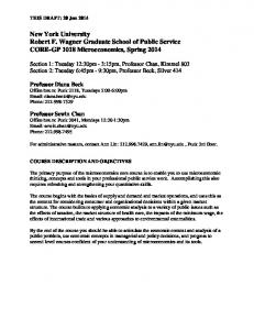
peritoneal cells was found. The in vitro labeling experiment thus clearly demonstrated that only a very small percentage of the peritoneal macrophages synthesize nuclear DNA. Labeling of Monocytes in the Peripheral Blood.--Mice labeled with 1 X 25 ~c thymidine-3H showed 2.9% labeled monocytes 1 hr after the injection (Fig. 7 c). After 12 hr 22.4% of the monocytes were labeled and at 48 hr the maximum of 57.0% labeled monocytes was found. At 168 hr there were still 14.0% labeled monocytes (Fig. 1). I00
o Peritoneal macrophages A Peripheral blood monocytes
8o
~ 6o cJ
~ 4o ck 20 o 0
I
I
i
6 12 24
I
I
I
48
72
96
I
120
I
144
168
Hours after t h y m i d i n e - 3 H ( I x l / ~ c / g weight) FIG. 1. Labeling of peritoneal macrophages and blood monocytesafter one intravenous injection of thymidine~H.
When 4 X 25/zc thymidine-3H was administered, 63 % of the monocytes were labeled 12 hr after the last injection and a maximum labding of 85 % was found at 60 hr. There were still 60.0% of the labeled monocytes 156 hr after the injection (Fig. 2). To check the counts of the labeled monocytes made from the blood smears, peripheral blood from labeled animals (1 X 25 #c/ml thymidine-~rI was cultured. After an incubation period of 24 hr the cells were processed for radioautography. The labeling percentage of the mononuclear phagocytes on the cover slip was 50.0% 24 hr after labeling, and 60.0% 48 hr after labeling. These results are in good agreement with the figures found from the bloodsmears. In vitro incubation of monocytes in the presence of thymidine-SH during 24 or 48 hr showed no labeled cells (Table I). This population of cells apparently does not synthesize nuclear DNA when present in the circulation.
420
ORIGIN A N D
KINETICS OF M O N O N U C L E A R
PHAGOCYTES
Previous experiments employing two different schedules of in vivo labeling showed that the maximum percentage of labeled macrophages in the peritoneal cavity was found at the same time point as the maximum percentage of the labeled monocytes in the peripheral blood. If the peritoneal macrophages are
o Peritoneal rnacrophages IOO
tO
z~ Peripheral blood monocytes
8O
O
-o
¢0 "G
60
O
4O 0~ O
IX. 201
0
I
|
12 24 36
I
60
8t4
'08
I
I
132
156
Hours after thymidine- 3H ( 4 x l F c / g weight) FIG. 2. Labeling of peritoneal macrophages and blood monocytes after four intramuscular injections of thymidine-3H. TABLE I In Vitro Labeling o] Mtmonudear Phagocytes* Labeled cells
Peritoneal macrophages Peripheral blood monocytes Bone marrow promonocytes
% 2.2 0 32.0
* In medium with 0.1 #c/m1 thymidine-SHfor 24 hr. derived from blood monocytes there appears to exist a rapid emigration of monocytes from the circulation into the peritoneal cavity. To obtain further evidence for this supposition the effect of an intraperitoneal injection of newborn calf serum was studied. This evokes a mild inflammatory response, represented by an increase of mainly mononuclear phagocytes and only a very few polymorphonuclear cells (Z. A. Cohn, unpublished observation).
421
R A L P H VAN F U R T H A N D ZANVIL A. COHN
The E(ect of Newborn Calf Serum on tke Percentage of Labeled Peritoneal Macropkagesand Blood Monocytes.--Mice were labeled with 4 X 25 uc thymidine-3H and 60 hr after the last injection, when the monocytes were maximally labeled, 1 ml newborn calf serum was injected intraperitoneally. During the first 48 hr after the serum injection the percentage of labeled peritoneal macrophages increased to 66 % (Fig. 3). The total number of peritoneal macrophages was doubled at this time point. Thereafter the percentage of labeled macrophages fell off. The mean grain count of the labeled macrophages of the serum-injected group was equal to the controls, at 108 hr. cO o
I00
o .,c o
• Normal
b 8o o E "S
o After stimulation with newborn calf serum /*(~ /"
eo 6 0 e-
\.
/ "o
/
4O
/ 0 - -
1./
20
C 0
%
./
0
a_
\.
1
I
2~4
i 56
260I
814
I 108
~ i 132
156
hours after thymidine-3H ( 4 x l p . c / g weight) FIo. 3. The effect of the intraperitoneal injection of 1 ml newborn calf serum, on the labeling pattern of peritoneal macrophages.
The percentage of labeled monocytes during the first 48 hr after the serum injection was within the range found in normal mice, however, the total number of labeled monocytes in the peripheral blood was tripled 24 hr after the injection, and fell off to normal during the following 48 hr (Table II). The incubation of the peritoneal macrophages, obtained 24 or 48 hr after an intraperitoneal injection of 1 ml newborn calf serum, in the presence of thymidine-rH for 24-120 hr, showed that 3.5% (range 1-5.8%) of the macrophages were labeled in vitro. This is no significant increase (P > 0.05) over the control group. From these experiments it can be concluded that the increase of peritoneal macrophages after an intraperitoneal injection of serum is not related to the rapid division of peritoneal cells. Rather, the serum evokes a rapid emigration of labeled monocytes from the circulation into the peritoneal cavity.
422
ORIGIN AND KINETICS OF MONONUCLEAR PHAGOCYTES
Additional experiments were then designed to elucidate the origin of the peripheral blood monocytes. The Effect of Splenectomy on the Percentage of Labeled Peritoneal Macropkages and Blood Monocytes.--The importance of the spleen as a source of monocytes was next examined. Groups of mice were splenectomized or sham operated. 25 days after surgery, 4 X 25 uc thymidine-~H was given to both groups of mice. TABLE II The Effect of an Intraperi~eal Injection of 1 ml Newborn Calf Serum on the Labeled Blood Monocytes* Time after thymidine-*H$
Labeled monocytes
Serum i.p.
Normal
Serum i.p.
Normal
hr
%
%
ram*
turn*
84 108 132
78 64 59
75.5 72 65
995 422 273
317 302 273
* 1 ml newborn calf serum intraperitoneaUy 60 hr after thymidine-3H. :~4 X 1 # c / g body weight intramuscularly. TABLE HI
The Effect of Splenectomy on the Percentage of Labded Blood Monocytes Labeled monocytes
Time after thymidine-*H* Splenectomy
Nomml
36
% 74
6O 84
81 65
% 76.5 85.5 75.5
]ir
Absolute number of monocytes3 wk after splenectomy:615/mma; normal 420/mm8. * 4 X 1 pc/g body weight intramuscularly. The labeling percentages of the monocytes of the splenectomized mice, at 36, 60, and 84 hr after the injections, was in the same range as found in normal mice (Table III). The total number of leukocytes of the mice, 25 days after splenectomy, was increased about 40 % in comparison with the normal group. However, the differential counts in both groups were equal. The peritoneal macrophages of splenectomized mice showed a higher labeling index than normal mice, but in the sham-operated group the labeling percentages were in the same range as the normal group (Table IV). The higher labeling index of the macrophages in the splenectomized mice might be due to an inflammatory reaction of the peritoneum, secondary to surgery, although
423
RALPH VAN I~URTH AND ZANVIL A. C0HN
no increase of polymorphonuclear cells were found in the peritoneal cavity. Since the total number of the labeled monocytes was increased, this may have accounted for a higher number of labeled peritoneal macrophages. These experiments have shown that the spleen is not a major source for the formation of blood monocytes. I t was next attempted to determine the origin of peripheral blood monocytes by studying the effect of X-irradiation. TABLE IV The E.ffe~t of S ~le~tomy on the Percentage of Labeled Peritoneal Macropkages Time after thymidine-tH*
Labeled peritoneal macrophag~
Spleaectomy:~
Sham operated:~
Normal
hr
%
%
%
36 60 34
33.3 32.9 31.0
7.8 20.1 14.0
13.4 17.0 8.3
* 4 X 1 p~/g body weight intramuscularly. Mean of two animals. TABLE V The Edffed of X-Irradlation, with and without Shielding of the Hind Limbs, on tke Labeled Blood Monocyte~* Labeled monocytes Time after thymidine-SH~ X-Irradiation kg
12
36 6O 84
X-Irradlation "t- shielding
mm 8
0 0.04 11 28
Normal mm!
lOO 165 305 475
267 321 359 317
* 650 R. 4 X 1 ~tc/g body weight intramuscularly 24 hr after X-irradiation. Tke Effect of Total-Body X-Irradiation on the Labeling of Blood Monecytes and Peritoneal Macropkages.--A group of mice received 435 rads total-body X-irradiation. 24 hr after the irradiation the mice were injected with 4 X 25/z¢ thymidine-SH. At 12, 36, 60, and 84 hr after the last injection peripheral blood monocytes and peritoneal macrophages were examined. During the first 36 hr after the injection apparently no labeled monocytes were present in the circulation; after that period a very small number, increasing to 10% of the normal value at 84 hr, were found (Table V). Labeled peritoneal macrophages were rarely found (one in 105-10e) at the 60- and 84-hr time points.
424
ORIGIN AND KINETICS OF MONONUCLEAR PHAGOCYTES
The Effect of Total-Body X-Irradiation, with Shielding of the Hind Limbs, on the Percentage of Labeled Blood Monocytes and Peritoneal Macrophages.IIn this group of animals, both the hind limbs and part of the pelvis were shielded with lead. The schedule of labeling was similar to the previous experiment. These studies, in which the peritoneum, liver, spleen, majority of the lymphatic tissues, and all bone marrow tissue except for both hind limbs were irradiated, showed that the total number of labeled monocytes during the first 36 hr was significantly lower than in the normal animals, but higher than in the unshielded irradiation experiment. At 84 hr the number of labeled monocytes was even higher than the controls (Table V). TABLE
VI
The Effect of X-Irradiation, with Skidding of Both Hind Limbs, on tke Percentage of Labeled Peritoneal Macrophages* Labeled peritoneal macrophages Time after thymldine-~H$
hr
12 36 6O 84 132 156
X-Irradiation + shielding
Normal
% 0.60 0.45 1.70 2.25 1.80 0.70
% 6.3 13.4
17.0 8.3 5.0 4.7
* 650 R. :~4 X 1 #c/g body weight intramuscularly. The labeling percentage of the peritoneal macrophages was significantly lower and the maximum percentage of labeled cells was found 24 hr later than in normal animals (Table VI). These experiments have thus shown an extreme decrease of the labeled monocytes and peritoneal macrophages after total-body X-irradiation. However, only a moderate diminuation of these labeled mononuclear phagocytes was found when a part of the bone marrow was shielded. These results point to the conclusion that the monocytes arise from the bone marrow. In the following experiments it was therefore attempted to label precursor cells in the bone marrow and to determine the turnover of these cells.
Labeling of Mononuclear Phagocytes of the Bone Marrmo---(Promonocytes).-Bone marrow cells cultured for 48 hr consisted of a pure population of macrophages, according to the previous criteria. The morphological events exhibited by the bone marrow ceils during this culture period will be discussed in a subsequent paper.
425
R A L P H VAN F U R T H A N D ZANVIL A. COHN
In vitro incubation of the bone marrow cells for 24, 48, or 72 hr in the presence of thymidine-SH showed that 32% (range 27-39%) of the mononuclear phagocytes were labeled (Table I) (Fig. 8 a, and b). Incubation of the bone marrow cells in a medium without thymidine-SH during the first 24 hr and thereafter during 24 hr in the presence of thymidine-SI-I, showed that 4% of the mononuclear phagocytes were labeled. When the incubation in nonradioactive medium was 48 hr, and was then followed by a 24 hr incubation with thymidine-SH, only 0.5 % of the mononuclear phagocytes were labeled. These results indicate that mononuclear phagocytes of the bone marrow belong to a I00/l'-
o Peritoneal macrophages
/
I80 / |
"~ Peripheral blood monocytes a Bone marrow promonocytes
/
6o O
40 ~. 20
6 12
4.8
7
96
120
14.4
168
Hours after thymidine-3H (IxlFc/g weight) FIt. 4. Labeling of mononuclear phagocytes of the bone marrow--promonocytes-monocytes,and peritoneal macrophagesafter one intravenous injection of thymidine-3H. pool of dividing cells. In vitro this property is present mainly during the first 24 hr of culture. In vivo labeling of mice with 1 X 25 #c thymidine-SH showed that 1 hr after injection 21.0% of the mononuclear phagocytes obtained from the bone marrow were labeled (Fig. 8 c). At 24 hr, 68.4 % of the cells were labeled, and there was a rapid decline thereafter (Fig. 4). Since no thymidine-SH was available during the 48-hr culture period in vitro, the labeling index at each time point represents the percentage of mononuclear phagocytes labeled in vivo. The peak of the labeled bone marrow cells is present 24 hr prior to the peak of labeled monocytes. This leads to the conclusion that the monocytes are derived from a progenitor in the bone marrow, which we will term a promonocyte.
ORIGIN AND KINETICS OF MONONUCLEAR PHAGOCYTES
426
I00
-
Q
o 8o 0
•E60
"[3
] _o 4O
> 2 grains
U
~ 2o o.
I 12
t 24
HOURS ofter
~ 48
5 72
>10 groins > 2 0 groins > 3 0 groins
96
thymidine-3H( I xl~,c/g
weight )
I00 --
b
~ _
> 2 grains
0 E 0
E 60--
"0
-~4o-~_._o~,~,.
>20 grains
2o-_-
> 6 0 grains >80 groins 12 24 Hours after
36 60 84 thymidine--3H ( 4 x I/~c/g
weight)
FIo. 5. Time sequence of labeled monocytes distributed in cohorts with di~erent grain counts, a, labeling I X 25 #c thymidine-SH intravenously; b, labeling 4 X 25/~c thymidine-IH intramuscularly.
427
RALPH VAN FURTH AND ZANVIL A. COHN
Kinetics of Perip~ral Blood Monocy~s.--The initial slope of the curve of labeled monocytes showed an hourly increase (N) of 1.7 %. If the initial influx ioo-
• Cells with 6 1 - 1 2 0 o C e l l s with 5 1 - 6 0
grains groins
g o O i,O
O
E
O
urs
io
¢.= o
I
I 12
I . 24
Hours
I
I
48
72
ofter
1 92
thymidine-3H
Fro. 6. Disappearance of heavily ~.be,]ed monocytes from the blood. ~ = 1.67 - 0.145 X for cohort with 31--60 ceils; ~ = 1.74 -- 0.135 × for cohort with 61-120 cells.
of monocytes from the bone marrow into the peripheral blood follows a strict "pipe line" principle (10), the maximal turnover time, which is the time to replace the total population of labeled and unlabeled monocytes in the peripheral blood compartment, is IO0/N = 100/1.7 = 59 hr (11-13). However, the
428
ORIGIN AND
KINETICS O F M O N O N U C L E A R
PHAGOCYTES
monocytes disappear randomly from the circulation, as will be discussed below. The minimal turnover rate will then be 1.7/0.693 = 2.45 % per hour and the maximal turnover time 40.8 hr for mice in the steady state. The life span of the monocytes in the circulation cannot be calculated from the disappearance rate of all labeled monocytes after the peak of labeling (Figs. 1 and 2). These figures, based on the decision that each cell with three grains or more over the nucleus is positive, represent a heterogeneous population. It includes heavily labeled ceils which have divided only once before entering the circulation, and lightly labeled cells which have divided several times in the bone marrow. When the labeled cells were classified in cohorts according to their grain count distribution (12, 14), clear differences in the rates of appearance and disappearance for the various cohorts were found (Fig. 5). The cohorts with the highest grain counts were determined by the heaviest labeled cells, so if these ceils would have divided once more, they would have been classified in the next cohort. According to this differentiation, the cohorts with 31-60 grains and with 61-120 grains represent a homogeneous population of cells. The rate of disappearance of the monocytes from the blood was determined by plotting the labeling percentages of the cohorts of heaviest labeled cells against time. On a semilogarithmic scale this relationship is linear (regression line y = 1.67 -- 0.0140 X for ceils with 31 -- 60 grains: ~ = 1.74 -- 0.0135 X for ceils with 61-120 grains), and therefore the disappearance of these ceils is exponential (Fig. 6). This indicated that the monocytes leave the peripheral blood by a random process. The half-time of the monocytes is 22 hr for both experiments, from which an average blood transit time of 22/0.693 = 32 hr can be calculated (15). DISCUSSION
The studies reported in this article demonstrate the interrelationship between mononuclear phagocytes in the peritoneal cavity, in the peripheral blood, and in the bone marrow. Mononuclear phagocytes were defined as mononuclear ceils which can attach to glass and can phagocytize. The peritoneal macrophages, which were chosen as an example of tissue macrophages, appear to derive from monocytes in the circulation. In vitro labeling studies of these cells obtained from normal mice and after the induction of a sterile inflammation, showed that only a very small percentage of the macrophages were labeled. This is correlated with the rare mitotic figures found in this population. The peritoneal macrophages apparently do not divide under these conditions and may therefore be regarded as end ceils. In vivo labeling of normal mice showed that the turnover rate of peritoneal macrophages is rather small, and amounts to about 0.1% per hour. The turnover time, which is the time required for the replacement of the total population
R A L P H VAN F U R T H A N D ZANVIL A . C O H N
429
of macrophages in the peritoneal cavity (16), can be estimated at about 40 days. However, after evoking an inflammatory reaction in the peritoneal cavity with newborn calf serum, a rapid entry of blood monocytes into this compartment was found. Recently similar data were reported for rats, in which an inflammatory reaction in the peritoneal cavity was induced with glycogen (7). The exact life span of mononuclear phagocytes in the tissues remains to be measured. The present experiments demonstrate, however, that about 1% of the peritoneal macrophages were still labeled 5 wk after four pulses of thymidine-SH, and after 8 wk an occasional positive peritoneal macrophage was found. Monocytes in the peripheral blood were shown to originate from the bone marrow. The possibility that other organs are a major source of monocyte precursors was ruled out from the results of the splenectomy and X-irradiation experiments. After X-irradiation of mice in which only a part of the bone marrow was shielded, but the liver, spleen, lymph nodes, peritoneal cavity, and the majority of the bone marrow tissue were irradiated, labeled monocytes and labeled peritoneal macrophages were found. These results indicated that the progenitor cells arise in the bone marrow. Bone marrow cultures then demonstrated, after in vitro or in vivo labeling with thymidine-SH, that the bone marrow contains a pool of rapidly dividing mononuclear phagocytes. The peak of labeling of these cells, 24 hr after one pulse of thymidine-3H, preceeded the maximum of labeled monocytes in the circulation by 24 hr. From these experiments it was concluded that the monocytes are derived from a progenitor cell in the bone marrow--promonocyte. A detailed discussion of the morphological characteristics of these cells, which fulfilled the forementioned criteria of a mononuclear phagocyte, will be given in a subsequent communication. The initial rate at which the monocytes appear in the peripheral blood amounted to 1.7 % per hour. This rate is about equal to the hourly increase of 2 % of the labeled promonocytes. From the first figure it was calculated that the maximal turnover time of the monocytes is 40 hr. Since the most heavily labeled monocytes disappear exponentially from the circulation, with a halftime of 22 hr, it was concluded that these cells leave the circulation at random and not as a consequence of senescence. This in general agrees with studies recently reported for rats (17). However, the half-time of 3 days of rat monocytes is probably an overestimation, since these calculations were based on a heterogeneous cell population. If the same type of classification had been employed as in the present study, in which the labeled monocytes were separated into cohorts, a lower half-time would have been found. For the promonocytes no exact data on the generation time and maturation time can be presented, as the fine structure of the different pools in the bone marrow are not yet known. I t can be assumed that the progenitor cells of this
430
ORIGIN A N D
KINETICS O~' M O N O N U C L E A R
PHAGOCYTES
cell line will go through one or more doubling divisions and then reside in a nonproliferating pool where the cells mature before they enter the circulation (IS). 1 hr after labeling, when about 20% of the promonocytes are labeled, there are already about 3 % labeled monocytes present in the peripheral blood. The minimal maturation time, which is the minimum time required after the last mitosis before the first labeled cell appears in the circulation (18) will thus be about 1 hr. The curves further showed that the labeled mononuclear phagocytes leave the bone marrow rapidly, which indicated that only a small pool of monocytes will be present. This has been confirmed by morphological studies of bone marrow smears. All evidence of the present studies point to a flow pattern of the mononuclear phagocytes. In the bone marrow the progenitor cells, promonocytes, proliferate and differentiate after which they enter the circulation. In the peripheral blood these cells, called monocytes, are shown to be end cells. In the normal steady state, the monocytes migrate randomly from the peripheral blood into the tissues, where they are called macrophages. This pathway holds not only for peritoneal macrophages but in general for macrophages in several tissues investigated so far. It was already demonstrated in studies using mouse radiation chimeras that peritoneal macrophages originate from bone marrow cells of the donor (19, 20). Recently the same pathway was reported for macrophages accumulating in glycogen-induced peritoneal exudates (7). In addition, the inflammatory macrophages of the skin and subcutaneous tissues, appear to originate from monocytes (4-6, 21). In preliminary studies, using the same technique of labeling, it was found that the Kupffer's cells of the liver and the lung alveolar macrophages originate from monocytes. These results would be in accordance with those investigations, which use a karyotypic marker (22-24). However, a major limitation of that technique is that only dividing cells can be assessed. The functional capacity of dividing cells cannot be studied as easily and the phagocytic function may well be absent in cells which are in division. For that reason the conclusioin drawn from these experiments, that a part of the alveolar macrophages and Kupffer's cells also originate from another precursor cell, for example the lymphocyte, should be reconsidered. So far only a unidirectional flow of mononuclear phagocytes from the bone marrow into the tissue has been discussed. The ultimate fate of the tissue macrophages has yet to be elucidated. It might be that these cells, which have a very low turnover rate, are eliminated by a process of senescence and death, but it may also be possible that these cells reenter the peripheral blood and thus recirculate. In the present studies no evidence was obtained that macrophages originate from lymphocytes, which is in accordance with other reports (4-7, 21). Throughout our experiments, the percentage of labeled lymphocytes remained
RALpIqr VAN ~'IYRTH A N D
Z A N V I L A. C O H N
431
low and showed a peak of 8 % at 72 hr after one pulse of thymidine-SI-I. Furthermore no transformation of lymphocytes into mononuclear phagocytes was observed in vitro. Discussions on the lymphocyte as a precursor cell of the mononuclear phagocytes are recurring, because cells which on morphological criteria would be characterized as lymphocytes, appear to be functionally phagocytes. SUMM'&Ry
The origin and turnover of different populations of mouse mononuclear phagocytes has been described. Mononuclear phagocytes were defined as mononuclear cells which are able to adhere to glass and phagocytize. In vitro labeling studies with thymidine-3H showed that monocytes in the peripheral blood and peritoneal macrophages do not multiply and can be considered end cells in a normal, steady state situation. However, the mononuclear phagocytes of the bone marrow appear to be rapidly dividing cells. This conclusion was supported by in vivo labeling experiments. A peak of labeled mononuclear phagocytes of the bone marrow was found 24 hr after a pulse of thymidine-SH. This was followed, 24 hr later, by a peak of labeled monocytes in the peripheral blood. From these experiments it was concluded that the rapidly dividing mononuclear phagocytes of the bone marrow, called promonocytes, are the progenitor cells of the monocytes. Labeling studies after splenectomy and after X-irradiation excluded other organs as a major source of the monocytes. Peak labeling of both the blood monocyte and peritoneal macrophages occurred at the same time. A rapid entry of monocytes from the blood into the peritoneal cavity was observed, after a sterile inflammation was evoked by an injection of newborn calf serum. These data have led to the conclusion that monocytes give rise to peritoneal macrophages. No indications have been obtained that mononuclear phagocytes originate from lymphocytes. In the normal steady state the monocytes leave the circulation by a random process, with a half-time of 22 hr. The average blood transit time of the monocytes has been calculated to be 32 hr. The turnover rate of peritoneal macrophages was low and estimated at about 0.1% per hour. On the basis of these studies the life history of mouse mononuclear phagocytes was formulated to be: promonocytes in the bone marrow, --> morwcytes in the peripheral blood, --* macrophages in the tissue. This study was made possible by a stipend from the Netherlands organization for the advancement of pure research (Z. W. O.), and by support from Research Grant AI 07212-02, United States Public Health Service, National Institute of Allergy and Infectious Diseases. The helpful advice and stimulating discussions with Dr. J. G. Hirsch are gratefully acknowledged. The discussions with Dr. V. P. Bond and Dr. E. P. Cronkite were most helpful. Skilful technical assistance has been provided by Miss J. Manlno and Miss J. van Plass.
432
ORIGIN AND KINETICS OF MONONUCLEARPHAGOCYTES BIBLIOGRAPHY
1. Cohn, Z. A., and J. G. Hirsch. 1965. Phagocytic cells. I n Bacterial and Mycotic Infections of Man. R. J. Dubos and J. G. Hirsch, editors. J. B. Lippincott Co., Philadelphia. 4th edition. 215. 2. Bennet, W. E., and Z. A. Cohn. 1966. The isolation and selected properties of blood monocytes. J. Exptl. Med. 19.3:145. 3, Ebert, R. H., and H. W. Florey. 1939. Extravascular development of monocytes observed in vivo. Brit. J. Exptl. Pathol. 98:342. 4. Spector, W. G., M. N. I. Waiters, and D. A. Willoughby. 1965. The origin of the mononuclcar cells in inflammatory excudates induced by fibrinogen. J. Pathol. Bacteriol. 90:181. 5. Spector, W. G., and E. Coote. 1965. Differentially labelled blood cells in the reaction to paraffin oil. J. Pathol. Bacteriol. 90:589. 6. Volkman, A., and J. L. Gowans. 1965. The production of macrophages in the rat. Brit. J. Exptl. Pathol., 46:50. 7. Volkman, A. 1966. The origin and turnover of mononuclear cells in peritoneal exudates in rats. J. Exptl. Med. 19.4:2411. 8. Cohn, Z. A., and B. Benson. 1965. The differentiation of mononuclear phagocytes. Morphology, cytochemistry, and biochemistry. J. Exptt. Meal. 121:153. 9. Caro, L. G., R. P. van Tubergen, and J. A. Kolb. 1962. High-resolution autoradiography. I. Methods. J. Cell Biol. 15:173. 10. Cronkite, E. P., and T. M. Fliedner. 1964. Granulocytopoiesis. New Engl. J. Med. 270, 1347, and 1403. 11. Bond, V. P., T. M. Fliedner, E. P. Cronkite, J. R. Rubini, and J. S. Robertson. 1959. Cell turnover in blood and bloodforming tissues studied with tritiated thymidlne. I n The Kinetics of Cellular Proliferation. F. Stokman, Jr., editor. Grune and Stratton, New York, 188. 12, Killman, S. A., E. P, Cronkite, J. S. Robertson, T. M. Fliedner, and V. P. Bond. 1963. Estimation of phases of life cycle of leukemic cells from labelling inhuman beings in vivo with titrated thymidine. Lab. Invest. 19.:671. 13. Bond, V. P., T. M. Fliedner, and J. O. Archambau. 1965. Mammalian Radiation Lethality. Academic Press, New York. 14. Patt, H. M., and M. A. Molony. 1959. Kinetics of neutrophil balance. I n The Kinetics of Cellular Proliferation. F. Stohlman, Jr., editor. Grnne and Stratton, New York. 201. 15. Cronkite, E. P. 1964. Enigmas underlying the study of hemopoiefic cell proliferation. Federation Proc. 9.3:649. 16. Trasher, J. D. 1966. Analysis of renewing epithelial cell populations. I n Methods in Cell Physiology. D. M. Prescott, editor. Academic Press, New York. 9.:323. 17. Whitelaw, D. M. 1966. The intravascular life span of monocytes. Blood. 98:445. 18. Fliedner, T. M., E. P. Cronkite, and V. P. Bond. 1962. Studies on myelocytic cell turnover in bone marrow and blood. J. Nucl. Meal. 9.(Suppl. I):279 19. Balner, H. 1963. Identification of peritoneal macrophages in mouse radiation chimeras. Transplantation. 217. 20. Goodman, J. W. 1964. On the origin of peritoneal fluid cells. Blood. 9.3:18.
R A L P H VAN F U R T H A N D Z A N V I L A. C O H N
433
21. Volkman, A., and J. L. Gowans. 1963. The origin of macrophages from bone marrow in the rat. Brit. J. Exptl. Pathol. 46:62. 22. Howard, J. G., J. L. Boak, and G. H. Christie. 1966. Further studies on the transformation of thoracic duct cells into liver macrophages. Ann. N. Y. Acad. Sci. 129:327. 23. Howard, J. G., J. L. Boak, and G. H. Christie. 1966. Macrophage-type cells in the liver derived from thoracic duct cells during graft-versus-lost reaction. In The Lymphocyte in Immunology and Haemopoiesis. Edward Arnold, Ltd., London. 216. 24. Pinkett, M. O., C. M. Cowdrey, and P. C. Nowell. 1966. Mixed hematopoietic and pulmonary origin of "alveolar macrophages" as demonstrated by chromosome markers. Am. J. Pathol. 48:859.
434
O R I G I N AND K I N E T I C S OF MONONUCLEAR PHAGOCYTES
FIG. 7. a. Cultivated peritoneal macrophages, labeled in vivo. Giemsa, X 1000. b. Cultivated peritoneal macrophages, labeled in vitro. Giemsa, × 1150. c. Bloodsmear containing in vivo-labeled monocyte and polymorphonuclear leukocyte. The lymphocyte is unlabeled. Giemsa, X 1000.
R A L P H VAN F U R T H A N D Z A N V I L A. C O H N
435
FIG. 8. a. Culture of mononuclear phagocytes obtained from the bone marrow. Cells were labeled in vitro. Giernsa, X 400. b. Cultivated mononuclear phagocytes obtained from the bone marrow. Cells were labeled in vitro and subsequently allowed to phagocytize staphylococci. The upper cell is labeled and its cytoplasm filled with bacteria. Giemsa, X 1150. c. Cultivated mononuclear phagocyte obtained from bone marrow and labeled in vivo. Giemsa, X 1000.