African Journal of Biotechnology Vol. 4 (7), pp. 611-614, July 2005 Available online at http://www.academicjournals.org/AJB ISSN 1684–5315 © 2005 Academic Journals
Short Communication
Genetic variation and relationship in Staphylococcus aureus isolates from human and food samples using random amplified polymorphic DNAs Olorunfemi, O.B.*, Onasanya, A.A. and Adetuyi, F.C. Microbiology Department, Federal University of Technology. P.M.B.704, Akure. Accepted 13 May, 2005
A genetic characterization of 18 different isolates of Staphylococcus aureus using random amplified polymorphic DNAs (RAPDs) was carried out. Out of one hundred primers tested, ten showed polymorphism. The amplification reactions with the 10 primers generated 88 bands, 51 of which is polymorphic with band size ranging between 200 and 3,000 bp. Variation and relatedness between different isolates were determined by converting RAPD data into a Jaccard similarity matrix and analysed by UPGMA (unweighted pair-group method, arithmetic average) to produce completely twelve different groups at 100% Jaccard similarity and at 50% coefficient of similarity. The isolates were classified into two major groups, the first comprises of mildly and weakly virulence, while the other group are the highly virulence Staphylococci. The results demonstrated that the RAPD technique may be of great use in the classification of S. aureus. Key words: Staphylococcus aureus, Genetic classification, RAPD technique. INTRODUCTION Staphylococcus aureus are Gram positive cocci in clusters. They cause a variety of superficial and deep infections, in most cases pus-forming in man. They are frequently found as contaminants in clinical specimens taken from the body surfaces, for example, swab from skin, nose, throat, wounds, burns and bed-sores. Occasionally, S. aureus acts as opportunistic pathogens and cause infections of the urinary tract, respiratory tract and intestinal tract (Coltman, 1981). The pathogenicity of S. aureus as indicated by Stokes and Ridgway (1980) include, abscesses, boils, conjunctivitis especially in newborn, cross-infections in hospitals septicaemia, mastitis and food poisoning (of meats, milk and milk products). S. aureus produces enzymes and toxins which include coagulase, an enzyme that clots plasma and coats staphylococcal cells, which prevents the cells from being phagocytosed and destroyed by macrophages.
*Corresponding author. E-mail:
[email protected].
Sng et al. (1981) presented those characteristics useful for identification of S. aureus, as golden colony, pigmentation, production of coagulase, deoxyribonulease, phosphatase, α-, β-, and -heamolytic toxins, leucocidin, fibrinolysin and hyaluronidase. All the identification and classification methods of characterizing S. aureus are complex, time-consuming and requiring basic knowledge of biochemistry or molecular biology of the species being studied (Thottappilly et al., 1999). Kloos and Schleifer (1981) described S. aureus as a variable bacterium with many morphological variants. It is of importance in epidemiology and ecology to be able to identify bacterial species and strains accurately. Rapid identification and classification of bacteria is normally carried out by morphology, nutritional requirements, antibiotic resistance, isoenzyme comparison, phage sensitivity (Eisenstein, 1990) and more recently DNA based methods, particularly rRNA sequences (Woese,1986) and strain-specific fluorescent oligonucleotides (Delong et al.,1990). Each of these methods has specific applications and advantages.
612
Afr. J. Biotechnol.
Table 1. S. aureus isolates used in this study.
S/N 1 2 3 4 5 6 7 8 9 10 11 12 13 14 15 16 17 18
Isolate Code 1SaUCH 5SaUCH 6SaUCH 17SaUCH 19SaUCH 20SaUCH 12SaUCH 13SaUCH 14SaUCH 25SaUCH 26SaUCH 28SaUCH 4SaUCH 7SaUCH 8SaUCH 33SaUCH 34SaUCH 14IT
Host Man Woman Man Woman Man Man Woman Woman Woman Man Woman Man Man Woman Man Man Man Soybean
Source Skin wound Skin wound Skin wound Nose swab Nose swab Nose swab Vagina swab Vagina swab Vagina swab Ear swab Ear swab Ear swab Urine Urine Urine Stool Stool Soy-milk
Virulence Mv Mv Mv Hv Mv Mv Hv Hv Mv Wv Mv Mv Hv Mv Hv Hv Hv Mv
Disease symptom Milky pus Milky pus Milky pus Yellowish mucus Yellowish mucus Yellowish mucus Whitish discharge Whitish discharge Whitish discharge Brownish pus Brownish pus Brownish pus Brownish pus Brownish pus Brownish pus Watery stool Watery stool Watery stool
Keyword: Hv---highly virulence. Mv---mildly virulence. Wv---weakly virulence.
However, closely related isolates are difficult to identify and differentiate using the biochemical methods. For effective chemotherapeutic treatments of infections or disease caused by this organism, the degree of virulence of different strains needed to be determined. The objective of this study is to carry out a genetic characterization of different isolates of S. aureus using random amplified polymorphic DNA polymerase chain reaction (RAPD-PCR). This RAPD procedure works with anonymous genomic markers, requires only small amounts of DNA and when compared with the biochemical methods, is simpler, very sensitive, cheaper, faster and less labour intensive than other DNA maker methodologies. MATERIALS AND METHODS Source of S. aureus Of the 18 pure isolates of S. aureus used in this work, 17 were human clinical samples obtained from the Microbiology Section of the University College Teaching Hospital (UCH) Ibadan, Nigeria and the 18th from the Seed Health Unit, International Institute of Tropical Agriculture (IITA) Ibadan, Nigeria (Table 1). They were stored according to the method of Gore and Walsh (1964). Cell Propagation S. aureus isolates were first propagated using a modified procedure developed by Kado and Heskett (1970). About 75 ml of nutrient
broth (pH 7.5) was prepared inside a conical flask. About 200 µl of nutrient broth containing the isolate was transferred into the freshly prepared broth and kept under constant shaking at 37ºC for 24 h for bacterial growth. The bacterial cell was removed by centrifugation, washed with 0.1 mM Tris- EDTA and kept at -20ºC for DNA extraction. Genomic DNA Extraction DNA extraction was according to Roeder and Broda (1987) with some modifications. Approximately 0.3 g of washed bacterial cell was suspended in 200 µl of 2X CTAB buffer (50 mM Tris, pH 8.0, 0.7 mM NaCl, 10 mM EDTA, 2% hexadecyltri-methylammonium bromide, 0.1% 2-mercaptoethanol), followed by 100 µl of 20% sodium dodecyl sulfate and incubated at 65ºC for 20 min. DNA was purified by two extraction with phenol: chloroform: isoamyl alcohol (24:25:1) and precipitated with -20ºC absolute ethanol. After washing with 70% ethanol, the DNA was dried and re-suspended in 200 µl of sterile distilled water. DNA concentration was measured using a DU-65UV spectrophotometer (Beckman Instruments Inc., Fullerto CA, USA) at 260nm. To check for degradation of the DNA, the samples were loaded on a 1% agarose gel 1X TAE (45 mM Tris-acetate, 1 mM EDTA, pH 8) and electrophoresed. RAPD-PCR analysis RAPD-PCR analysis was according to Guthrie et al. (1992). DNA primers tested in this study were purchased from Operon Technologies (Alamada, California, USA) and each is 10 nucleotides long. Two concentrations of each template DNA (24 ng and 96 ng per reaction) were used to test reproducibility and eliminate sporadic amplification products from the analysis. One hundred (100) primers OPA, OPY, OPQ, OPX, and OPW series
Olorunfemi et al.
613
Table 2. Oligonucleotide primers that showed genetic discrimination among the isolates.
S/N
Operon code
1 2 3 4 5 6 7 8 9 10
OPX-04 OPX-12 OPX-17 OPX-20 OPY-01 OPW-07 OPW-09 OPW-10 OPW-11 OPW-13
Nucleotide sequence (5’ to 3’)
No of fragments amplified
No of polymorphic bands
CCGCTACCGA TCGCCAGCCA GACACGGACC CCCAGCTAGA GGTGGCATCT CTGGACGTCA GTGACCGAGT TCGCATCCCT CTGATGCGTG CACAGCGACA Total
12 14 15 7 8 5 7 6 6 8 88
6 9 9 5 3 3 5 2 3 6 51
were screened with two isolates (1SaUCH and 4IT) for their to amplify the S. aureus DNA. Ten of these primers were found useful and gave polymorphism. These were used in amplifying the DNA from all the isolates. Amplifications were performed in 25 µl reaction mixture consisting of genomic DNA, 1X reaction buffer (Promega), 100 µM each of dATP, dCTP, dGTP, and dTTP, 0.2 µM primer, 2.5 µM MgCl2 and 1 U of Taq polymerase (Boehringer, Germany). A single primer was used in each reaction. The reaction mixture was overlaid with 50 µl of mineral oil to prevent evaporation. Amplification was performed in a thermowell microtiter plate (Costa Corporation) using a Perkin Elmer programmable Thermal Controller (Model 4800 and 9600). The cycing program was (i) 1 cycle of 94ºC for 3 min; (ii) 45 cycles of 94ºC for 1 min for denaturation, 35ºC for 1 min for annealing of primer and 72ºC for 2 min for extension; and (iii) a final extension at 72ºC for 7 min. Amplification products were maintained at 4ºC until electrophoresis. The reaction products were resolved by electrophoresis in a 1.4% agarose gel using 1X TAE buffer at 150 V for 2 h. A 1-kb ladder (Life Technologies,Gaithersburg, MD, USA) was included as molecular size marker. Gels were visualized by staining with ethidium bromide solution (0.5 µg/ml) and banding patterns were photographed over UV light using a red filter. Data analysis Position of unequivocally scorable RAPD bands were transformed into a binary character matrix (“1” for the presence and “0” for the absence of a band at a particular position). Pairwise distance matrices were compiled by the NTSYS-PC 2.0 software packages (Rohlf, 1993), using the Jaccard coefficient of similarity (Jaccard, 1908). Dendorgrams were created by UPGMA cluster analysis.
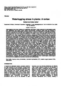
classified in two major groups. The first major group comprised of mildly and weakly virulent isolates while the second group comprised of highly virulent ones. It was hypothesized that the frequent occurrence of mutants might be responsible for the high level of variation among the isolates. The results demonstrated that the RAPD technique may be of great use for the classification of S. aureus. Obviously, for these patterns to have a practical meaning in the areas of medicine, population biology and epidemiology, specific DNA bands must be related with the virulence genes (Welsh et al., 1990). This could be accomplished by a systematic comparison of DNA band patterns among bacteria contrasting for the different virulence genes. Similar approach has been used to differentiate aggressive from non-aggressive isolates of the oilseed rape pathogen Phoma lingam (Schafer and Wostmeyer, 1992). The DNA fingerprint defined for each race of S. aureus could be useful for the surveillance of epidemiological revolution strategies, medical diagnoses and in the identification of new strains and isolates. The next step would to clone the isolates specific bands (bands with different virulence genes), sequence them and design specify primers for the development of sequence characterized amplified regions which would be much more specific and easier to apply. More research involving additional molecular techniques is needed to confirm if further sub-grouping would be appropriate.
RESULTS AND DISCUSSION The amplification reactions with the 10 primers generated 88 bands, 51 of which was polymorphic (Table 2) with size range of between 200 and 3,000 bp. According to the pairwise genetic distances among the isolates analyzed at 100% similarity, all the isolates were classified completely into twelve different groups (Figure 1). At 50% coefficient of similarity, all the isolates were
REFERENCES Coltman K (1981). Urinary tract infections. New thoughts on an old subject. The Practitioner 223: 351-355. Delong E F, Wickham GS, Pace NR (1989). DNA fingerprinting using very short random primers. J. Sci. 243: 1360-1363. Eisenstein BI (1990). Bacterial diseases and control. J. Infectious Dis. 161: 595-602. Gore LF, Walsh P (1964). Preservation and storage of bacterial cultures
614
Afr. J. Biotechnol.
J. Med. Lab. Technol. 21: 244-246. Guthrie PAI, Magill CW, Frederiksen RA, Odvody GN (1992). Random amplified polymorphic DNA markers: A system for identifying and differentiating isolates of Colletotrichum graminicola. Phytopathology 82: 832-835. Jaccard P (1908). Nouvelles recherches sur la distribution florale. Bull. Soc. Vaud. Sci. Nat. 44: 223-270. Kado CI, Heskett MG (1970). Selective media for isolation of Agrobacterium,Corynebacterium, Erwinia, Pseudomonas, Xanthomonas. Phypathology .60: 969-976. Kloos WE, Schleifer KH (1981). The genus Staphylococcus in the prokaryotes : A handbook on habitat, isolation and identification of bacteria. Vols. 1 and 2. Roeder V, Broda P (1987). Rapid preparation of DNA from filamentous fungi.Letters in Appl. Microb. 1: 17-20. Rohlf FJ (1993). NTSYS-PC. Numerical taxonomy and multivariate analysis system Version 2.0. Exeter, New York.
Schafer E, Wostmeyer J (1992). Random primer dependent PCR differentiates aggressive and non-aggressive isolates of the oilseed rape pathogen Phoma lingam (Leptosphaeria maculans). J. Phytopathol. 136:124-136. Sng EH, Yeo KL, Rajan VS (1981). Simple method for detecting penicillinase-producing Neisseria gonorrhoeae and Staphylococcus aureus. Br. J. Veneral Dis. 57: 141-142. Stokes JE, Ridgway GL (1980). Clinical Bacteriology. Edward Arnold.5th Edition. pp. 35-50. Welsh J, McClelland M (1990). Fingerprinting genomes using PCR with arbitrary primers. Nucleic acids Res. 18: 6531-6535. Woese CR (1986).Evolution in Prokaryotes. Schleifer KH, Stackebrandt E(ed).Academic Press, London.