Functions of Huntingtin in Germ Layer Specification and Organogenesis Giang D. Nguyen1,2,4,6, Aldrin E. Molero1,2,3,6, Solen Gokhan1,2,3,6, Mark F. Mehler1,2,3,4,5,6,7,8,9,10* 1 Roslyn and Leslie Goldstein Laboratory for Stem Cell Biology and Regenerative Medicine, Albert Einstein College of Medicine, Bronx, New York, United States of America, 2 Institute for Brain Disorders and Neural Regeneration, Albert Einstein College of Medicine, Bronx, New York, United States of America, 3 Department of Neurology, Albert Einstein College of Medicine, Bronx, New York, United States of America, 4 Department of Neuroscience, Albert Einstein College of Medicine, Bronx, New York, United States of America, 5 Department of Psychiatry and Behavioral Sciences, Albert Einstein College of Medicine, Bronx, New York, United States of America, 6 Rose F. Kennedy Center for Research on Intellectual and Developmental Disabilities, Albert Einstein College of Medicine, Bronx, New York, United States of America, 7 Einstein Cancer Center, Albert Einstein College of Medicine, Bronx, New York, United States of America, 8 Ruth L. and David S. Gottesman Institute for Stem Cell Biology and Regenerative Medicine, Albert Einstein College of Medicine, Bronx, New York, United States of America, 9 Center for Epigenomics, Albert Einstein College of Medicine, Bronx, New York, United States of America, 10 Institute for Aging Research, Albert Einstein College of Medicine, Bronx, New York, United States of America
Abstract Huntington’s disease (HD) is a neurodegenerative disease caused by abnormal polyglutamine expansion in the huntingtin protein (Htt). Although both Htt and the HD pathogenic mutation (mHtt) are implicated in early developmental events, their individual involvement has not been adequately explored. In order to better define the developmental functions and pathological consequences of the normal and mutant proteins, respectively, we employed embryonic stem cell (ESC) expansion, differentiation and induction experiments using huntingtin knock-out (KO) and mutant huntingtin knock-in (Q111) mouse ESC lines. In KO ESCs, we observed impairments in the spontaneous specification and survival of ectodermal and mesodermal lineages during embryoid body formation and under inductive conditions using retinoic acid and Wnt3A, respectively. Ablation of BAX improves cell survival, but failed to correct defects in germ layer specification. In addition, we observed ensuing impairments in the specification and maturation of neural, hepatic, pancreatic and cardiomyocyte lineages. These developmental deficits occurred in concert with alterations in Notch, Hes1 and STAT3 signaling pathways. Moreover, in Q111 ESCs, we observed differential developmental stage-specific alterations in lineage specification and maturation. We also observed changes in Notch/STAT3 expression and activation. Our observations underscore essential roles of Htt in the specification of ectoderm, endoderm and mesoderm, in the specification of neural and non-neural organ-specific lineages, as well as cell survival during early embryogenesis. Remarkably, these developmental events are differentially deregulated by mHtt, raising the possibility that HD-associated early developmental impairments may contribute not only to region-specific neurodegeneration, but also to non-neural co-morbidities. Citation: Nguyen GD, Molero AE, Gokhan S, Mehler MF (2013) Functions of Huntingtin in Germ Layer Specification and Organogenesis. PLoS ONE 8(8): e72698. doi:10.1371/journal.pone.0072698 Editor: Domingos Henrique, Instituto de Medicina Molecular, Portugal Received May 22, 2013; Accepted July 12, 2013; Published August 13, 2013 Copyright: © 2013 Nguyen et al. This is an open-access article distributed under the terms of the Creative Commons Attribution License, which permits unrestricted use, distribution, and reproduction in any medium, provided the original author and source are credited. Funding: This work was supported by NIH grants to M.F.M. (NS071571 Funding institution NINDS, HD071593 Funding institution NICHD, MH66290 Funding institution NIMH) and to A.E.M. (NS073758 Funding institution NINDS), as well as by the F.M. Kirby, Alpern Family, Harold and Isabel Feld and Roslyn and Leslie Goldstein Foundations (M.F.M.). The funders had no role in study design, data collection and analysis, decision to publish, or preparation of the manuscript. Competing interests: The authors have declared that no competing interests exist. * E-mail:
[email protected]
Introduction
focused on interrogating disease stages exhibiting overt clinical signs and symptoms. Indeed, the functional pleiotropism of the huntingtin protein (Htt), which ranges from transcriptional regulation to anti-apoptotic functions, as well as the adverse effects of mHtt have been thoroughly studied in adult life [2–4]. However, several recent studies have suggested that Htt also plays essential roles during early embryogenesis. Targeted deletion of htt in mice (KO) resulted in excessive cell death in the epiblast and severe developmental defects such as head-fold involution, a shortened primitive streak and
Huntington’s disease (HD) is an autosomal dominant genetic disorder caused by abnormal CAG expansion in exon 1 of the huntingtin gene (mhtt) and pathologically characterized by progressive degeneration of striatal and cortical neurons [1]. Clinical hallmarks of HD include the adult onset of characteristic neuropsychiatric and motor abnormalities. Since the first published characterization of HD by George Huntington in 1872, studies of HD pathogenesis have predominantly
PLOS ONE | www.plosone.org
1
August 2013 | Volume 8 | Issue 8 | e72698
Roles of Huntingtin in Early Embryogenesis
absence of the embryonic organizer, culminating in embryonic lethality as early as embryonic day 6.5 (E6.5) [5–8]. In addition, silencing of htt in progenitor cells of the ventricular zone from E14.5 has also been shown to alter their lineage commitment associated with enhanced cell death [9]. In addition, analysis of aggregation chimeras with htt-/- ESCs revealed that Htt is essential for neural development in selective brain structures, particularly the striatum [10]. The findings of the in vivo ablation studies suggest that Htt may play critical roles in germ layer specification and region-specific neurogenesis. However, it remains unclear whether the pathogenic HD mutation may impair these early developmental events. Indeed, our group has recently demonstrated an array of developmental impairments in the specification and maturation of striatal medium spiny neurons (MSNs) in a mhtt knock-in mouse model (Q111) as early as E13.5 [11]. Therefore, it is plausible that mHtt may also impair not only germ layer specification, but also organogenesis, and thus contribute to HD-associated systemic co-morbidities. In this study, we examined the roles of Htt and the potential adverse effects of mHtt during early embryonic development. We analyzed huntingtin knock-out (KO) and Q111 ESCs utilizing well established ESC culture paradigms to recapitulate early developmental events [12]. We hypothesized that Htt plays important spatial and temporal roles during embryogenesis and that mHtt differentially alters these key developmental events.
regulation of the core pluripotency factors, and mHtt does not alter these earliest developmental functions. We next investigated whether Htt and mHtt are involved in ESC differentiation, by analyzing spontaneously differentiating ESCs obtained after removal of leukemia inhibitory factor (LIF), a critical factor for ESC maintenance [17]. Four days in vitro (DIV) following LIF removal, both KO ESCs and Q111 ESCs, respectively, exhibited constitutive cellular expression of the pluripotency factors in concert with persistent expression of BrdU, KI67 and pHisH3, as compared to the progressive downregulation of these developmental factors and cell cycle parameters in CTL ESCs and Q18 ESCs, respectively (all comparisons are statistically significant with p-values < 0.0001; Figure 1B, C, E and F; Figure S2A–J). These observations indicate that both the absence of Htt and the presence of mHtt may alter the capacity of ESCs to differentiate spontaneously by suppressing the downregulation of pluripotency factors and the active modulation of cell cycle progression. To further examine the possibility that Htt regulates the subsequent specification of the three cardinal germ layers, we assessed the specification of the derivatives of the three germ layers in ESC-derived embryoid bodies (EBs), which have been shown to partially recapitulate the process of gastrulation in vivo [18]. Compared to CTL, we observed a significant proportion of TUNEL+ dying cells in KO EBs as early as 4DIV (29.6% vs 9.2%, p-values < 0.0001), with severe reductions in both the number and size of KO EBs (DIV4: 6.0 vs 35.7, p-value = 0.00064; 0.028 mm2 vs 0.096 mm2, p-value < 0.0001; DIV6, 4 vs 34.3, p-value = 0.00063; 0.039 mm2 vs 0.227 mm2, p-value < 0.0001; DIV8, 1.0 vs 33.0, p-value < 0.0001; 0.032 mm2 vs 0.292 mm2, p-value < 0.0001; DIV10, 1.0 vs 32.0, p-value < 0.0001; 0.121 mm2 vs 0.554 mm2, p-value < 0.0001; respectively, Figure 2A–D). In contrast, there were no differences in the size and number of Q111 EBs as well as in the proportion of TUNEL+ cells as compared to Q18 EBs (Figure 2E–H). Further, gene expression analysis of FGF5 (ectoderm), Nodal (endoderm) and Brachyury (mesoderm) revealed that both KO- and Q111-EBs, as compared to their respective controls, have significantly higher expression of Nodal (Fold change [Fc]: 1.533, p-value < 0.001; Fc: 2.204, pvalue < 0.001; respectively) and lower expression of Brachyury (Fc: 0.131, p-value < 0.001; Fc: 0.596, p-value = 0.001; respectively). However, FGF5 expression was significantly lower in KO EBs while it was increased in Q111 EBs (Fc: 0.138, p-value < 0.001; Fc: 1.216, p-value < 0.001; respectively; Figure 2I and J). These findings suggest that Htt plays important roles in maintaining the integrity of germ layer specification, and mHtt disrupts these developmental events. Previous studies have shown that Htt has anti-apoptotic functions [19,20]. Given the fact that cell death was significantly increased in KO EBs, it is possible that impairments in germ layer specification may stem from differential profiles of cell survival of ESC-derived progenitor species. To investigate this possibility, we knocked down BAX, an upstream regulator of the caspase 3/9 apoptosis signaling cascade to attempt to enhance cell survival in KO EBs. Using this approach, there was a relative rescue of the proportion of TUNEL+ dying cells (KO-shSCR: 46.6%; KO-shBAX: 30.3%; CTL-shSCR: 26.0%;
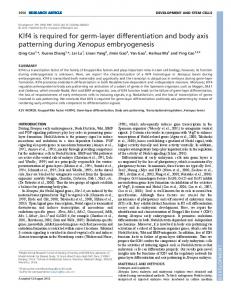
Results Htt is not required for the maintenance of undifferentiated ESCs, but is important for specification and survival of ectoderm, endoderm and mesoderm, whereas mHtt impairs spontaneous ESC differentiation and differentially alters derivatives of these germ layers Our group recently reported developmental alterations in the expression profiles of Nanog and Sox2 in the striatal generative zone and mantle region of the Q111 mouse brain [11]. These factors, together with Oct4 and Klf4, form the core pluripotency network that is critical for the maintenance and differentiation of ESCs [13]. To determine whether Htt is required for the regulation of pluripotency factors and consequentially for the maintenance of undifferentiated ESCs, we compared Hdhex4,5/ ex4,5 ESCs [7,14,15], hereby referred to as KO ESCs, with wildtype ESCs (CTL ESCs). To further investigate the effects of the pathogenic HD mutation on these functions, we compared mhtt knock-in ESCs, hereby referred to as Q111 ESCs, which carries an expanded polyglutamine tract (111 glutamines), with wild type htt knock-in ESCs, hereby referred to as Q18, which conversely carries a normal polyglutamine tract (18 glutamines) [15,16]. There were no differences in the expression profiles of the pluripotency factors, Nanog, Oct4, Sox2 and Klf4, and the ESC marker, SSEA1, as well as KI67 and phosphorylated histone H3 (pHisH3), markers for dividing cells and the G2/Mphase of the cell cycle, respectively, in KO ESCs versus CTL ESCs and in Q111 ESCs versus Q18 ESCs (Figure 1A, D; Figure S1A–F). These observations indicate that Htt is not required for maintenance of undifferentiated ESCs and the
PLOS ONE | www.plosone.org
2
August 2013 | Volume 8 | Issue 8 | e72698
Roles of Huntingtin in Early Embryogenesis
Figure 1. mHtt impairs the spontaneous differentiation of ESCs in ways analogous to Htt ablation. (A, D) Immunofluorescence analysis of the ESC marker (SSEA1) and the pluripotency factors (Nanog, Oct4, Sox2) in undifferentiated ESC maintained with LIF. (B, E) Quantification of Nanog+, Oct4+, Sox2+ (n=1203, 995, 1138 and 608 for CTL, KO, Q18 and Q111, respectively) and Klf4+ (n=1228, 592, 1113 and 1112 for CTL, KO, Q18 and Q111, respectively) cells present in ESCs after 4 DIV following removal of LIF. (C, F) ESC cultures pulsed with BrdU for 4 hrs in media without LIF. The ESCs were then fixed at 1DIV, 2DIV, and 4DIV and quantification of BrdU+ cells was assessed at these three time points (n=1426, 1162, 1571 and 2033 for CTL, KO, Q18 and Q111, respectively). All error bars represent ±95% CI; *p-values < 0.0001 unless otherwise noted. Scale bar = 20 µm. doi: 10.1371/journal.pone.0072698.g001
progenitors, whereas mHtt promotes precocious specification of neuroectodermal fate
CTL-shBAX: 26.5%) and of the size of KO-shBAX EBs as compared to control EBs expressing a scrambled shRNA (CTLshSCR) or BAX shRNA (CTL-shBAX) (Figure 2K and Figure S3). However, Brachyury gene expression in KO-shBAX EBs remained unchanged from KO-shSCR EBs and significantly reduced as compared to CTL-shSCR EBs (Fc: 0.012, p-value < 0.001). In addition, there was further enhancement of Nodal expression as compared to both CTL-shSCR and KO-shSCR EBs (Fc: 2.670, p-value < 0.001; Fc: 1.61, p-value < 0.001; respectively). Although FGF5 expression showed a significant upregulation in KO-shBAX EBs as compared to KO-shSCR EBs (Fc: 2.12, p-value < 0.001), it remained significantly lower as compared to CTL-shSCR EBs (Fc: 0.62, p-value = 0.006; Figure 2L). The rescue of cell viability together with partial rescue of germ layer-associated gene expression suggests that Htt plays primary roles in cell survival during both ESC maintenance and germ layer specification.
During gastrulation, the posterior region of primitive ectoderm generates mesendodermal progenitors that subsequently give rise to definitive endoderm and mesoderm, whereas the anterior region generates neuroectodermal progenitors that give rise to the developing nervous system [21]. In vitro inductive paradigms using Wnt3A and retinoic acid (RA) in the absence of EB formation have been utilized to generate early mesendodermal and neuroectodermal cell types, respectively [22]. We employed this instructive experimental protocol to examine whether Htt plays a role in the early program of neuroectodermal and mesendodermal specification. Prior to Wnt3A induction, KO ESCs exhibited a significantly higher percentage of TUNEL+ dying cells as compared to CTL ESCs (0.9% vs 10.6%, p-value < 0.0001; Figure 3A and B). Following Wnt3A induction, KO ESCs contained a significantly lower percentage of Brachyury+ mesendodermal progenitors and a higher percentage of TUNEL+ cells as compared to CTL ESCs (TUNEL: 15.9% vs 8.2%, p-value < 0.0001; Brachyury: 16.7% vs 32.6%, p-value