Nov 11, 1988 - sulin release from the insulinoma cells. Both the galanin- evoked hyperpolarization and inhibition of insulin release were abolished in cells ...
The EMBO Journal vol.8 no.2 pp.413-420, 1989
Galanin activates nucleotide-dependent K+ channels in insulin-secreting cells via a pertussis toxin-sensitive G-protein Mark J.Dunne, Marion J.Bullett, Guodong Li1, Claes B.Wollheim1 and Ole H.Petersen MRC Secretory Control Research Group, Physiological Laboratory, University of Liverpool, PO Box 147, Liverpool L69 3BX, UK, and 'Institut de Biochimie Clinique, Centre Medical Universitaire, University of Geneva, 1211 Geneva 4, Switzerland Communicated by M.Lazdunski
The effects of galanin (7-70 nM) on ATP-sensitive K+ channels (KATP channels), membrane potential and the release of insulin have been studied in the insulinoma cell line, RINm5F. Single-channel currents have been recorded from excised outside-out membrane patches as well as intact insulin-secreting cells and it is shown that galanin, added to the outside of the membrane, specifically activates KATP channels. Studies carried out using the fluorescent probe bisoxonol demonstrate that galanin hyperpolarizes RINm5F cells. Galanin was also found to abolish glyceraldehyde-stimulated immunoreactive insulin release from the insulinoma cells. Both the galaninevoked hyperpolarization and inhibition of insulin release were abolished in cells pre-exposed to pertussis toxin. The possibility that the gating of KATP channels could be mediated by a G-protein was studied in patch-clamp experiments by adding F to the solution bathing the inside of the cell membranes (open-cell), in order to generate the alumino-fluoride complex AIF4-. F (1- 10 mM) evoked dose-dependent activation of KATP channels and this effect was fully reversible. F was also able to activate K+ channels inhibited by ATP. That the fluoride activation of KATP channels is mediated by the complex AIF4- was indicated by experiments in which AICI3 (10 nM) was found to enhance further the activation of K+ channels evoked by 1 mM F and by results showing that F-stimulation of KATP channels was (i) abolished in the continued presence of F by the Al3+ chelator deferoxamine (0.5 mM) and (ii) could be mimicked by V043- which has a structure similar to that of the AIF4- complex. Key words: KATP channel/ATP/galanin/F-/G-protein
Introduction ATP-sensitive K + channels (KATP channels) (Cook and Hales, 1984; Findlay et al., 1985) play a pivotal role in the regulation of insulin secretion since carbohydrate secretagogues close these pores (Ashcroft et al., 1984; Rorsman and Trube, 1985; Dunne et al., 1986) thereby evoking depolarization and opening of voltage-gated Ca2+ channels (Petersen and Findlay, 1987; Petersen, 1988). Galanin is a 29 amino acid neuropeptide that inhibits insulin secretion under a variety of experimental conditions both in vivo and in the isolated perfused pancreas (Tatemoto et al., 1983; Ahren etal., 1986, 1988; Dunning etal., 1986; ©IRL Press
Hramiak et al., 1988). De Weille et al. (1988) have recently shown that galanin hyperpolarizes the cell membrane and reduces spontaneous electrical activity in the rat insulinoma cell line RINm5F as well as stimulating 86Rb efflux. In single-channel current recording experiments on excised inside-out membrane patches exposed to ATP on the inside, opening of KATP channels was only observed in those cases where galanin was present in the pipette solution in contact with the membrane outside and abolished by the sulphonylurea glibenclamide (De Weille et al., 1988), a specific inhibitor of KATP channels in these cells (SchmidAntomarchi et al., 1987a,b). De Weille et al. (1988) therefore concluded that galanin activates KATP channels by a mechanism not involving a soluble cytoplasmic messenger. GTP-binding proteins (G-proteins) play a crucial role in coupling receptors for hormones or neurotransmitters to effectors such as enzymes and ion channels (Dolphin, 1987; Iyengar and Birnbaumer, 1987; Brown and Birnbaumer, 1988; Neer and Clapham, 1988). In cardiac cells both acetylcholine and adenosine activate the same K+ channel via a G-protein (Kurachi et al., 1986). Similarly, the muscarinic-gated K+ channel of cardiac atrial myocytes (Pfaffinger et al., 1985; Logothetis et al., 1987), the dopamine-, histamine- and acetylcholine-gated K+ channel of dorsal root ganglion cells (Sasaki and Sato, 1987), the somatostatin- and muscarinic-gated K+ channels of clonal rat anterior pituitary cells (Yatani et al., 1987b) as well as angiotensin II stimulation of voltage-gated Ca2+ channels in an adrenal cortical cell line (Hescheler et al., 1988) all involve G-proteins. One tool available to study the possible involvement of a G-protein is AlF4-. The influence of fluoride ions on hormone-regulated systems has long been recognized. In 1958 Rall and Sutherland noted that F- activated the adenylate cyclase enzyme, by a mechanism later found to be dependent upon the presence of trace amounts of AJ3+ ions, etched from glassware by the millimolar concentrations of NaF or KF used (Hewitt and Nicholson, 1963; Sternweiss and Gilman, 1982). AlF4- is now recognized as a potent activator of guanine-nucleotide binding proteins for a number of hormone-regulated systems, including the activation of the adenylate cyclase enzyme (Howlett et al., 1979; Sternweiss and Gilman, 1982; Katada et al., 1984), a number of polyphosphoinositide phospholipase C enzymes (Blackmore et al., 1985; Guillon et al., 1986) and the cGMP phosphodiesterase enzyme of retinal rod outer segments (Bigay et al., 1985, 1987; Stein et al., 1985). The mechanism of activation of AMF4- has been studied in detail by Bigay et al. (1985, 1987). A1F4 appears to be structurally analogous to the terminal phosphate residue of GTP-the -y-phosphate. Bigay et al. (1985, 1987) have therefore proposed that the alumino-fluoride complex, acting as a highly specific analogue of -y-phosphate, binds next to the $-phosphate of the GDP molecule on the guaninenucleotide binding domain. In effect AlF4- fools the G413
M.J.Dunne et al.
protein into thinking that GTP is bound (Cockcroft, 1987), and transforms the protein into its 'active' state, a conformation normally only occurring if GTP is bound (Bigay et al., 1987). Another tool that can be useful for the study of G-protein involvement is pertussis toxin (PTX). Many receptoractivated events can be blocked by pre-treating cells with PTX which acts by catalysing ADP-ribosylation of the asubunit of certain G-proteins, in particular Gi (Graziano and Gilman, 1987; Gilman, 1987). The most direct evidence for a receptor-operated ion channel is achieved by using excised outside-out membrane patches (Hamill et al., 1981; Petersen and Petersen, 1986) since an acute and reversible opening of ion channels can be demonstrated in experiments on individual patches of cellfree surface membrane. We now show directly that galanin activates nucleotide-dependent K+ channels (KATP channels) in outside-out membrane patches from RINmSF cells and also demonstrate in cell-attached patches that there are many more and frequent KATP channel openings when galanin is present in the pipette solution than in its absence. F- acting on the membrane inside in single-channel current experiments on open (permeabilized) cells (Maruyama and Petersen, 1984; Petersen and Petersen, 1986) reversibly activates the KATP channels, an effect that is blocked by the A13+ chelator deferoxamine. Galanin-evoked membrane hyperpolarization in intact RINmSF cells is demonstrated using the fluorescent probe bisoxonol, an effect abolished by pre-treatment with PTX. Galanin is shown to inhibit glyceraldehyde-evoked insulin release and this effect of galanin is also blocked by PTX.
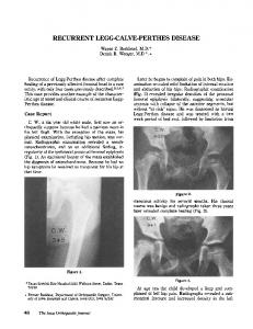
Results Although insulin-secreting cells, including the RINmSF cells, possess both ATP-sensitive and Ca2+- and voltage-activated K+-selective channels (Petersen and Findlay, 1987) all records presented in this paper are derived exclusively from currents passing through the ATP-sensitive channels. Ca2' and voltage-activated K+ channel currents have been eliminated by keeping tCa2+]j low. In the intact cells all results have been obtained at the resting membrane potential where the Ca2+-activated K+ channels are never operational and in addition no Ca2+ was present in the pipette solution. In the excised patches or open-cell-attached patches [Ca2+] in the solution in contact with the membrane inside was always very low, since no Ca2+ was added and EGTA (1 mM) was present. Under such conditions openings of Ca2+- and voltage-activated K+ channels cannot be observed except at extreme positive membrane potentials (Petersen and Findlay, 1987). In our experiments only membrane potentials of 0 mV were employed and the presence of Ca2+-activated K+ channel currents can therefore be excluded. The conductance properties of the two channel types are also markedly different (Petersen and Findlay, 1987) so that it is very east to distinguish between openings of ATPsensitive and Ca +-activated K+ channels. The effects of galanin on the nucleotide-dependent K + channel Figure 1 shows the direct effect of 70 nM galanin on KATP channels recorded in intact RINm5F insulin-secreting cells. The current records were obtained from two separate intact cells, galanin (70 nM) being added to the pipette solution 414
CONTROL closed
- - - -
-
1,
--,.AJjN1-&Jh jj. 71"FW"f 11, I"' ,I, .-
-11W
j-"-
I
200 msec
7OnM GALANIN closed
- -
"A--
L--L
5 pA
L-LL
LikLi-
-
-.1
Fig. 1. The effect of galanin
on KATP channels in intact RINm5F insulinoma cells. The current records shown, upper panel and lower panel, were obtained at a pipette potential equal to the bath potential (Vp = 0) (i.e. the potential difference across the patch membrane was the normal membrane potential) and come from two separate cells. Galanin (70 nM) was present in the pipette solution in the test
experiment.
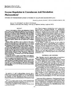
in the test experiment. In control experiments the maximum number of coincident channel openings was two, and the apparent average number of operational channels per patch was estimated to be 0.5 + 0.25 (SEM) (n = 11) (Figure 1 upper panel). In contrast, when galanin (7-70 nM) was present in the pipette solution the maximum number of coincident channel opening events was four and the apparent average number of operational channels per patch estimated to be 2.5 i 0.25 (n = 15) (Figure 1, lower panel). On average, galanin enhanced the KATP patch current recorded in cell-attached membrane patches to 2128 i 300% (n = 15) of that observed in the control situation (100%). Galanin (7-70 nM) failed to activate the K+ channels when it was added to the bath solution (n = 21). Figure 2 shows the direct effect of galanin on single KATP channels in an outside-out membrane patch with 0.5 mM ATP, 0.5 mM ADP and 100 AM GTP bathing the cytosolic side of the membrane. Galanin increased the KATP patch current from 100 to 694% of the control level, and this effect was completely reversible, as the current decreased to 91 % of the original control value after galanin was washed away. In four experiments the galanin-evoked KATP channel activation was abolished by tolbutamide (1 mM), a specific inhibitor of KATP channels in these cells (Dunne et al., 1987). The data presented in Figure 2A is typical of five separate experiments on outside-out membrane patches. Three of these were sufficiently long to be analysed quantitatively for galanin-evoked changes in the KATP patch current and the results are shown in Figure 2B. On average 70 nM galanin was found to enhance the patch current from 100%, in the control situation, to 889 169% (n = 3) returning to 110 4I 44% of the original control value, upon removal of galanin. The effect of galanin on membrane potential and insulin secretion Figure 3 shows the results of a series of experiments in which qualitative changes in the membrane potential of RINm5F cells were studied with the fluorescent dye bisoxonol. Galanin (100 nM) was consistently found to hyperpolarize the surface membrane of intact RINmSF cells (n = 5 experiments, in duplicate).
Activation of KATP channels by galanin
A.
B.
7OnM GALANIN
ii
1000o GALANIN
[II
800'
llikii b
a
IlOpA
Au, I
0
4 sec
c
c
0
600
U
a
a
b
C
200
0 -A-
- -i- --
400
A.
J
-
----
]
o0 I lOpA 400 msec
Fig. 2. Direct stimulation of KATP channels by galanin. Panel A shows a continuous current record taken from an excised outside-out membrane patch. During the period indicated the outside of the membrane (facing the bath solution) was exposed to 70 nM galanin. The inserts (a), (b) and (c), showing current traces obtained at a higher time resolution, come from the periods indicated in the upper trace. Panel B shows a histogram summarizing the effect of galanin on the average KATP patch current recorded from three separate outside-out patches. Relative changes in the apparent open-state probability (P) of K+ channels have been expressed as a percentage of the pre-control level of activity (100%), see Materials and methods. SEM bars have been included.
However, if RINm5F cells were incubated for 3-24 h in a solution containing pertussis toxin (PTX) (100 ng/ml), the effects of galanin were abolished (n = 5 experiments, in duplicate). As a control it was shown that diazoxide (50 zM) evoked hyperpolarization both in the control and PTXtreated cells (Figure 3) (n = 3). Table I summarizes the data concerning the effects of galanin on the secretion of immunoreactive insulin. Glyceraldehyde (10 mM) evoked a secretory response and this was blocked by galanin (100 nM). In the PTX-pretreated cells glyceraldehyde evoked the normal increase in insulin secretion, but this could not be blocked by galanin.
N
4
4c
cc
0. z
GAL1KCI KCI
GAL
1
KCI
GAL
2 min z
0 4 N
4
The effects of F on the gating of nucleotidedependent K+ channels The direct effect of adding 10 mM KF to the solution in contact with the inside of the plasma membrane of a RINmSF open-cell is illustrated in Figure 4A. KF evokes a pronounced and reversible activation of the KATP channels. In the record shown F- activation enhanced the KATP patch current from 100 to 607 % of the control level, a value that was subsequently lowered to 138 % upon removal of KF. Vanadate (VO43-) (1 AM) had effects similar to those of F- (Figure 4B). In the record shown V043- activated the KATP patch current from 100 to 216% of the control value, which was lowered to 76% upon removal of Vo43 -. The reversible activation of K+ channels by KF (1-10 mM) was dose-dependent (Figure 5) and seen in every one of 74 separate open-cell records. Such activations were also observed in five excised inside-out patches. KF (10 mM) had no effect on K+ channels when it was added to the outside of the membrane (n = six separate patches). GTP and GDP can also activate KATP channels (Dunne and Petersen, 1986b) (Figure 5) but unlike F-, GTP could not activate the channels when they were closed down by ATP (Figure 6B). Interestingly a second F- application in
PTX
CONTROL z
0
CONTROL
0 0.
z
GAL GAL
TIDIAZ
PTX GAL
DIAZ
Fig. 3. Effect of galanin on the membrane potential of control (left) and pertussis toxin (PTX) treated (right) RINm5F cells. Qualitative changes in potential have been assessed using the probe bisoxonol from cells exposed to galanin (100 nM), diazoxide (50 yM) and KCI (24 mM). Each trace is representative of at least five independent experiments.
the continued presence of ATP always evoked a larger response than the first stimulation (Figure 6A). Figure 6C summarizes the results of F- and GTP activation in patches continuously exposed to ATP. Figure 7A shows a typical result from a series of experiments in which the effects of the A13+ chelator deferoxamine (0.5 mM) (Blackmore et al., 1985) were studied on K+ channels activated by F-. The deferoxamine-evoked inhibition of F- (1-10 mM)stimulated K+ channels was seen in every one of 15 separate open-cell patches where this experiment was carried out. Eleven of these experiments, involving 5 mM KF, were
415
M.J.Dunne et al.
Table I. The effects of galanin (100 nM) on the release of immunoreactive insulin from unstimulated and glyceraldehyde (10 mM)-stimulated RINm5F cells Control
Unstimulated Galanin Glyceraldehyde Galanin and glyceraldehyde
PTX-treated
4.13 L 0.3 (n 4.73 ± 0.4 (n 8.86 4 0.9 (n 4.54 4 0.3 (n
= = = =
10) 10) 9) 10)
4.36 5.82 8.34 9.38
i 0.4 i 0.3 ± 0.4 + 0.6
(n (n (n (n
= 9) = 10) = 10) = 10)
Data has been expressed in ng/106 cells/10 rnin (+ SEM), from control and pertussis toxin (PTX)-treated cells (100 ng/ml for 3 h), as described in Materials and methods.
analysed quantitatively and the results are shown in Figure 7B. Deferoxamine (0.5 mM) had no direct effect on K+ channels in the absence of KF (n = 4 separate patches). As the activation of G-proteins by KF requires the presence of trace amounts of Al3 + ions, relatively ineffective concentrations of fluoride can be made effective by adding AiC13 (micromolar concentrations), thereby potentiating the effects of KF (Blackmore et al., 1985). It was therefore interesting to note that the activating effect of 1 mM KF, sub-maximal for the stimulation of K+ channels (Figure 5), was enhanced by the addition of 10 A.tM AlCl3 in the continued presence of KF (n = 4 separate open-cell records). On average AlCl3 enhanced the KATP patch current evoked by 1 mM KF (100%) to 290 4 5 % of the prestimulus value. This effect was completely reversible, as upon removal of AlCl3 the average KATP patch current decayed to 106 i 14% of the original control value. In a further five open-cell membrane patches AlCl3 (10 ,uM) was found not to evoke further activation of K+ channels stimulated by 10 mM KF. AlCl3 had no direct effect on the gating of KATP channels in the absence of KF (n = 3 patches).
A.
10mM KF
|i _ -~ ~ ~JP
I5pA
4 sec
B.
l1jM V043-
i ta2; SpA ;*±A I*Ps{ sec L~
I
Fig. 4. The activation of KATP channels by fluoride and vanadate ions. Records A and B come from two separate RINm5F open (permeabilized) cells. In (A) 10 mM KF is present in the bath (in contact with inside of patch membrane) for the period indicated whereas in (B) the inside of the patch membrane is in contact with 1 /tM Na3VO4 for the time indicated. Both drugs evoke a reversible activation of KATP channels. ATP was not present in the bath (internal) solution.
Discussion Figure 8 summarizes our conclusions in the context of what is already known about the regulation of KATP channels in insulin-secreting cells. The KATP channel dominates the electrical properties of the resting cell membrane and an increase or decrease in the degree of channel opening will result in hyperpolarization or depolarization, respectively, of the cell membrane (Petersen and Findlay, 1987; Petersen, 1988). The level of membrane potential in turn determines the degree of Ca2+ channel opening, in this way governing Ca2+ influx and therefore [Ca2+],, which is a key regulator of insulin secretion (Wollheim and Sharp, 1981; Wollheim and Biden, 1986a; Petersen, 1988). We have now shown directly that galanin reversibly activates KATP channels in excised cell-free membrane patches (Figure 2) and that the presence of galanin on the outside of the cell membrane in the intact cell is associated with a markedly increased degree of channel opening relative to control conditions (Figure 1). These findings confirm the conclusions reached by De Weille et al. (1988) that galanin activates KATP channels without the involvement of a soluble cytosolic messenger. We have also shown that galanin hyperpolarizes the cell membrane (Figure 3), confirming previous findings of Ahren et al. (1986) and De Weille et al. (1988) and demonstrated that this effect, like the inhibitory effect of galanin on glyceraldehyde-evoked insulin secretion (Table I) is blocked by pre-treating the cells with PTX (Figure 3). This is 416
6001
500pM
500pM
GDP
GTP
5mM KF
1mM KF
10mM KF
1pM
Vo.j-
5001 -
400-
z 0 Dp
300 -
0
I
200-
100-
0-
LE
I
7k
L-i
Fig. 5. Comparison between the activating effects of GDP, GTP, KF and V043- on KATP channels in RINm5F open cells. Relative changes in P have been expressed as a percentage of the pre-control value for membrane patches exposed to: 500 ,M GDP (n = 3 separate patches), 500 4M GTP (n = 8), 1 mM KF (n = 6), 5 mM KF (n = 11), 10 mM KF (n = 13) and 1 jiM Na3VO4 (n = 6). SEM bars have been included. ATP was not present in the bath (internal) solution in any of these experiments.
Activation of KATP channels by galanin
A. 10mM 1mM ATP
lOmM KF
II
1LLJL
KF
,. til
1.11 Al iiil I I, I. -..2s 5pA
~
C.
11
200.
ATP KF
20sec
ATP
G3TP
KFr
150-j 0
z 0
100-
r
0)
B.
lmM ATP
50-
KF
GTP 0-
215PA Fig. 6. The effect of KF and GTP on ATP-inhibited K+ channels. The current records shown in A and B come from two separate open-cell membrane patches. In (A) ATP (1 mM) almost completely inhibited K+ channels activated upon permeabilization of the cell. Adding KF (10 mM) in the continued presence of ATP evoked a reversible activation of K+ channels. A second application of KF was found to further enhance the stimulation of K+ channels. In contrast 0.5 mM GTP, as shown in (B), had no effect of KATP channels inhibited by ATP. The maximal number of coincident KATP channel open events observed in these two patches immediately after permeabilization (not shown) was 28 and 11 respectively. Panel C summariz the averaged data relating to the effects of KF (n = 3 separate patches) and GTP (n = 3) on ATP-inhibited K+ channels. SEM bars have been included.
A.
5mM
fI
KF
B.
DEFEROXAMINE
SmM KF
I~~~~~~~~~~~~~ ~
500.
I!
DEFEROXAMINE
'lI 400
I5pA b
a
d
c
I
sec
e
-
m z
300
0
*
d
bw
200.
e
100I -
j
J--- -,I
0-
J5pA 1 sec
Fig. 7. Deferoxamine-induced inhibition of F--stimulated KATP channels in open (permeabilized) cells. In the continued presence of 5 mM KF, the A13+ chelator deferoxamine reversibly closed K+ channels activated by KF. The inserts (a-e), showing current traces at a higher time resolution, come from the periods indicated in the upper trace. Panel B summarizes the data relating to the averaged KATP patch current recorded from F--stimulated K+ channels in the presence and absence of deferoxamine (n = 11 separate patches). ATP was not present in the bath (internal) solution in these
experiments.
evidence for the involvement of a G-protein in the coupling of galanin-receptor interaction to KATP channel opening. De Weille et al. (1988) observed opening of ATP-sensitive K+
channels when galanin was in contact with the outside of excised inside-out membrane patches in a situation where no GTP was present in the bath solution in contact with the 417
M.J.Dunne et al.
to do with the G-protein activation caused by external galanin or internal F- stimulation, but is more likely to represent interaction with another site sensitive to guanosine and
Fig. 8. The KATP channel and its physiological regulation in insulinsecreting cells. Channel activation, resulting in a membrane hyperpolarization, evoked by galanin is mediated by the receptor (R) activating a pertussis toxin (PTX)-sensitive G-protein (G) coupled to the KATP channel. Conversely, K+ channel inhibition resulting in a membrane depolarization, evoked by either glucose or glyceraldehyde, is mediated either by a metabolic increase in the ATP:ADP ratio and/or through the activation of a C-kinase protein, coupled to the channel, via the metabolic generation of 1,2 diacylglycerol (DG).
membrane inside. This finding may seem to contradict our conclusion that a G-protein is involved, but it should be borne in mind that ATP (2.5 mM) was present in the bath solution in the experiments of De Weille et al. (1988) and that therefore phosphorylation of GDP to GTP in the binding domain of the G-protein could easily have occurred. We have also directly shown that AlF4- activates KATP channels when added to the inside of the cell membrane (Figures 4-7) providing further evidence for a stimulatory link via a G-protein to the KATP channels. The interpretation of our results shown in Figure 8 is the simplest one accounting for all the data, but we cannot exclude the possibility that AIF4- acts by inhibiting an ATPase (Missiaen et al., 1988), although it is by no means clear that such an effect, if it occurred, would cause channel activation. As shown in Figure 8 there are now known to be many different agents capable of influencing the degree of KATP channel opening, directly or indirectly. The physiologically important closure of the channel evoked by glucose or glyceraldehyde metabolism may be partly mediated by an increase in the intracellular free ATP concentration combined with a concomitant fall in the ADP level (Dunne and Petersen, 1986a; Kakei et al., 1986; Misler et al., 1986; Dunne et al., 1988b) and partly by metabolic generation of diacylglycerol (Peter-Riesch et al., 1988; Wollheim et al., 1988) (Figure 8). The tight control of channel opening by relatively small changes in ATP/ADP levels is now clear (Dunne et al., 1988b) and it is also evident that the channels are sensitive to both guanosine (Dunne and Petersen, 1986b) and pyridine nucleotides (Dunne et al., 1988a). The somewhat puzzling acute and acutely reversible channel opening effects of GTP, GTP--y-S, GDP-,B-S and GDP (Figure 5) (Dunne and Petersen, 1986b) have probably little 418
perhaps also adenosine and pyridine nucleotides. It is significant in this context that GTP, unlike F-, could not activate ATP-inhibited channels (Figure 6). The immediate stimulatory effects of GTP, GTP--y-S, GDP and GDP-3-S on KATP channels seem to be of a completely different nature from the very much slower stimulation by, for example, GTP--y-S on K+ channel currents in pituitary (GH3) plasma membranes that take -30 min to reach maximal activation (Yatani et al., 1987b). The somatostatinactivated K+ channels in the pituitary cells (Yatani et al., 1987b) like the ACh-activated K+ channels in cardiac cells (Yatani et al., 1987a) are not ATP-sensitive. Finally it should be mentioned that although the galaninevoked K+ channel opening and the associated hyperpolarization could explain the inhibition of insulin secretion (Petersen, 1988) it is possible that there are also other targets for the action of this neuropeptide. Thus, galanin has been shown to inhibit insulin secretion evoked by secretagogues with different modes of action, not necessarily affected by membrane hyperpolarization (Hramiak et al., 1988). This might suggest an additional action of galanin exerted at a point distal to the regulation of K+ channels in the sequence of events leading to exocytosis. Such an action has clearly been demonstrated for another inhibitor of insulin secretion, epinephrine. It has recently been shown in RINm5F cells that activators of a2 adrenoceptors inhibit Ca2+-stimulated insulin secretion in permeabilized cells and that this effect was mediated by a G-protein (Ullrich and Wollheim, 1988). Such a distal effect on secretion is also likely to be involved in somatostatin inhibition of insulin secretion, as this peptide inhibits Ca2+ ionophore-induced insulin secretion (Wollheim and Sharp, 1981). It is of interest that a2 adrenoceptor agonists, somatostatin and galanin inhibit the generation of cyclic AMP in insulin secreting cells, an effect also involving a pertussis toxin sensitive G-protein (Wollheim and Sharp, 1981; Ulirich and Wollheim, 1984; Amiranoff et al., 1988). Further studies are required to clarify whether the various actions of the inhibitory neuropeptides and neurotransmitters are mediated by the same or different members of the growing family of G-proteins.
Materials and methods Cell isolation and maintenance All experiments were carried out on the insulin-secreting cell-line RINm5F (Halban et al., 1983; Praz et al., 1983) maintained as previously described (Dunne et al., 1986).
Media for patch-clamp experiments The standard Na+-rich solution contained 140 mM NaCl, 4.7 mM KCI, 1.13 mM MgC12, 2.5 mM glucose and 10 mM Hepes. In patch-clamp experiments where this solution was used as the bathing solution (cell-attached and outside-out membrane patches) 2 mM CaCI2 was added. However, in experiments where this solution was added to the recording pipette (opencell patches) CaCI2 was omitted and 1 mM EGTA was added. The pH of all Na+-rich solutions was set at 7.2 (NaOH). The standard K+-rich intracellular bathing solution had the following composition: 140 mM KCI, 10 mM NaCl, 1.13 mM MgCl2, 2.5 mM glucose, 10 mM Hepes and I mM EGTA. No CaCl2 was added and the pH was set at 7.2 (KOH). Al3+ is present in trace amounts in all our solutions since no special precautions were taken to avoid this (Bigay et al., 1987; Stemweiss and Gilman, 1982). When F- is added in millimolar concentrations (1 -10 mM) stable
Activation of KATP channels by galanin complexes of aluminium with fluoride are formed and according to the published stability constants the predominant species would be A1F4-. The overall equilibrium constant 3 = [A1F4-] / ([Al3+] [F- ]4) is lol8 (Sillen and Martell, 1964). Al can also bind to EGTA (equilibrium constant - 1014) whereas the affinity of HEGTA for Al3+ is very much less (- 104) (Martell and Smith, 1974). In some experiments Al3" was removed from the solution by adding 0.5 mM of the iron chelator deferoxamine (Keberle, 1964) which has an extremely high affinity for Al3+ (1022) (Weast, 1984). In other experiments AIC13 was added to the intracellular F--containing solutions. Since there was always excess F-, an increase in the much lower A13+ concentration would cause formation of more AIF4-. _
Single-channel current recording and analysis Single-channel current records from membranes of RINmSF cells, obtained from cell-attached membrane patches (K+ pipette/Na+ bath) and outsideout membrane patches (K+ pipette/Na+ bath) were carried out according to the principles described by Hamill et al. (1981), and from open-cells (Na+ pipette/K+ bath) as described by Maruyama and Petersen (1984) and Dunne et al. (1986, 1987, 1988a,b). Patch-clamp recording pipettes (Type 101-PB, Ceebee Glass, Denmark), coated with SylgardR (Dow Coming, Corp, USA) had a final resistance of - 5 M Ql when filled with the appropriate pipette solution. Gigaohm seal formation was achieved in the continuous flow of the Na+-rich bathing solution from one of a series of outlet pipes, the other reservoirs containing various test solutions (Dunne et al., 1987). Exchange from control to test solution was achieved manually under visual control. Analysis of single-channel currents was performed on stretches of continuous current records (filtered at 1 KHz, low pass), lasting between 20 and 40 s. All data were digitized at 8 KHz (CED 1401, Cambridge, UK) and analysed with a Tandon microcomputer in conjunction with a software package supplied by CED Cambridge, UK. Idealized current traces were obtained from computerized threshold analysis of the data, using a pre-selected current threshold level. Comparative values of open-state probability (P) have been presented, expressed as a percentage of the control (pre-test) value (100%). This method of quantification is preferred to that of expressing absolute open-state probability values as the number of operational K+ channels per patch of membrane is often unknown in cellattached and outside-out membrane patches. The pipette voltage was held at 0 mV throughout all experiments. In all single-channel current records illustrated, upward deflections represent outward current flow (i.e. from the inside to the outside of the membrane patch). All current traces, photographed directly from the oscilloscope screen, have been filtered at 400 Hz (low pass).
Measurement of membrane potential and insulin secretion For the measurement of membrane potential with fluorescent probes, as well as insulin secretion, suspensions of RINm5F cells were used after a 3-h spinner culture (Wollheim and Pozzan, 1984; Wollheim and Biden, 1986b). Batches of - 1.5 x 10⁶ cells were suspended in a cuvette containing 2 ml of a modified Krebs-Ringer-bicarbonate buffer (KRBH) with 5 mM NaHCO3, 1 mM CaCl2 and 2.8 mM glucose (Wollheim and Pozzan, 1984; Wollheim and Biden, 1986b). Membrane potential fluctuations were measured by adding the fluorescent probe bisoxonol (100 nM) to the cuvette from a 1000-fold concentrated stock solution. The excitation wavelength was 540 nm and the emission wavelength was 580 nm (Wollheim and Pozzan, 1984; Wollheim and Biden, 1986b). Immunoreactive insulin secretion was measured during a 10-min incubation using -0.5 x 10⁶ cells/mi, exactly as described previously (Wollheim and Pozzan, 1984).
Acknowledgements We wish to thank Alan Higgins, Tim Underwood and Mark Houghton for the technical assistance and maintenance of the RINm5F cells. We also wish to thank Beverley Houghton for typing this manuscript. This work was supported by grants from the MRC (UK) and the Swiss National Science Foundation.
References Ahren,B., Arkhammer,P., Berggren,P.-O. and Nilsson,T. (1986) Biochem. Biophys. Res. Commun., 140, 1059-1063. Ahren,B., Rorsman,P. and Berggren,P.-O. (1988) FEBS Lett., 229, 233-237.
Amiranoff,B., Lorinet,A.-M., Lagny-Pourmir,I. and Laburthe,M. (1988) Eur. J. Biochem., 177, 147-152. Ashcroft,F.M., Harrison,D.E. and Ashcroft,S.J.H. (1984) Nature, 312, 446-448. Bigay,J., Deterre,P., Pfister,C. and Chabre,M. (1985) FEBS Lett, 191, 181-185. Bigay,J., Deterre,P., Pfister,C. and Chabre,M. (1987) EMBO J., 6, 2907-2913. Blackmore,P.F., Bocckino,S.B., Waynick,L.E. and Exton,J.G. (1985) J. Bio. Chem., 260, 14477-14483. Brown,A.M. and Birnbaumer,L. (1988) Am. J. Physiol., 254, H401 -H410. Cockcroft,S. (1987) Trends Biochem. Sci., 12, 75-78. Cook,D. and Hales,C.N. (1984) Nature, 311, 271-273. De Weille,J., Schmid-Antomarchi,H., Fosset,M. and Lazdunski,M. (1988) Proc. Natl. Acad. Sci. USA., 83, 517-521. Dolphin,A.C. (1987) Trends Neurosci., 101, 53-57. Dunne,M.J. and Petersen,O.H. (1986a) FEBS Lett., 208, 59-62. Dunne,M.J. and Petersen,O.H. (1986b) Pflugers Arch., 407, 564-565. Dunne,M.J., Findlay,I., Petersen,O.H. and Wollheim,C.B. (1986) J. Membr. Biol., 93, 271-279. Dunne,M.J., Ilott,M.C. and Petersen,O.H. (1987) J. Membr. Biol., 99, 215-224. Dunne,M.J., Findlay,I. and Petersen,O.H. (1988a) J. Membr. Biol., 102, 205-216. Dunne,M.J., West-Jordan,J., Abraham,R.J., Edwards,R.H.T. and Petersen,O.H. (1988b) J. Membr. Biol., 104, 165-177. Dunning,B.E., Ahren,B., Veith,R.C., Bottcher,G., Sundler,F. and Taborsky,G.J.,Jr. (1986) Am. J. Physiol., 251, E127-E133. Findlay,I., Dunne,M.J. and Petersen,O.H. (1985) J. Membr. Biol., 88, 165-172. Gilman,A.G. (1987) Annu. Rev. Biochem., 56, 617-649. Graziano,M.P. and Gilman,A.G. (1987) Trends Pharm. Sci., 8, 478-481. Guillon,G., Mouillac,B. and Balestre,M.N. (1986) FEBS Leti., 204, 183-188. Halban,P.A., Praz,G.A. and Wollheim,C.B. (1983) Biochem. J., 212, 439-443. Hamill,O.P., Marty,A., Neher,E., Sakmann,B. and Sigworth,F.J. (1981) Pflugers Archiv., 391, 85-100. Hescheler,J., Rosenthal,W., Hinsch,K.-D., Wulfern,M., Trautwein,W. and Schultz,G. (1988) EMBO J., 7, 619-624. Hewitt,E.J. and Nicholson,D.J.D. (1963) In Hochster,R.M. and Quastel,J.H. (eds), Metabolic Inhibitors. Academic Press, New York, Vol. 2, pp. 311-421. Howlett,A.C., Sternweiss,P.C., Macik,B.A., Von Arsedale,P.M. and Gilman,A.G. (1979) J. Biol. Chem., 254, 2287-2295. Hramiak,I.M., Dupre,J. and McDonald,T.J. (1988) Endocrinology, 122, 2486-2491. Iyengar,R. and Birnbaumer,L. (1987) ISI Atlas of Science: Pharmacology 1, 213-221. Kakei,M., Kelly,R.P., Ashcroft,S.J.H. and Ashcroft,F.M. (1986) FEBS Lett., 208, 63-66. Katada,T., Northup,J.K., Bokoch,M., Ui,M. and Gilman,A.G. (1984) J. Biol. Chem., 259, 3578-3585. Keberle,H. (1964) Ann. N.Y Acad. Sci., 119, 758-768. Kurachi,Y., Nakajima,T. and Sujimoto,T. (1986) Pflugers Archiv., 407, 264-274. Logothetis,D.E., Kurachi,Y., Galler,J., Neer,E.J. and Clapham,D.E. (1987) Nature, 325, 321-326. Martell,A.E. and Smith,R.M. (1974) Critical Stability Constants, Vol. 1: Amino Acids. Plenum, New York, pp. 269-271. Maruyama,Y. and Petersen,O.H. (1984) J. Membr. Biol., 81, 83-87. Misler,S., Falke,L.C., Gillis,K. and McDaniel,M.L. (1986) Proc. Natl. Acad. Sci. USA., 83, 7119-7123.
Missiaen,L., Wuytack,F., DeSmedt,H., Vrolix,M. and Casteels,R. (1988) Biochem. J., 253, 827-833. Neer,E.J. and Clapham,D.E. (1988) Nature, 333, 129-134. Peter-Riesch,B., Fathi,M., Schlegel,W. and Wollheim,C.B. (1988) J. Clin. Invest., 81, 1154-1161. Petersen,O.H. (1988) ISI Atlas of Science: Biochemistry. 1, 144-149. Petersen,O.H. and Petersen,C.C.H. (1986) News Physiol. Sci., 1, 5-8. Petersen,O.H. and Findlay,I. (1987) Physiol. Rev., 67, 1054-1116. Pfaffinger,P.J., Martin,J.M., Hunter,D.D., Nathanson,N.M. and Hille,B. (1985) Nature, 317, 536-538. Praz,G.A., Halban,P.A., Wollheim,C.B., Blondel,B., Strauss,A.J. and Renold,A.E. (1983) Biochem. J., 210, 345-352. Rall,T.W. and Sutherland,E.W. (1958) J. Biol. Chem., 232, 1065-1067.
419
M.J.Dunne et al. Rorsman,P. and Trube,G. (1985) Pflugers Archiv., 405, 305-309. Sasaki,K. and Sato,M. (1987) Nature, 325, 259-262. Schmid-Antomarchi,H., De Weille,J., Fosset,M. and Lazdunski,M. (1987a) J. Biol. Chem., 262, 15840-15844. Schmid-Antomarchi,H., De Weille,J., Fosset,M. and Lazdunski,M. (1987b) Biochem. Biophys. Res. Commun., 146, 21-25. Sillen,L.G. and Martell,A.E. (1964) Stability Constants of Metal-ion Complexes. Special Publication No. 17, The Chemical Society, London, pp. 264-265. Stein,P.J., Halliday,K.R. and Rasenick,M.M. (1985) J. Biol. Chem., 260, 9081 -9084. Sternweiss,P.L. and Gilman,A.G. (1982) Proc. Natl. Acad. Sci. USA., 79, 4888-4891. Tatemoto,K., Rokaeus,A., Jomvall,H., McDonald,T.-J. and Mutt,V. (1983) FEBS Lett., 164, 124-128. Ullrich,S. and Wollheim,C.B. (1984) J. Biol. Chem., 259, 4111-4115. Ullrich,S. and Wollheim,C.B. (1988) J. Biol. Chem., 263, 8615-8620. Weast,R.C. (ed.) (1984) Handbook of Chemistry and Physics, 64th edn. CRC Press, Boca Raton, Florida, p. B-219. Wollheim,C.B. and Biden,T.J. (1986a) Ann. N. Y Acad. Sci., 488, 317-333. Wollheim,C.B. and Biden,T.J. (1986b) J. Biol. Chem., 261, 8314-8319. Wollheim,C.B. and Pozzan,T. (1984) J. Biol. Chem., 259, 2262-2267. Wollheim,C.B. and Sharp,G.W.G. (1981) Physiol. Rev., 61, 914-973. Wollheim,C.B., Dunne,M.J., Peter-Riesch,B., Bruzzone,R., Pozzan,T. and Petersen,O.H. (1988) EMBO J., 7, 2443-2449. Yatani,A., Codina,Y., Brown,A.M. and Birnbaumer,L. (1987a) Science, 235, 207-211. Yatani,A., Codina,J., Sekura,R.D., Birnbaumer,L. and Brown,A.M. (1987b) Mol. Endocrinol., 1, 283-289.
Received on October 3, 1988; revised on November 11, 1988
420