to pathological fibrosis ofthe skin in diseases such as scleroderma. (2), keloid formation (3, 4), or pathological fibrosis of internal organs such as the liver or lungs ...
Gamma-interferon Inhibits Collagen Synthesis In Vivo in the Mouse Richard D. Granstein,* George F. Murphy,t Randall J. Margolis,* Michael H.
Byrne,1 and Edward P. Amento
*Wellman Laboratory, Department ofDermatology, and lArthritis Unit, Department ofMedicine, Massachusetts General Hospital and Harvard Medical School, Boston, Massachusetts 02114; and tDermatopathology Laboratory, Brigham and Women's Hospital and Harvard Medical School, Boston, Massachusetts 02115
Abstract Subcutaneous implantation of osmotic pumps into CAF1 mice resulted in the formation of thick fibrous capsules around the pumps. When pumps were loaded with recombinant murine gamma-interferon (rMuIFN-'y) to deliver 2 X 10³ U/h for 14 d, there was a marked decrease in thickness and collagen content of the capsules from rMuIFN-'y-treated animals compared with capsules from animals receiving diluent alone. The collagen content of the capsules was estimated by hydroxyproline analysis of the tissue and by quantitative electron microscopy of collagen bundles. Heat-inactivated rMuIFN-'y failed to reduce the fibrotic response in this assay. These results provide compelling evidence that gamma-interferon can down-regulate collagen synthesis in vivo and suggest the possibility that this lymphokine may be useful in the treatment of disease states characterized by excessive fibrosis.
Introduction Collagen, as the major fibrous protein in connective tissue, is the primary structural component of the organs and tissues in the human body. The amount of collagen in tissues is maintained by control of the balance between synthesis and degradation of collagen and is transiently altered in repair processes such as wound healing (1). Abnormalities in collagen turnover may lead to pathological fibrosis ofthe skin in diseases such as scleroderma (2), keloid formation (3, 4), or pathological fibrosis of internal organs such as the liver or lungs. Studies from several laboratories have demonstrated that products of inflammatory cells may function in the regulation of collagen synthesis and degradation (5), particularly in cell culture systems (6-13). The monocyte product, interleukin 1 (IL- 1), increases collagen and fibronectin synthesis by dermal and synovial fibroblasts and chondrocytes (13, 14) and murine mammary epithelial cells (15). The lymphokine, gamma-interferon (IFN-'y),' in contrast, inhibits types I and III collagen and fibronectin synthesis by dermal and syThis work was presented in part at the combined Association ofAmerican Physicians/American Society for Clinical Investigation/American Federation for Clinical Research meeting, May 1986, in Washington, DC and was published in abstract form (1984. Clin. Res. 34:617A). Address reprint requests to Dr. Granstein, Department of Dermatology, Massachusetts General Hospital, Wellman 2, 50 Blossom Street, Boston, MA 02114. Receivedfor publication 18 September 1986. 1. Abbreviations used in this paper: IFN-'y, gamma-interferon; rMuIFNrecombinant murine gamma-interferon.
'y,
J. Clin. Invest. © The American Society for Clinical Investigation, Inc.
0021-9738/87/04/1254/05 $1.00 Volume 79, April 1987, 1254-1258 1254
Granstein, Murphy, Margolis, Byrne, and Amento
novial fibroblasts and type II collagen by chondrocytes in a dosedependent manner (16-18). The decrease in collagen synthesis is associated with decreased levels of cellular messenger RNA (mRNA) for these proteins (19-21). IFN-y has been shown to influence the function ofa variety of cells in vitro and in vivo. This lymphokine alters the phenotype of macrophages, several effector functions such as the increased expression of Fc receptors and enhanced killing of tumor cells, and increased macrophage H202-releasing capacity and antiprotozoal activity (22-25). The expression of the major histocompatibility complex class II antigens on many normal and tumor cells is also enhanced or induced by IFN-'y both in vitro and in vivo (17, 26, 27). In order to examine the physiological relevance of our observations on collagen synthesis, an in vivo model has been developed to assess the potential modulation by IFN-y of collagen synthesis in the fibrotic reaction of a murine host to an implanted foreign body. In studies of the actions of several different drugs, osmotic pumps (28) have been implanted subcutaneously or intraperitoneally to deliver soluble substances into rats or mice. We noted that the subcutaneous implantation of these pumps into mice is followed by the formation of a thick fibrous capsule in 1-2 wk. We took advantage of this model of new connective tissue matrix formation to study the in vivo influence of recombinant murine IFN-y (rMuIFN-T) on connective tissue remodeling. We reasoned that the influence of rMuIFN-'y could be more readily seen with the elicitation of new fibrous tissue formation than by observation of the normal process of connective tissue remodeling.
Methods Mice. Female CAF, (BALB/c X A/J) mice were obtained from the Jackson Laboratory (Bar Harbor, ME). The animals were 8-14 wk old at the start of each experiment and the age of the animals did not vary by > 2 wk within an experiment. The mice had free access to Purina Mouse Chow (Ralston Purina Co., St. Louis, MO) and chlorinated water and were housed in a facility where ambient light was regulated on a 12-h light/dark cycle. IFN--y. Biologically active rMuIFN-'y (specific activity, 10.3 X 10⁶ U/mg) was kindly provided by Dr. H. M. Shepard (Genentech, Inc., South San Francisco, CA), and was stored at 4°C in concentrated form and diluted immediately before use. Units of activity were determined by a cytopathic effect inhibition assay using L929 murine fibroblasts challenged with encephalomyocarditis virus. Titers were expressed in international units on the basis of the murine IFN-a/,B research reference standard G002-904-5 11. This preparation contained < 0.25 EU/ml by the Limulus amebocyte lysate test where I EU is the amount ofLimulus amebocyte lysate-reactive material in 2 ng of U. S. Pharmacopeia reference standard endotoxin (Escherichia coli lipopolysaccharide). Pump implantation. Small osmotic pumps were obtained from Alza Corp. (model 2002, Palo Alto, CA). These cylindrical pumps, when loaded and assembled, measure 3.0 cm along the long axis and 0.7 cm in diameter and deliver 0.5,ul/h for 14 d. They have an outer membrane of cellulose ester and have a cap at the exit port composed of ethylene copolymer.
The pumps are lucent for a portion of the distance from the exit port to the distal end of the pump (Fig. 1). Pumps were loaded with rMuIFN-y in phosphate-buffered saline containing 0.1% mouse serum or medium alone. A small, full-thickness cut was made on the caudal side of the dorsum of ether-anesthetized mice and a subcutaneous pouch prepared by blunt dissection. A pump was then placed in the pouch with the exit port at the cranial end of the pouch. Each mouse received only one pump. The skin was closed with one or two autoclips (Becton, Dickinson & Co., Parsippany, NJ). Fibrous capsules. Fibrous capsules were noted to form around pumps in days to weeks after implantation. At indicated times, animals were killed and an area containing a pump excised en bloc. Capsules were then carefully dissected off of the pumps. Transmission electron microscopy. Small specimens of capsule were minced into cubes I mm3, fixed in Karnovsky's II solution for 5 h, and rinsed in 0.1 M sodium cacodylate buffer (pH 7.4) for 2 h. Postfixation was achieved with 2% osmium tetroxide for 2 h. After two, 15-min rinses with 0.1 M sodium cacodylate buffer, the tissue was dehydrated in graded ethanol solutions and embedded in an Epon-Araldite mixture. Ultrathin sections subsequently cut for electron microscopy were stained with uranyl acetate and lead citrate and examined with a JEOL JEM I00s electron microscope (JEOL USA, Peabody, MA). Computerized image analysis of randomly selected cellular fields was performed as previously described (29, 30). Hydroxyproline content determination. Tissue was hydrolyzed in 5 ml of 6 N HCI and dried, and hydroxyproline content was determined as previously described (31). Statistical analysis. The significance of differences in hydroxyproline content between groups was assessed by the Student's t test. All experiments were performed at least twice.
Results Capsule formation. Initial studies were designed to compare capsule formation surrounding pumps containing rMuIFN-'y (6.7 X lIO U/ml) with control pumps containing medium alone. These pumps were loaded to deliver 2 X 10³ U/h of rMuIFN-y. 7 d after implantation of pumps into each group of three CAF mice, the area of the pumps was excised en bloc and the
Figure 1. Fully assembled osmotic pump with exit port on the right. Note the lucent area of the pump (right).
capsule was examined. At this time point, no grossly observable capsule was present around the pumps loaded with rMuIFN-,y although some thin strands of tissue did adhere to the pumps. In contrast, the control pumps had readily observable capsules. In a second experiment, pumps loaded with rMuIFN-'y or medium alone were implanted into each of three CAF1 mice and were not disturbed for 14 d. At 14 d, the animals were killed and the capsules surrounding the pumps were carefully removed. Capsules surrounding pumps containing rMuIFN-'y appeared to be thinner and smaller compared with control capsules. Fig. 2 compares the gross appearance of a capsule dissected from a pump delivering rMuIFN-y with that from around a control group. In an additional experiment, pumps loaded with media alone or media containing rMuIFN-,y were implanted into CAF, mice and the mice were weighed at 0, 5, 10, and 15 d. No significant differences in weight were noted between the groups at any time point, and no individual mouse varied in weight by > 5% between any time points in either group. In all experiments, mice
Figure 2. Capsules were removed from a pump containing rMuIFN--y (left), and one containing media alone (right).
Gamma-Interferon Inhibits Collagen Synthesis In Vivo
1255
;1 -, ,v;.. he s
e *
f@'S
t
;v _s
vA'~~~~~~~~~~~~~~~~~~~~~,
s
,,
s i}*
fir
a,;
blue
%Ng.iS;
'
5
l. t
o
for IdAn; .zw *!
e so 9
;>3 t
;
t
*
~~~-.1 ~ ~ ~ ~ *.
r v
e
{
I.
w1,
He
S_ 1C
A\.
.,. R.,
2.
xv
0
;
,...
.,
._
ii MW
,~~Af
',~~~~~4
_
As
w >
._-;) **
Jr em ,,, .,>,. , t..;
-An
2
-PI
a
As
do *
*
m
>
.> t 3zf >
:'
4:.
,
*
,.
;
ssnf:'
;
e_ x
-..
I-,.' i.
j; T
-
,
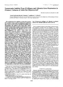
7.Wr Figure 3. Transmission electron micrographs of capsules from a pump containing medium alone (A) or containing rMuIFN-y (B) after 14 d. In the control specimen, fibroblasts (F) contain dilated endoplasmic reticulum (inset); the extracellular matrix is composed predominantly
of closely aggregated mature collagen bundles (C). In the rMuIFN-7treated specimens, fibroblasts appear inactive and strands of loosely packed collagen are observed within an electron-lucent background (A, X 3,000; inset, X 10,000; B, X 3,000).
appeared to eat normally, groomed themselves well, and did not have diarrhea. Electron microscopy. In subsequent experiments, tissue specimens from capsules surrounding pumps loaded with rMuIFN-'y and from controls were processed for transmission electron microscopy. The fibrous capsules from control animals contained numerous thick collagen bundles (Fig. 3 A) and fibroblasts containing endoplasmic reticulum dilated by granular material, an appearance consistent with active collagen synthesis (inset). In contrast, capsules that formed around pumps containing rMuIFN-y contained thin collagen bundles within an electron-lucent matrix and fibroblasts which did not show evidence ofactive collagen synthesis (Fig. 3 B). Computerized image analysis of randomly selected fields (30,328 jzm2 evaluated in 50 randomly selected fields) demonstrated collagen fibers to occupy between 65.4% and 93.8% (mean 81.5%) of the surface area in capsules from control animals compared with 3.2%26.7% (mean 13.5%) in capsules from animals implanted with rMuIFN-y-loaded pumps.
Hydroxyproline content. Specimens were obtained by dissecting the one-quarter of the capsule close to and surrounding the exit port of each pump. This was done easily by utilizing a landmark present on each pump. These specimens were assayed for hydroxyproline content (31). As shown in Table I, the control specimens had much greater hydroxyproline content than the rMuIFN-y-treated specimens. Even when normalized to dry weight, a greater collagen content was found in the control specimens compared with specimens from rMuIFN-y-treated animals. Normalizing to sample weight minimizes the difference in hydroxyproline content observed, because much of the weight of specimens from either group is due to collagen content. Marked differences were also seen when the collagen content of an entire capsule was analyzed (control of 955 jtg of hydroxyproline compared to 205 mg of hydroxyproline for rMuIEFN-"y). The ability of heat-inactivated (80'C for 30 min) rMuIFN-'y (32) to affect collagen formation was also compared with controls treated with medium alone. The hydroxyproline content of the fibrous capsules surrounding pumps filled with heat-inactivated rMuIFN-'y did not vary significantly from controls implanted
Table L Hydroxyproline Content of Capsule Tissue Content of
pumps*
Mean
Mean
hydroxyproline content per 1/4 capsule
hydroxyproline P value
51.3±18.4
*
395.0±40.0
Mean hydroxyproline content per 1/4 capsule
Medium plus heat-treated IFN-y
187±14
Medium alone
235±21
0.964
14.8±1.7
Granstein, Murphy, Margolis, Byrne, and Amento
P value
pg±SEM