Nov 11, 2006 - associated with the accumulation of sulfatide in glial cells and neurons. Despite the fact that the enzymatic deficiency is systemic, disease ...
Research article
Related Commentary, page 2857
Gene therapy of metachromatic leukodystrophy reverses neurological damage and deficits in mice Alessandra Biffi,1,2 Alessia Capotondo,1,2 Stefania Fasano,3 Ubaldo del Carro,4 Sergio Marchesini,5 Hisaya Azuma,6 Maria Chiara Malaguti,4 Stefano Amadio,4 Riccardo Brambilla,3 Markus Grompe,6 Claudio Bordignon,1,2 Angelo Quattrini,4 and Luigi Naldini1,2 1San
Raffaele Telethon Institute for Gene Therapy, 2Vita-Salute San Raffaele University, 3Department of Molecular Biology and Functional Genomics, and Unit, San Raffaele Scientific Institute, Milan, Italy. 5Department of Biomedical Science and Biotechnology, University of Brescia, Brescia, Italy. 6Department of Medical and Molecular Genetics, Oregon Health and Science University, Portland, Oregon, USA.
4Neurology
Metachromatic leukodystrophy (MLD) is a demyelinating lysosomal storage disorder for which new treatments are urgently needed. We previously showed that transplantation of gene-corrected hematopoietic stem progenitor cells (HSPCs) in presymptomatic myeloablated MLD mice prevented disease manifestations. Here we show that HSC gene therapy can reverse neurological deficits and neuropathological damage in affected mice, thus correcting an overt neurological disease. The efficacy of gene therapy was dependent on and proportional to arylsulfatase A (ARSA) overexpression in the microglia progeny of transplanted HSPCs. We demonstrate a widespread enzyme distribution from these cells through the CNS and a robust cross-correction of neurons and glia in vivo. Conversely, a peripheral source of enzyme, established by transplanting ARSAoverexpressing hepatocytes from transgenic donors, failed to effectively deliver the enzyme to the CNS. These results indicate that the recruitment of gene-modified, enzyme-overexpressing microglia makes the enzyme bioavailable to the brain and makes therapeutic efficacy and disease correction attainable. Overall, our data provide a strong rationale for implementing HSPC gene therapy in MLD patients. Introduction Metachromatic leukodystrophy (MLD) is an autosomal recessive lipidosis caused by the deficiency of the lysosomal enzyme arylsulfatase A (ARSA) and can be considered a lysosomal storage disorder (LSD) with predominant neurological involvement (1, 2). The enzymatic defect results in the accumulation of the ARSA substrate galactosylceramide I3-sulfate (sulfatide), a major sphingolipid of myelin. The disease is characterized by myelin degeneration in both the CNS and peripheral nervous system (PNS), associated with the accumulation of sulfatide in glial cells and neurons. Despite the fact that the enzymatic deficiency is systemic, disease manifestations are restricted to the nervous system. Children affected by MLD display progressive neurologic symptoms, including ataxia, seizures, and quadriplegia, culminating in decerebration and eventual death early in infancy (1, 2). As is true for other LSDs affecting the CNS, there is currently no effective treatment for MLD, and the benefits of allogenic hematopoietic stem cell transplantation (HSCT) are still controversial Nonstandard abbreviations used: ARSA, arylsulfatase A; ARSA-HA, HA-tagged ARSA enzyme; BBB, blood-brain barrier; CA2–3, cornu Ammonis, areas 2–3; CCT, central conduction time; CpC, copies per cell; DRG, dorsal root ganglia; EM, electron microscopy; FAH, fumarylacetoacetate hydrolase; GFAP, glial fibrillar acidic protein; GT, gene therapy; HSCT, hematopoietic stem cell transplantation; HSPC, hematopoietic stem progenitor cell; Lamp1, lysosome-associated membrane protein 1; LRh, N-lissamine rhodaminyl; LSD, lysosomal storage disorder; LV, lentiviral vector; MBP, myelin basic protein; MEP, motor-evoked potential; MLD, metachromatic leukodystrophy; M6P, mannose 6-phosphate; NeuN, neuron-specific nuclear protein; NTBC, 2-(2-nitro-4-trifluoromethylbenzoyl)-1,3-cyclohexanedione; p-NC, para-nitrocatechol sulfate; PFA, paraformaldehyde; PNS, peripheral nervous system; RV, retroviral vector; SC, Schwann cell. Conflict of interest: The authors have declared that no conflict of interest exists. Citation for this article: J. Clin. Invest. 116:3070–3082 (2006). doi:10.1172/JCI28873. 3070
(3, 4). Allogenic HSCT corrects the metabolic deficiency in hematopoietic lineages. Furthermore, some of the progeny of engrafted hematopoietic stem progenitor cells (HSPCs) migrate extravascularly and release some functional enzyme in the affected tissues (5). As cells can take up extracellular enzyme by the mannose 6-phosphate (M6P) receptor and correct their defect in vitro (6, 7), it is thought that a similar process of cross-correction may occur in vivo. Several preclinical and clinical studies support the notion that enzymatic cross-correction of the liver and other visceral organs is substantial after HSCT or enzyme replacement therapy in LSDs (8). However, a similar bioavailability of functional enzyme in the CNS is more difficult to achieve. Consistent with this limitation, HSCT has generally had a poor outcome in neurodegenerative storage disorders. Recent clinical studies in globoid cell leukodystrophy report clinical benefits of transplanting BM (4) and umbilical cord blood (9) HSPCs if the procedure is performed soon after birth, at a presymptomatic stage. The same treatment was ineffectual when applied to symptomatic patients (9), suggesting either that recruitment of the HSPC progeny to the CNS is too slow to reverse the disease once it is established or that unique permissive conditions for CNS cross-correction are in place in the perinatal period. Besides its limited efficacy, allogenic HSCT is associated with substantial morbidity and mortality and may adversely affect disease outcome per se. Moreover, recent in vitro studies have raised the concern that macrophages and microglia, which are the HSPC progeny recruited to the CNS, may release lysosomal enzymes lacking the M6P recognition marker (10) and thus represent an inadequate source of enzyme for in vivo cross-correction. Regarding enzyme replacement therapy, studies conducted in murine models indicate that enzyme bioavailability is poor in the CNS when the blood-brain barrier (BBB) integrity is preserved, as is the case in MLD (11).
The Journal of Clinical Investigation http://www.jci.org Volume 116 Number 11 November 2006
research article
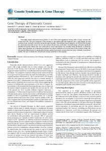
Figure 1 Enzymatic reconstitution and correction of neurological defects in Arsa–/– mice. (A) ARSA activity in PBMC (left y axis) and LV content in BM (right y axis) of untreated (Arsa–/–), mock-treated (GFP), and GT-treated (pool and groups A and B) Arsa–/– mice and WT controls. ARSA activity is expressed as fold increase compared with WT levels and LV content in CpC. (B) ARSA activity (left y axis) and LV CpC (right y axis) from liver samples from the same groups as in A. (C) ARSA activity of brain extracts is expressed as percentage of WT values. For statistical analysis, see Table 1. (D) Representative TLC gel from Rh-sulfatide test on liver and brain extracts from the indicated mice groups. (E) Assessment of central motor conduction in untreated and mock-treated Arsa–/– mice, 12-month-old GT mice (pool), and age-matched WT controls (n = 15 mice per group). The GT group showed significantly lower CCT as compared with 6-month-old and age-matched Arsa–/– mice; comparison with WT mice showed normalization of CCT (*P < 0.05, **P < 0.01). (F) Behavioral evaluations of GT mice. Mean latencies on rotarod ± SEM for each day are indicated. The GT group was indistinguishable from age-matched WT controls (left panel). Twelve-month-old GT mice in group B had a significantly improved performance compared with 6-month-old Arsa–/– mice, demonstrating correction of the neurological deficit present at the time of treatment (right panel). For statistical analysis, see Table 3 (n = 15–30 mice per group.). GalCer, galactosylceramide.
Thus, several biological and clinical factors severely limit the therapeutic potential of HSCT and enzyme replacement therapy for CNS storage disorders, and the development of new effective strategies is badly needed. We and others have proved that HSCT combined with gene therapy (GT) may represent one such candidate strategy, as it combines
advantages of an autologous HSPC source with the benefits of enzyme overexpression in transplanted cells (12–18). We recently demonstrated progressive and extensive reconstitution of well-differentiated microglia in the CNS by the transgene-expressing progeny of transplanted HSPCs in Arsa–/– mice (14). Taking advantage of lentiviral vectors (LVs) to achieve efficient gene transfer into
The Journal of Clinical Investigation http://www.jci.org Volume 116 Number 11 November 2006
3071
3072
We analyzed the following groups of mice: untreated (UT) or mock-treated (MT) Arsa–/– mice, the latter transplanted with Arsa–/– HSPCs transduced with GFP-LV; GT-treated mice (GT, pool and groups A and B; for details, see text); UT and MT WT animals at the indicated age in months. ARSA activity was quantified by p-NC assay on total PBMCs and by Rh-sulfatide test for liver and brain extracts and is expressed as fold increase compared with WT (fold to WT) or percentage (%) of WT levels. LV CpC was quantified by TaqMan on bone marrow DNA from transplanted mice. For rotarod test, the mean latency on rod measured at the ninth trial is reported. For histopathology, the semiquantitative score for white matter deposits and the percentage of degenerated neurons in hippocampal CA2–3 and Purkinje cell layer (neu damage) are reported. For statistical analysis, Student’s t test and 2-way ANOVA were used for CpC, ARSA activity, and neurophysiology, and for behavior, respectively. AP < 0.05, BP < 0.01 for comparison with age matched WT groups; CP < 0.05; DP < 0.01 for comparison with 6-month-old UT Arsa–/–; EP < 0.01 for comparison with 12-month-old MT Arsa–/–; FP < 0.05 for comparison with group A.
1.7 ± 0.5 41 ± 18 2.9 ± 0.7D 62 ± 20C 1.8 ± 0.8E 32 ± 15E 2.2 ± 0.7 45.5 ± 14 1.5 ± 0.8E,F 25.7 ± 8D,E,F – – – – 1.5 ± 0.5 17 ± 2.8 2.7 ± 0.6D 57 ± 9.8D 1.5 ± 0.7E 26.5 ± 15E 1.8 ± 0.6E 40.9 ± 18 1.2 ± 1E 18.4 ± 7E,F – – – – 36 45 61 36 25 29 20 UT Arsa–/–, 6 mo MT Arsa–/–, 12 mo GT, pool, 12 mo GT, group A, 12 mo GT, group B, 12 mo UT WT, 6 mo MT WT, 12 mo
– 4.5 ± 2 6 ± 3 4.1 ± 1.8 7.8 ± 1.4F – 5.1 ± 1.6
0.3 ± 0.3 0.3 ± 0.2 3.5 ± 1.0E 2.8 ± 0.8 4.2 ± 1.0F 1 ± 0.5 1 ± 0.5
0.1 0.1 2.1 ± 1E 1.2 ± 0.3 2.5 ± 0.9F 1 ± 0.3 1 ± 0.3
0.1 0.1 10.1 ± 5.3E 7 ± 2.5 12.6 ± 4.2E 100 ± 2.5 100 ± 2.5
3.4 ± 0.7A 3.9 ± 0.7B,C 3.0 ± 0.3C,E 3.2 ± 0.45E 2.92 ± 0.1C,E 3.0 ± 0.4 2.9 ± 0.5
232 ± 10B 162 ± 12B,D 248 ± 7E 223 ± 10E 263 ± 10D,E,F 262 ± 9 272 ± 9
Deposits, Neu damage, white matter Purkinje (score) (% deg neu) Deposits, Neu damage, fimbria CA2–3 (score) (% deg neu) Latency (s) CCT (ms) PBMC Liver Brain (fold to WT) (fold to WT) (% of WT)
CpC n Treatments
Table 1 Dose-effect relationships in treated Arsa–/– mice
ARSA activity
Neurophysiology
Rotarod
Hippocampus
Cerebellum
research article HSPCs and long-term overexpression of the ARSA gene in their cellular progeny, we prevented the development of major disease manifestations in mice treated at the presymptomatic stage (14). Interestingly, transplantation of WT cells did not achieve significant clinical benefit, suggesting a critical role of enzyme overexpression in the success of our strategy. Unfortunately, in most cases, and unless a family history is available, the diagnosis of MLD is made after the onset of symptoms. Thus, in order for a clinical benefit to be achieved in patients with an established neurologic disease, a new therapeutic strategy should be devised (19). Here we report complete normalization of established behavioral abnormalities, motor conduction deficits, and neuropathological alterations of Arsa–/– mice after HSPC GT. We demonstrate that microglia is the exclusive source of ARSA in the CNS and that enzyme transfer and robust cross-correction of neural cell targets occur in vivo. Thus, our results strongly support the use of HSPC GT in clinical trials for MLD patients. Results Correction of established neurological disease in GT-treated MLD mice. The mouse model of MLD, generated by targeted disruption of the murine ARSA gene Arsa, is characterized by slowly progressive CNS and PNS disease (20) and allows longitudinal evaluation of disease evolution. By 5–6 months of age, Arsa–/– mice already display motor coordination impairment, delayed motor conduction in the CNS and PNS, neuronal degeneration, and widespread storage of metachromatic material; further deterioration along these lines occurs with age (see below). We transplanted HSPCs from Arsa–/– donors transduced with LV expressing either ARSA or GFP into a total of 106 lethally irradiated 6-month-old Arsa–/– recipients (61 with ARSA and 45 with GFP in 8 rounds of transplantation), according to a previously optimized protocol (14). To permit in situ immunodetection of ARSA, in all experiments, we used a C terminal–tagged transgene, in which the gene was fused in frame with the sequence encoding the HA peptide from the HA protein of the human influenza virus. The HA-tagged enzyme had a specific activity comparable to that of the unmodified enzyme and was properly sorted to the lysosomal compartment (21). By measuring ARSA activity with the para-nitrocatechol sulfate (p-NC) assay in PBMCs of treated mice 6 months after transplantation, we demonstrated a complete reconstitution of enzymatic activity and ARSA overexpression with respect to WT levels (Figure 1A). Molecular analysis demonstrated efficient engraftment of transduced cells, with 80%–100% of BM CFCs positive in LV-specific PCR (not shown) and a mean of 6 ± 3 vector copies per cell (CpC), as assessed by quantitative PCR on total BM DNA (Figure 1A). These data indicate that enzyme overexpression in hematopoietic cells is associated with multiple vector integrations. We noticed that the average ARSA activity in circulating hematopoietic cells and average LV content in BM in mice in the first 4 transplantation groups were lower than those for animals in the last 4 transplantation groups. This difference could possibly be due to variations in the infectivity of the vector batch used or in the extent of myeloablation achieved. Based on this difference, we retrospectively divided all treated animals (pool) into 2 groups (A, with lower average ARSA activity, and B, with higher average ARSA activity) and investigated dose-effect relationships for all functional parameters analyzed (Figure 1A and Table 1).
The Journal of Clinical Investigation http://www.jci.org Volume 116 Number 11 November 2006
research article Table 2 Statistical analysis of neurophysiology data Comparison variables F wave latency MCV s-MEP latency c-MEP latency CCT CCT/F
Arsa–/– (6 mo) versus WT (6 mo)
Arsa–/– (6 mo) versus Arsa–/– (12 mo)
Arsa–/– (12 mo) versus WT (12 mo)
GT (12 mo) versus Arsa–/– (6 mo)
GT (12 mo) versus Arsa–/– (12 mo)
GT (12 mo) versus WT (12 mo)
Group A versus group B
< 0.002 < 0.001 0.001 0.006 0.046 0.045
0.30 0.09 0.12 0.014 0.012 0.006
< 0.001 < 0.001 < 0.001 < 0.001 < 0.001 < 0.001
< 0.001 < 0.001 < 0.001 < 0.001 0.019 0.010
< 0.001 < 0.001 < 0.001 < 0.001 < 0.001 < 0.001
0.38 0.56 0.91 0.53 0.68 0.27
0.94 0.44 0.063 0.031 0.49 0.86
Data obtained from neurophysiological tests were analyzed by Student’s t test (2-tail distributions, 2 samples with equal variance, α = 0.05). P value for each examined variable has been reported, considering the mentioned pairs for comparison. c-MEP, cortical MEP; MCV, motor conduction velocity; s-MEP, spinal MEP.
By challenging tissue extracts with the natural ARSA substrate N-lissamine rhodaminyl–sulfatide (LRh-sulfatide), we observed enzyme overexpression in the liver of transplanted mice (Figure 1B). Supranormal ARSA activity likely reflected the extensive liver infiltration by Kupffer cells and macrophages derived from donor-transduced HSPCs, which overexpress the enzyme. Reconstitution of sulfatide metabolism was also detected in the brains of treated mice (Figure 1D) at levels corresponding to an average 10% of WT activity in the pool of treated mice (Figure 1C). As expected, whereas ARSA reconstitution was observed in the tissues of all treated mice, its average level was significantly higher in group B versus group A (Table 1). Functional studies were performed 6 months after the transplant and before sacrifice for all treated and control mice. Neurophysiological evaluations demonstrated complete correction of the previously established defective motor conduction upon GT. Motor-evoked potentials (MEPs) showed correction to normal range of central conduction parameters (central conduction time [CCT], CCT normalized over F wave latency, and cortical MEP latency), indicating not only prevention of further deterioration that occurs with aging but also a reversal of the already established impairment (Figure 1E). Likewise, electroneurographic recordings demonstrated normalization of motor conduction velocity and F wave and spinal MEP latencies as compared with 6month-old untreated Arsa–/– animals (Table 2). Importantly, both group A and group B mice showed normalization of neurophysiological abnormalities. A rotarod test showed protection of treated mice from further impairment of motor coordination and from the development of ataxia, which occurs with aging (P = 0.72, comparing the GT pool at 12 months with age-matched WT controls) (Figure 1F and Table 3). Remarkably, when considered separately, group B
mice showed significantly improved latency on the rod also with respect to the 6-month-old untreated Arsa–/– controls (Figure 1F and Table 3), indicating near complete correction of the previously established behavioral deficit. Histopathological analysis on the CNS and PNS was performed by toluidine blue staining on semithin sections of the brain, cerebellum, dorsal root ganglia (DRG), and sciatic nerve, applying a double-blind scoring system for metachromatic deposit accumulation, neurodegeneration, and demyelination. At the time of transplantation, Arsa–/– mice already showed signs of neuronal damage and lipid storage in the CNS and PNS (see Figures 2 and 3 and Table 1). Small lipid storage deposits were found throughout the white matter, particularly in the fimbria and corpus callosum, and in the cerebellar white matter. Cells with pathological features were detectable in the pyramidal cell layer of the cornu Ammonis, areas 2–3 (CA2–3) regions of the hippocampal cortex and in the Purkinje cell layer of the cerebellum. In the PNS, small metachromatic deposits were detectable in Schwann cell (SC) cytoplasm and in sensory neurons of DRG. By 12 months, the neuronal damage in the CNS and PNS was more severe, and the number and size of metachromatic deposits increased significantly. Furthermore, demyelination of nerve fibers in the PNS became progressively more evident, as confirmed by electron microscopy (EM) studies. Remarkably, in GT–treated mice, we observed a marked reduction of all of the neuropathological features described above (Figure 2). In the hippocampus CA2–3 and in the Purkinje cell layer of the cerebellum, the total number of neurons was higher and the fraction of neurons showing signs of degeneration (percentage over the total number of neurons) was reduced as compared with age-matched mock-treated Arsa–/– mice (Figure 2). Interestingly, when group B mice were considered separately, the
Table 3 Statistical analysis of behavior data Comparison
Arsa–/– (6 mo) versus WT (6 mo)
Arsa–/– (6 mo) versus Arsa–/– (12 mo)
Arsa–/– (12 mo) versus WT (12 mo)
Two-way ANOVA