KAREN L. O'MALLEY*t, ALEX MAURON*t, JOACHIM RAESEt, JACK D. BARCHASt, AND LARRY KEDES*t§ .... as described by Kemp and Cowman (16).
Proc. NatL Acad. Sci. USA Vol. 80, pp. 2161-2165, April 1983 Biochemistry
Genes for catecholamine biosynthesis: Cloning by expression and identification of the cDNA for rat dopaminee,-hydroxylase (recombinant DNA/antigenic determinants/in situ immunoassay/hybrid-selected translation)
KAREN L. O'MALLEY*t, ALEX MAURON*t, JOACHIM RAESEt, JACK D. BARCHASt, AND LARRY KEDES*t§ *The MEDIGEN Project, Department of Medicine, and *the Nancy Pritzker Laboratory of Behavioral Neurochemistry, Department of Psychiatry and Behavioral
Sciences, Stanford University School of Medicine, Stanlbrd, California 94305; and tVeterans Administration Medical Center, Palo Alto, California 94304
Communicated by Richard F. Thompson, January 10, 1983
but also a crossreacting enzyme's mRNA. This can be achieved by translating fractionated mRNAs in an in vitro protein-translating system and identifying the products by immunoprecipitation with the crossreacting antisera. The mRNA fraction enriched in the species of interest can then be used to prepare complementary DNA clones encoding the crossreacting protein. We have used such a scheme to isolate mRNA from the PC 12 clonal cell line and have used TyrOHase antisera for the cloning and screening procedure by which a DBH clone was selected. We made positive identification of the cDNA clone by its ability to selectively hybridize with DBH mRNA.
mRNA for dopamine 3-hydroxylase [3,4-dihydroxyphenylethylamine, ascorbate:oxygen oxidoreductase (fl-hydroxylating), EC 1.14.17.1] has been partially purified from poly(A)+ mRNA isolated from a rat pheochromocytoma cell line. Shared antigenic determinants between tyrosine hydroxylase and dopamine f-hydroxylase allowed us to obtain enriched fractions of dopamine 13hydroxylase mRNA by immunoprecipitating translated mRNA products with tyrosine hydroxylase antisera. The enriched dopamine (-hydroxylase mRNA was used to synthesize the corresponding cDNAs, which were then cloned in the Pst I site of pBR322. Recombinant colonies were characterized by an in situ colony immunoassay and hybrid-selected translation. In vitro translation of the mRNA selected from one recombinant clone produced a protein of 75,000 daltons that comigrated with authentic dopamine 3-hydroxylase. Partial proteolysis of both authentic dopamine 3-hydroxylase and the protein encoded by the recombinant clone produced identical peptide patterns.
ABSTRACT
METHODS Materials. Avian myeloblastosis virus reverse transcriptase, terminal deoxyribonucleotidyltransferase, and Pst I were purchased from P-L Biochemicals. Oligo(dT)-cellulose and oligo(dG)12-18were obtained from Collaborative Research (Waltham, MA). Staphylococcus aureus protein A and CNBr-activated Sepharose 4B were obtained from Pharmacia. BA-85 nitrocellulose filters were obtained from Schleicher and Schuell. Restriction endonucleases were purchased from New England BioLabs. Rabbit reticulocyte lysate, Bolton-Hunter reagent (2,000 Ci/mmol; 1 Ci = 3.7 x 101" Bq) and [35S]methionine (600 Ci/mmol) were purchased from Amersham. EN3HANCE was obtained from New England Nuclear. Purified DBH was provided by S. Watson, University of Chicago. TyrOHase was purified from rat pheochromocytoma (9), and specific antibodies were raised against the enzyme. Antibody specificity against TyrOHase was established by immunoelectrophoresis, and localization of TyrOHase was established by immunohistochemistry in catecholamine-containing neurons in rat brain (unpublished data). The rat pheochromocytoma cell line PC 12 was supplied by E. Shooter, Stanford University. Cell lines were maintained in Dulbecco's modified Eagle's tissue culture medium supplemented with 5% horse serum and 10% fetal calf serum. Cultures were maintained in 12% C02/88% 02 at 370C. Cultures were stimulated by the addition of dexamethasone to the medium at 10 ,uM final concentration. Fresh medium and dexamethasone were added daily. Cells were harvested after 3 days of treatment. Isolation of Poly(A)+ mRNA and Enrichment for DBH Messages. Extraction of RNA from the PC 12 cell line was by the procedure of Strohman et al. (10). Total poly(A)-containing RNA was centrifuged through a 5-20% linear sucrose gradient. Frac-
The catecholamines dopamine, epinephrine, and norepinephrine are formed from their amino acid precursor tyrosine in brain, chromaffin cells,and sympathetic nerves and ganglia. Enzymes involved in this pathway include tyrosine hydroxylase (TyrOHase), DOPA decarboxylase, dopamine-,B3hydroxylase [DBH;
3,4-dihydroxyphenylethylamine, ascorbate:oxygen oxidoreduc-
tase (3-hydroxylating), EC 1.14.17.1], and phenylethanolamine methyltransferase (PhEtnMeTase). Although little is known about the cellular and molecular mechanisms governing the long-term regulation of these enzymes, glucocorticoids (1-3), neural stimulation (4), and nerve growth factor (2, 5-6) can all influence the expression of TyrOHase. Such studies have been facilitated by the development of a rat pheochromocytoma cell line PC 12, which produces relatively large amounts of the catecholamine pathway enzymes (5, 7). Preliminary data from Joh et al. (8) suggested considerable homology between TyrOHase, DBH, and PhEtnMeTase based on peptide analysis and antisera crossreactivity. In particular, Joh et al. demonstrated that antibodies raised against TyrOHase precipitated DBH and PhEtnMeTase as well. Peptide mapping by partial proteolysis and gel electrophoresis indicated that DBH, TyrOHase, and PhEtnMeTase had identical peptides in common, but no primary amino acid sequence data has yet been
reported.
Shared antigenic determinants and peptide patterns not only suggest similar primary structure among these proteins but also structural homologies at the nucleotide level. Such interrelationships might be exploited to isolate specific mRNAs for the catecholamine pathway enzymes. For example, available antisera for one enzyme can be used to purify not just its own mRNA
Abbreviations: TyrOHase, tyrosine hydroxylase; DBH, Dopamine (3 hydroxylase; PhEtnMeTase, phenylethanolamine methyltransferase. § To whom correspondence should be addressed at: The MEDIGEN Project, 151M, Veterans Administration Medical Center, Palo Alto,
The publication costs of this article were defrayed in part by page charge payment. This article must therefore be hereby marked "advertisement" in accordance with 18 U. S. C. §1734 solely to indicate this fact.
CA 94304.
2161
2162
Proc. Natl. Acad. Sci. USA 80 (1983)
Biochemistry: O'Malley et al.
tions were assayed for DBH mRNA by in vitro translation with a rabbit reticulocyte lysate system (11). Labeled translation products were immunoprecipitated (12), electrophoresed on NaDodSO4 gels (13), and fluorographed (14). Construction of cDNA Clones. Sucrose gradient fractions containing maximum DBH mRNA activity were pooled, and the mRNA was used for the synthesis of double-stranded cDNA by the procedure of Land et al. (15). After a dCTP-tailing step, the double-stranded cDNA was hybridized to pBR322 tailed with dGTP at the Pst I site. The inserted plasmid was used to transform Escherichia coli C600rk mk . Ampicillin-sensitive, tetracycline-resistant clones were transferred to nitrocellulose filters on agar plates. All recombinant DNA procedures followed the pertinent National Institutes of Health guidelines. In Situ Colony Immunoassay. Colonies grown on nitrocellulose filters were prepared for colony immunoassay essentially as described by Kemp and Cowman (16). Bacterial colonies were lysed in situ, and proteins were bound to activated CNBr paper (17). Covalently bound antigens were detected by incubating with antisera followed by '25I-labeled protein A from S. aureus. In order to remove antibacterial antibodies, rabbit TyrOHase antiserum was passed over a column of E. coli C600/pBR322 protein extract bound to a CNBr-Sepharose 4B column (18). S. aureus protein A was iodinated with '25I-labeled Bolton-Hunter reagent (19) to a specific activity of 1 X 10⁷ cpm/,ug. Positive mRNA Selection. Plasmid DNA was isolated from immunoreactive clones, CsCl-purified, and bound to nitrocellulose filters by modifications of the method of Ricciardi et al. (20). Plasmid DNA (10 ,ug) was denatured in 2 M NaCl/0.2 M NaOH, briefly heated at 90°C, chilled, and immediately loaded onto nitrocellulose filters. Prior to loading the DNA, filters were 1
2
3
4
wetted with 0.3 M NaCl/2 mM EDTA/0.1% NaDodSO4/10 mM Tris, pH 7.5/0.1% diethyl pyrocarbonate. After DNA application, the filters were baked for 2 hr at 80°C in a vacuum oven. Filters were again wetted in the filter binding solution and allowed to air dry. Total mRNA prepared from dexamethasone-treated cells was hybridized to the filter-bound DNA in 30% formamide/500 mM NaCl/2 mM EDTA/50 mM Pipes, pH 7.5/0.5% NaDodSO4. Hybridization continued for 16 hr at 42°C. Nonspecific RNA bound to the filters was removed by extensive washes in 75 mM NaCl/7.5 mM Na citrate at 42°C. Hybridized RNA was eluted by boiling the filters for 1 min in 300 1,l of sterile H20. Eluted RNAs were precipitated with ethanol, dried, resuspended in 2 ,ul of H20, and translated as described. Peptide Mapping by Partial Proteolytic Digestion of Authentic DBH. The [3S]methionine-labeled protein was synthesized in vitro from mRNA that had been eluted after filter hybridization to a putative DBH cDNA clone. The radiolabeled protein band was cut out of the NaDodSO4 gel and placed in the sample well of a second NaDodSO4 gel. Partial proteolytic digestion of both the translated protein and of authentic DBH was performed as described by Cleveland et al. (21) with S. aureus V8 protease. Peptide patterns were revealed by the silver staining procedure described by Oakley et al. (22) followed by autoradiography. RESULTS Total poly(A)+ mRNA was prepared from dexamethasone-stimulated PC 12 cells and translated in vitro in the rabbit reticulocyte system. Translation products were immunoprecipitated with a rabbit serum directed against rat pheochromocytoma TyrOHase. Analysis of the translation pattern indicated a major band at 60,000 daltons corresponding to the subunit molecular weight of TyrOHase (3) and a 75,000-dalton protein corre1
92
2
3
4
5
6
7
8
-
DBH -
DBH-
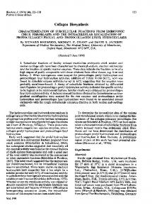
:311 FIG. 1. Immunoprecipitation of DBH from rat PC 12 cells. Total poly(A)+ mRNA from dexamethasone-treated cells was translated in a reticulocyte cell-free system. An aliquot was precipitated with antisera raised against TyrOHase. [35S]Methionine-labeled products were separated on a NaDodSO4/10% polyacrylamide gel. Lanes: 1, Coomassie blue staining pattern of authentic DBH (25 jig); 2, immunoprecipitation of DBH and TyrOHase with TyrOHase antisera from total translated products of PC 12 poly(A)+ mRNA; 3, total translated products; 4, products seen when no mRNA is added to the system. Molecular mass (shown x 10-3) was calculated by reference to the mobilities of standard proteins: phosphorylase b (92,000), bovine serum albumin (68,000), ovalbumin (45,000), and carbonic anhydrase (31,000).
92
- 68
..........;
- 45
-31
FIG. 2. Sucrose gradient fractionation of poly(A)+ mRNA from dexamethasone-treated PC 12 cells. Aliquots of the indicated fractions were translated in vitro, and translation products were analyzed by NaDodSO4/10% polyacrylamide electrophoresis. Fractions enriched for DBH mRNA (6,7) were pooled and used to prepare complementary DNA. Daltons are shown x 103.
Biochemistry: O'Malley et al. sponding to the subunit molecular weight of DBH (23) (Fig. 1). One hundred micrograms of poly(A)+ mRNA was then fractionated on a 5-20% sucrose gradient. An aliquot of each fraction was translated in vitro, and fractions 6 and 7, enriched in DBH mRNA activity, were pooled (Fig. 2). Analysis of the optical density profile (data not shown) indicated that these fractions were somewhat larger than the 18S peak. Molecular Cloning of the cDNA for DBH. Enriched mRNA (15 ug) was transcribed into cDNA; subsequently double strands were formed and inserted into the Pst I site of pBR322 by the d(G&C)n-tailing method. Five hundred tetracycline-resistant, ampicillin-sensitive clones were obtained. Transformants were picked onto nitrocellulose filters, grown overnight, and then lysed on gauze pads saturated with 0.1 M NaHCO3/1% Triton X-100/2 mg of lysozyme per ml in an atmosphere saturated with CHC13. Filters were subsequently placed on gauze pads saturated with 0.1 M NaHCO3/0. 1% Tri-
A
B
Proc. Natl. Acad. Sci. USA 80 (1983)
2163
ton X-100 and then onto filter paper soaked in the same buffer. Activated CNBr paper that had been dampened with binding buffer was placed over the lysed colonies, and antigen binding was carried out for 4 hr at 40C. Remaining functional groups on the paper were then blocked with 1 M ethanolamine, pH 9.0, for 1 hr at room temperature. Treatment with 2% glycine as suggested by Kemp and Cowman (16) proved insufficient in our hands to prevent high filter backgrounds after autoradiography. After inactivation, immobilized proteins were allowed to react with pretreated TyrOHase antisera. Without removing antibacterial antibodies, all colonies appeared to give a positive signal. Similarly, the appropriate dilution of antisera was necessary to prevent filter backgrounds from obscuring positive signals. A dilution of 1:300 of the presorbed antibody was used in all experiments shown. Of the 500 colonies, 12 gave a positive signal of varying intensity (Fig. 3). From these 12 colonies, 3 gave a strong positive signal in each of four experiments, 6 had weaker signals in all experiments, and 3 failed to immunoreact in subsequent experiments. All of the immunoreactive clones could be displaced by sera presorbed with native TyrOHase, indicating the reaction was specific for this enzyme or crossreacting species, or both. Analysis of plasmid DNA from the nine immunoreactive clones indicated that all nine were different based on restriction endonuclease digestions. Insert size of the immunoreactive clones ranged from 300 base pairs to 3.5 kilobases, with an average size of 500 base pairs. With the exception of the 3.5-kilobase transformant, the remaining clones would not be long enough to code for a subunit of either TyrOHase or DBH. Apparently, even a short insert sequence covalently linked to the E. coli (3
1 2
3
4
5
6
-
92 68
C
-45
31
_ FIG. 3. Detection of immunoreactive clones by colony immunoassay. Colonies were grown on nitrocellulose filters and then lysed in situ. Proteins from the lysates were blotted onto CNBr-activated filter papers. Antigens covalently bound to the CNBr paper were detected by reaction with TyrOHase antiserum, followed by 125I-labeled protein A and autoradiography. (A) Colonies (100) were incubated with TyrOHase antiserum (1:300). (B) Duplicate colonies were incubated with antiserum presorbed with 500 ug of cold TyrOHase; cold TyrOHase (100 ng) was also spotted next to the colonies. (C) Cold TyrOHase (100 ng) was spotted onto activated filters and processed with untreated TyrOHase antiserum.
FIG. 4. Autoradiograms of [35S]methionine-labeled in vitro translation products synthesized with mRNA complementary to pDBH-1. CsCl-purifled plasmid DNA was denatured and bound to nitrocellulose filters. Filters were hybridized with total PC 12 RNA. RNA bound to the filters was eluted thermally and subsequently analyzed by cell-free translation. [3S]Methionine translation products were separated by electrophoresis on NaDodSO4 gels and autoradiographed. Lanes: 1, translation products of total PC 12 RNA; 2, products immunoprecipitated with TyrOHase antisera; 3, immunoprecipitation of the translated product synthesized with RNA selected by pDBH-1; 4, total translation products; 5, total translation products when no RNA was added to the system; 6, authentic DBH (25 pg) stained with Coomassie blue.
2164
Biochemistry: O'Malley et al.
Proc. Natl. Acad. Sci. USA 80 (1983)
lactamase gene produces enough of the fused polypeptide to be detected, at least by a polyvalent antibody. Filter Hybridization and Translation. The plasmids from the nine immunoreactive clones were individually purified and used to select complementary RNA in the filter hybridization-positive selection procedure. Selected RNAs were translated in vitro in the rabbit reticulocyte system; translated products were displayed on NaDodSO4 polyacrylamide gels. From the nine clones, RNA selected from one revealed a unique band of 75,000 daltons (Fig. 4). This protein which could be immunoprecipitated with TyrOHase antisera comigrated with authentic DBH. This plasmid contained an insert of 330 base pairs and was designated pDBH-1. Peptide Mapping of the Protein Encoded by pDBH-1 and Native DBH. In order to unequivocally identify this clone as containing nucleotide sequences specific for DBH, we analyzed peptide patterns obtained from the protein encoded by pDBH1. The [35S]methionine-labeled protein obtained from the positive selection procedure was removed from the gel and subjected to partial proteolysis with S. aureus V8 protease. Simultaneously we digested a sample of authentic DBH that had similarly been cut out of a NaDodSO4 gel. Digestion products were displayed on 15% NaDodSO4 gels that had been stained with silver. After autoradiography, methionine-labeled bands were compared with those generated by the partial proteolysis of the authentic protein. As seen in Fig. 5, the labeled pattern corresponded with that of the authentic DBH. To verify this result, gel slices from the translated protein and authentic DBH were incubated also with "I in order to label all of the peptides, not just those containing methionine groups. Again, in this experiment, all of the labeled bands observed in the authentic DBH peptide pattern matched those of the plasmidspecific protein. In the latter case, additional bands also were observed, presumably corresponding to a comigrating reticulocyte lysate protein (data not shown). 1
2
3
4
5
.-V8 PROTEASE
FIG. 5. Peptide mapping of authentic DBH and the translated product obtained from RNA selected by pDBH-1. Peptide mapping was done with gel slices obtained from the electrophoresis of either authentic DBH (25 ,ug) or the translated product from RNA selected by pDBH-1. Samples were incubated with 1 ,ug of V8 protease in a 4-cm stacking gel prior to resolution in a 15% NaDodSO4/polyacrylamide gel. One experiment (lanes 1-3) shows the silver-stained peptide patterns from one gel slice of authentic DBH (lane 1) and one gel slice of the hybrid-selected translation product (lane 2); lane 3 shows the [35S]methionine pattern of lane 2 after 2 wk of autoradiographic exposure. In another experiment (lanes 4 and 5), five gel slices of the hybridselected translation product and one gel slice of the authentic DBH were incubated with 5 ,ug of V8 protease. By increasing the amount of the translated product of pDBH-1, silver staining alone was sufficient to demonstrate the similarity in banding patterns of authentic DBH (lane 5) and the hybrid-selected product (lane 4).
DISCUSSION What appear to be shared antigenic sites of the catecholamine pathway enzymes enabled us to obtain from PC 12 cells a messenger RNA fraction enriched for DBH mRNA by using TyrOHase antiserum. The enriched DBH mRNA was then used to prepare a complementary DNA library from which a DBH cDNA clone was isolated. In the absence of a reported amino acid sequence for DBH, the following criteria were used to establish the identity of the DBH clone. (i) Clones were picked that synthesized peptides crossreacting with TyrOHase antiserum. Fused polypeptides from expressing transformants could be displaced by using antiserum presorbed with native TyrOHase. (ii) One such clone selected a mRNA that, when translated in vitro, formed a protein of 75,000 daltons, the size of DBH. (iii) The translated protein could be immunoprecipitated with TyrOHase antiserum and comigrated with authentic DBH. (iv) Partial proteolysis of both authentic DBH and the protein encoded by pDBH-1 produced identical peptide patterns. The results described here demonstrate the effectiveness of the colony immunoassay procedure. A general disadvantage of the method is that only one in six clones would contain an insert in the right orientation and reading frame to code for a fused polypeptide. However, enriching for the desired messenger population, inserting double-stranded cDNA as close to the full length as possible, and screening additional transformants would increase the chances of detecting an expressing transformant. Kemp and Cowman (16) detected even greater proportions of positive transformants when full-length inserts were screened by colony immunoassay, probably due to peptide initiation within the insert itself (24, 25). But as demonstrated in the case of pDBH-1, even small inserts can generate a fused peptide carrying immunoreactive determinants. As with all immunological procedures, antibody specificity is crucial. Of the other eight immunoreactive clones described here, two proved to be clones of a noncatecholamine-related protein slightly recognized by the TyrOHase antibody. Affinitypurified antibodies should increase sensitivity further and minimize selection of false positives. This raises another issue. Although the preliminary data of Joh et al. (8) suggests that the catecholamine biosynthetic enzymes, TyrOHase, DBH, and PhEtnMeTase share antigenic determinants, it is also possible that the antigens used to generate antisera are cross-contaminated because the antigens are derived from tissues in which all three enzymes are present. The availability of a rat cDNA probe for DBH will allow investigation of the transcriptional regulation of the DBH gene in the PC 12 cell line. The clonal cell line not only possesses the highly differentiated properties of typical adrenergic cells but also exhibits responses typical of early cells in the neural crest (5, 6). As such, the PC 12 cell line should be a good model for additional studies examining mechanisms responsible for differentiation of the neural crest in vivo. Recently, it has been reported that rat cDNA clone for TyrOHase cross-hybridized with mRNA from human tissue (26). Presumably, this also will be true for DBH, so that the rat cDNA clone could be used to identify corresponding sequences in clones from a human genomic DNA library. Then human probes could be used to investigate atypical catecholamine function at the molecular level in humans. We thank George Makk for excellent technical assistance. This work was supported in part by National Institute of Mental Health Grant MH 23861 and Office of Naval Research Grant SRO-001 to J. D. B., and by grants from the National Institutes of Health and Veterans Administration to L.K.
Biochemistry: O'Malley et al. 1. Otten, U. & Thoenen, H. (1976) Brain Res. 111, 438-441. 2. Goodman, R. & Herschman, H. R. (1978) Proc. Nati. Acad. Sci. USA 75, 4587-4590. 3. Baetge, E. E., Kaplan, B. B., Reis, D. J. & Joh, T. H. (1981) Proc. Nati. Acad. Sci. USA 78, 1269-1273. 4. Thoenen, H. (1974) Life Sci. 14, 223-235. 5. Greene, L. A. & Tischler, A. S. (1976) Proc. Nati. Acad. Sci. USA 73,.2424-2428. 6. Greene, L. A. & Rein, G. (1977) Brain Res. 129, 247-263. 7. Warren, S. & Chute, R. N. (1972) Cancer 29, 327-331. 8. Joh, T. H., Baetge, E. E., Kaplan, B. B., Ross, M. E., Brodsky, M. J., Albert, V. R., Park, D. H. & Reis, D. J. (1981) Neurosci. Abstr. 7, 206. 9. Raese, J. D., Edelman, A. M., Laar, M. A. -& Barchas, J. D. (1977) in Structure and Function of Monoamine Enzymes, eds. Usdin, E., Weiner, N. & Youdim, M. B. H. (Dekker, New York), pp. 383-421. 10. Strohman, R. C., Moss, P. S., Micou-Eastwood, J., Spector, D., Przybyla, A. E. & Paterson, B. (1977) Science 196, 1313-1319. -11. Pelham, H. R. B. & Jackson, R. J. (1976) Eur.j. Biochem. 67, 247256. 12. Kessler, S. W. (1976)J. hnmunol. 117, 1482-1490. 13. Douglas, M., Finkelstein, D. & Butow R. (1976) Methods Enzynol. 56, 58-66. 14. Bonner, W. M. & Laskey, R. H. (1974) Eur. J. Biachern. 46, 8388.
Proc. Natl. Acad. Sci. USA 80 (1983)
2165
15. Land, H., Grez, M., Hauser, H., Lindenmaier, W. & Schutz, G. (1981) Nucleic Acids Res. 9, 2251-2266. 16. Kemp, D. J. & Cowman, A. F. (1981) Proc. Natl. Acad. Sci. USA 78, 4520-4524. 17. Clarke, L., HIitzeman, R. & Carbon, J. (1979) Methods Enzymol. 68, 436-442. 18; Erlich, H. A., Cohen, S. N. & McDevitt, H. 0. (1978) Cell 13, 681-689. 19. Bolton, A. E. & Hunter, W. M. (1973) Biochem.J. 133, 529-534. 20. Ricciardi, R. P., Miller, J. S. & Roberts, B. C. (1979) Proc. Nati. Acad; Sci. USA 76, 4927-4931. 21. Cleveland, D. W., Fischer, S. G., Kirschner, M. W. & Laemmli, U. K. -(1977) J. Biol. Chein. 252, 1103-1106. 22. Oakley, B. R., Kirsch, D. R. & Morris, N. R. (1980) Anal. Biochemn. 105, 361-363. 23. Ciaranello, R. D., Wooten, G. F. & Axelrod, J. (1975)J. Biol. Chern. 250, 3204-3211. 24. Emtage, J. S., Tacon, W. C. A., Catlin, G. H., Jenkins, B., Porter, A. G. & Carey, N. H. (1980) Nature (London) 283, 171-174. 25. Chang, A. C. Y., Nunberg, J. H., Kaufman, R. J., Ehrlich, H., Schimke, R. T. & Cohen,.S. N. (1978) Nature (London) 275, 617624. 26. Lamouroux, A., Biguet, N. F., Samolyk, D., Privad, A., Salomon, J. C., Pujol, J. F. & Mallet, J. (1982) Proc. Natl. Acad. Sci. USA 79, 3881-3885.