mechanisms of lung cancer to paclitaxel. ... tivity of lung cancer cells to paclitaxel and the expres sion of a ..... Alley, M.C., Scudiero, D.A., Monks, A., et al., Cancer.
ISSN 1607�6729, Doklady Biochemistry and Biophysics, 2011, Vol. 437, pp. 105–108. © Pleiades Publishing, Ltd., 2011. Original Russian Text © K.N. Kashkin, E.A. Musatkina, A.V. Komelkov, D.A. Sakharov, E.V. Trushkin, E.A. Tonevitsky, T.V. Vinogradova, E.P. Kopantzev, M.V. Zinovyeva, O.V. Kovaleva, K.A. Arkhipova, I.B. Zborovskaya, A.G. Tonevitsky, E.D. Sverdlov, 2011, published in Doklady Akademii Nauk, 2011, Vol. 437, No. 6, pp. 837–841.
BIOCHEMISTRY, BIOPHYSICS AND MOLECULAR BIOLOGY
Genes Potentially Associated with Resistance of Lung Cancer Cells to Paclitaxel K. N. Kashkina, E. A. Musatkinab, A. V. Komelkovb, D. A. Sakharovc, E. V. Trushkinc, E. A. Tonevitskyc, T. V. Vinogradovaa, E. P. Kopantzeva, M. V. Zinovyevaa, O. V. Kovalevab, K. A. Arkhipovab, I. B. Zborovskayab, Corresponding Member of the RAS A. G. Tonevitskyc, and Academician E. D. Sverdlova Received December 24, 2010
DOI: 10.1134/S1607672911020153
Paclitaxel is a natural substance from the group of taxanes and one of the most widely used drugs in che� motherapy of malignant tumors, including the non� small cell lung cancer (NSCLC). Treatment of patients with advanced NSCLC with paclitaxel alone or in combination with other drugs led to complete or partial tumor regression in at most 41% of patients [1]. Individual selection of drugs that are effective for a particular patient is one of the hottest approaches in the strategy to improve the results of chemotherapeu� tic treatment of cancer patients. This work is dedicated to finding new markers and studying the resistance mechanisms of lung cancer to paclitaxel. The main mechanism of action of paclitaxel is the blockade of cell division due to its specific binding to β�3 tubulin and stabilization of microtubules. An increased expression of some β�tubulin isotypes corre� lates with resistance to paclitaxel and the degree of malignancy of tumors of the lung, prostate, ovary and other tissues. The same result can be caused by muta� tions of β�tubulins, although the clinical significance of mutations is controversial (see reviews [2, 3]). It is known that the resistance of tumor cells to paclitaxel may be associated with an increased expres� sion or polymorphism of some ABC�transporters responsible for multidrug resistance [4] as well as with activity of growth factor receptors (EGFR and HER� 2), proteins LIMK1, LIMK2, TGFB1, STMN1, spin� dle checkpoint proteins, apoptosis regulators, and others [2, 3].
a
Shemyakin–Ovchinnikov Institute of Bioorganic Chemistry, Russian Academy of Sciences, ul. Miklukho�Maklaya 16/10, Moscow, 117997 Russia b Blokhin Cancer Research Center, Russian Academy of Medical Sciences, Kashirskoe sh. 24, Moscow, 115478 Russia c All�Russia Institute of Physical Culture and Athletics, Elizavetinskii per. 10, Moscow, 105005 Russia
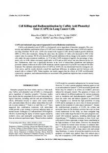
The use of DNA microchips [5–10] made it possi� ble to reveal the sets of genes whose expression corre� lates with chemoresistance of lung cancer cells. How� ever, the sets of genes that, with a certain probability, may be involved in resistance development, which were proposed by different authors, differ. In addition, the genes detected in cell cultures may be uninforma� tive for predicting the response of patients to chemo� therapy [11]. At the same time, studies of clinical specimens only may give ambiguous results due to polymorphisms, which lead to a high interindividual variability of systems involved in the development of chemoresistance of cancer patients, as well as because of chemotherapy regimens that usually include several drugs. To search for new informative markers and to reveal the molecular mechanisms of drug resistance in tumors, we studied the relationship between the sensi� tivity of lung cancer cells to paclitaxel and the expres� sion of a wide range of genes using the new microchip platform Affymetrix Human GeneChip ST1.0, which contained probes for over 28000 human mRNAs. As a biological model, we used lung cancer cells of six lines derived from ATCC (A549, NCI�H292, NCI� H460, and NCI�H1299) and ECACC (NCI�H322 and NCI�H358). Although the sensitivity of certain lines to various drugs is known (http://dis� cover.nci.nih.gov/cellminer/), we redefined the IC50 of paclitaxel for all cells using the MTT technique [12] in order to eliminate the effect of subculturing of cells on their sensitivity to the drug. For each cell line, we analyzed the results of at least three significant mea� surements (Fig. 1). Hybridization of labeled samples prepared from total cell RNA with microchips was performed using the equipment and method of Affymetrix. The results of hybridization were processed by the RMA algo� rithm using the xps library (Christian Stratowa; http://www.bioconductor.org) in R system. Fluores� cence signals were filtered using I/INI algorithm [13]; signals with random variation were ignored.
105
106
KASHKIN et al. Cells with intermediate sensitivity
Paclitaxel, IC50, µM
0.04
Resistant cells
Sensitive cells
0.03 0.02 0.01 0
A549
NCI�H1299NCI�H358 NCI�H322 NCI�H292 NCI�H460
IC50 concentrations of paclitaxel for lung cancer cells. The mean IC50 value (diamond sign), the standard error of the mean (SEM), and conditional separation of cells into groups according to sensitivity to paclitaxel are shown. The abscissa axis shows cell lines.
According to the results of IC50 measurements, cells were divided into three groups: resistant cells (lines A549, NCI�H1299, and NCI�H358), cells of intermediate sensitivity (line H322), and sensitive cells (lines NCI�H292 and NCI�H460). According to the results of hybridization with the microchip, we selected the probes for which the difference between the threshold fluorescence values (expression index) in the first and third groups differed by at least twice (Table 1). In addition, we determined the Pearson cor� relation coefficient (R) for the gene expression rate in cells of all six lines with IC50 and selected genes with R ≥ 0.9 and R ≤ –0.9. Having combined the genes selected by the two methods, we obtained two sets of genes whose expression is associated with paclitaxel� resistant (56 genes) and paclitaxel�sensitive (52 genes) phenotypes. The obtained panels of genes were used to pre� dict possible mechanisms that determine the sensi� tivity or resistance of tumor cells to paclitaxel. For this purpose, we identified the biological pro� cesses that involve the identified genes using NCI Pathway Interaction Databases (NCI PID, http://pid.nci.nih.gov/index.shtml), Gene Ontology database (http://www.geneontology.org), and the Table 1. Genes differing in the expression level between groups of resistant and sensitive cells* Gene predominantly ex� pressed in resistant cells XAGE1D, FLJ35848, UCP2, VCAN, PEG10, GEM, TNNT1, ARL4C, CCDC144A, C8orf37
Gene predominantly ex� pressed in sensitive cells SERPINB2, CDH13, NID2, IL1A, SORBS2, FAM134B, C4orf18, ASS1, SMOC1, ADAMTS1
* Ten genes that most significantly differed in the level of expression are shown.
Kyoto Encyclopedia of Genes and Genomes (KEGG, http://www.genome.jp/kegg/kegg2.html) (Table 2). The study of the processes showed that the resistant cells actively expressed the genes that are involved in signal transduction cascades mediated by SMAD2/3, TGF�β, and C�MYC. These signaling pathways are responsible for the passage of cell�cycle checkpoints by the cell in the G1 phase and are not associated with interleukins. Probably, these genes and signal trans� duction pathways allow cells to stop in the G1 phase and survive at low concentrations of paclitaxel [14]. Note an increased expression of certain target genes of C�MYC, a transcriptional regulator of a broad spec� trum of genes that control pivotal cell functions, including DNA replication and repair and the cell cycle passage. In addition, paclitaxel�resistant cells are characterized by an increased expression of some genes whose products are involved in signal transduc� tion and genes encoding proteins with different trans� port functions: ENY2 (a transcription activator that ensures the transport of mRNA to the cytoplasm), SMAD3 (protein involved in nuclear translocation of β�catenin), GABRB3 (a member of the GABA�A family receptors, which are components of ion chan� nel), and UCP2 (mitochondrial regulator of proton transport). The specific role of these processes in drug resistance of cells should be investigated in further studies. An increased expression of genes encoding inter� leukins and other regulators of apoptosis was detected in the paclitaxel�sensitive cells. This indicates a possi� ble role of interleukin�dependent signaling cascades in cell response to paclitaxel and their relation to certain diseases and interleukin�mediated reactions of the body (e.g., prion diseases, inflammation, etc.). This opens up new ways to boost cell response to the drug. In particular, it was shown that the prion protein PrPc interacts with P�glycoprotein during the development of multidrug resistance and that it is involved in the regulation of doxorubicin�induced apoptosis by mod� ulating the expression of Bcl�2 and Bax genes [15]. Interestingly, the group of genes identified in the pacli� taxel�sensitive cells includes both the genes preventing cell proliferation (ADAMTS1 and CDH13) and the genes with the opposite function, which inhibit apop� tosis (SERPINB2/PAI�2 and SERPINB9/PI�9). Apparently, the paclitaxel�sensitive cells also differ from the paclitaxel�resistant cells in some metabolic characteristics (oxidation–reduction, fatty acid metabolism, and the respiratory electron transport chain), which should also be studied in the future. One of the key regulators of the cell cycle and apo� ptosis that may determine the degree of cell resistance to drugs, in particular to antimitotic drugs, is p53 [3]. We noted that the group of paclitaxel�sensitive cell lines included the cell lines with the wild�type p53 gene (p53wt, lines NCI�H292 and NCI�H460) and that the group of paclitaxel�resistant cell lines included two cell lines with the p53 gene deletion
DOKLADY BIOCHEMISTRY AND BIOPHYSICS
Vol. 437
2011
GENES POTENTIALLY ASSOCIATED WITH RESISTANCE
107
Table 2. Biological processes associated with different sensitivity of lung cancer cells to paclitaxelTable 2. Pathways and in� teractions associated with different sensitivity of lung cancer cells to paclitaxel Gene KPNA2, SMAD3 PPP2CB, SMAD3 PEG10, SMAD3 ADRA1B, F2RL1, GABRB3 ENY2, GABRB3, SMAD3, UCP2 ADRA1B, PPP1R14A IL1A, IL1B IL1A, IL1B, SERPINB2, SERPINB9 ADAMTS1, CDH13, IL1A, IL1B HADH, SDHD, PRDX6 BACE2, IL1B, SDHD IL1A, IL1B
Process* Paclitaxel resistant phenotype Regulation of cytoplasmic and nuclear SMAD2/3 signaling TGF�beta receptor signaling Validated targets of C�MYC transcriptional activation GO:0007165 Signal transduction; ko04080 Neuroactive ligand�receptor interaction GO:0006810 Transport (of molecules and ions) ko04270 Vascular smooth muscle contraction Paclitaxel sensitive phenotype IL1�mediated signaling events GO:0006916 Anti�apoptosis GO:0008285 Negative regulation of cell proliferation ko01100 Metabolic pathways ko01110 Biosynthesis of secondary metabolites ko05010 Alzheimer’s disease ko04210 Apoptosis; ko04010 MAPK signaling pathway; ko04060 Cytokine�cytokine receptor interaction; ko05332 Graft�versus�host disease; ko04640 Hematopoietic cell lineage; ko04940 Type I diabetes mellitus; ko05140 Leishmaniasis; ko05020 Prion diseases
* Processes without indexes are specified by NCI PID (if two or more genes from the set are involved), with “GO” index by Gene Ontol� ogy (if three or more genes from the set are involved, Hyp*