Jul 4, 2007 - Enterococcus faecalis, a ubiquitous member of mammalian gastrointestinal flora, is a leading cause of ... aggregation substance, a hyaluronidase, and a bile salt hydrolase .... equal proportions, and 50 ul spotted onto non-selective BHI agar ...... for cellobiose, fructose, glucitol, glucose, maltose, mannitol,.
Genetic Diversity among Enterococcus faecalis Shonna M. McBride1, Vincent A. Fischetti2, Donald J. LeBlanc3¤, Robert C. Moellering, Jr.4, Michael S. Gilmore1* 1 Schepens Eye Research Institute, Harvard Medical School, Boston, Massachusetts, United States of America, 2 The Laboratory of Bacterial Pathogenesis and Immunology, The Rockefeller University, New York, New York, United States of America, 3 Antibacterial Molecular Sciences, Global Research and Development, Pfizer, Inc., Ann Arbor, Michigan, United States of America, 4 Department of Medicine, Beth Israel Deaconess Medical Center, and Harvard Medical School, Boston, Massachusetts, United States of America
Enterococcus faecalis, a ubiquitous member of mammalian gastrointestinal flora, is a leading cause of nosocomial infections and a growing public health concern. The enterococci responsible for these infections are often resistant to multiple antibiotics and have become notorious for their ability to acquire and disseminate antibiotic resistances. In the current study, we examined genetic relationships among 106 strains of E. faecalis isolated over the past 100 years, including strains identified for their diversity and used historically for serotyping, strains that have been adapted for laboratory use, and isolates from previously described E. faecalis infection outbreaks. This collection also includes isolates first characterized as having novel plasmids, virulence traits, antibiotic resistances, and pathogenicity island (PAI) components. We evaluated variation in factors contributing to pathogenicity, including toxin production, antibiotic resistance, polymorphism in the capsule (cps) operon, pathogenicity island (PAI) gene content, and other accessory factors. This information was correlated with multi-locus sequence typing (MLST) data, which was used to define genetic lineages. Our findings show that virulence and antibiotic resistance traits can be found within many diverse lineages of E. faecalis. However, lineages have emerged that have caused infection outbreaks globally, in which several new antibiotic resistances have entered the species, and in which virulence traits have converged. Comparing genomic hybridization profiles, using a microarray, of strains identified by MLST as spanning the diversity of the species, allowed us to identify the core E. faecalis genome as consisting of an estimated 2057 unique genes. Citation: McBride SM, Fischetti VA, LeBlanc DJ, Moellering RC Jr, Gilmore MS, et al (2007) Genetic Diversity among Enterococcus faecalis. PLoS ONE 2(7): e582. doi:10.1371/journal.pone.0000582
Recently, multi-locus sequence typing (MLST) schemes have been developed to facilitate analysis of a number of bacterial species [21], including E. faecalis [22–24]. The development of a facile means for characterizing the genetic background of a strain makes possible the study of the flow of mobile elements within the species. Previous studies using MLST to differentiate isolates of E. faecalis at a subspecies level centered on identifying virulent clusters and outbreak isolates, as well as plotting the emergence of antibiotic resistance elements in the population [22–24]. In this study, we utilize MLST to define the diversity of the E. faecalis species and to determine the core genome content. A collection of 21 E. faecalis strains were previously assembled by Maekawa and coworkers [25] from approximately 1,000 isolates and from pre-existing collections, to represent the diversity of the E. faecalis species as judged by serotyping. Since the genetic basis for serological identity was unknown, it was of interest to determine the extent to which serologic diversity reflected genetic diversity, and to determine the genetic relationships among these strains. The diversity of this collection was expanded by examining
INTRODUCTION Enterococcal species are core constituents of the intestinal flora of many animal species ranging from humans to flies [1]. Enterococci have gained notoriety over the past few decades as frequent causes of multiple antibiotic resistant, hospital-acquired bloodstream, urinary tract and surgical wound infections; and because of their capacity to transfer antibiotic resistances to other microbes [2–5]. Although more than a dozen different enterococcal species have been associated with human disease, the majority of human enterococcal infections are due to the species Enterococcus faecalis [4–8]. The ability of E. faecalis isolates to cause serious infections has been linked to the intrinsic ruggedness of the bacterium, which allows the organism to persist in the hospital environment and survive many host defenses, compounded by the acquisition of a variety of variable virulence traits by horizontal transfer from other organisms [9–12]. Examples of variable traits that are known or suspected of enhancing the virulence of the organism, include the cytolysin toxin, a gelatinase, enterococcal surface protein Esp, aggregation substance, a hyaluronidase, and a bile salt hydrolase [4,7,10,13–16]. Enterococci employ mechanisms, such as pheromone-induced plasmid exchange, and contact dependent plasmid and transposon exchange, to disseminate these traits [4]. Treatment of E. faecalis infections is often confounded by antibiotic resistance. Beyond the comparatively high level of resistance that is intrinsic to the species [17], acquired genes confer resistance to chloramphenicol, clindamycin, erythromycin, tetracycline, high-level aminoglycosides, beta-lactamase, and vancomycin [7]. E. faecalis strain V583 represented the first vancomycinresistant enterococcal isolate in the U.S. [18]. Its genome sequence consisted of more than 25% mobile or foreign DNA elements [19]. In 2002 a transposon well documented in enterococci, was discovered in a vancomycin-resistant clinical isolate of Staphylococcus aureus [12,20], strongly implicating E. faecalis in the dissemination of resistances to other species of clinical importance. PLoS ONE | www.plosone.org
Academic Editor: Leah Cowen, University of Toronto, Canada Received May 4, 2007; Accepted May 21, 2007; Published July 4, 2007 Copyright: ß 2007 McBride et al. This is an open-access article distributed under the terms of the Creative Commons Attribution License, which permits unrestricted use, distribution, and reproduction in any medium, provided the original author and source are credited. Funding: The authors have no support or funding to report. Competing Interests: The authors have declared that no competing interests exist. * To whom correspondence should be addressed. E-mail: michael.gilmore@ schepens.harvard.edu ¤ Current address: Saline, Michigan, United States of America
1
July 2007 | Issue 7 | e582
Diversity of E. faecalis
additionally 85 isolates from outbreaks, clinical strains of special interest (i.e. by the discovery of novel traits), commensal strains, animal isolates, and strains collected from the pre-antibiotic era. To determine the extent to which traits associated with virulence and antibiotic resistance had penetrated into the species, these strains were examined for elements of the E faecalis pathogenicity island [10], antibiotic resistance, capsule locus polymorphism, and other traits associated with E. faecalis strains of increased pathogenic potential. To determine the approximate size and composition of the core E. faecalis genome, and to comprehensively assess the penetrance of variable traits identified within the genome of vancomycin resistant strain V583 into the rest of the species lineages, strains representing the deepest nodes spanning the unrooted cladogram derived from MLST data, were compared by microarray.
Dot blot hybridization Total DNA from enterococcal strains was isolated from overnight cultures grown in BHI, and 500 ng was spotted onto Hybond-N+ nylon membranes (Amersham). DNA was fixed by UV crosslinking with 70,000 mJ/cm2. Membranes were washed in 26SSC buffer [27] and blotted dry. Hybridization was carried out using the DIG-High Prime DNA Labeling and Detection Starter Kit I (Roche Diagnostics), per manufacturer’s instructions. PCR products used for probes included amplified internal fragments of a putative bile acid hydrolase, cbh; capsule locus cpsF; cytolysin locus cylB; biofilm-related protein encoding esp; gelatinase, gelE; putative stress regulator, gls-24-like (EF0117); putative glycosyl hydrolase (EF0077); putative nuclease (EF0031); S. pneumoniae psaA Mn transporter homolog (EF0095); bifunctional aminoglycoside inactivating gene aac69-aph20; and the chloramphenicol acetyltransferase gene, cat. Each was amplified using primers listed Table S1. To sample genomes for the presence of portions of the E. faecalis pathogenicity island (PAI), genes from across the pathogenicity island were selected as shown in Fig S1; sampled regions of the PAI did not include the 59 most region that contained high homology to plasmid pAM373, due to the extrachromosomal and highly variable nature of this DNA in isolates. Genes blaZ and ermB were detected by PCR using primer pairs ermB-1/ermB-2 and blaZ-1/blaZ-2, respectively. b-lactamase activity was confirmed by colorimetric assay. Due to the large diversity of tetracycline resistance determinants, genotyping for tetracycline resistance was performed only by PCR for the most common resistance determinants, tetL, and tetM [29].
MATERIALS AND METHODS Bacterial strains and culture methods E. faecalis strains used in this study are listed in Table 1. These isolates were selected for analysis based on the following criteria: prior use in E. faecalis diversity studies (i.e., development of serotyping methods for strain identification), diverse dates of isolation, association with disease, occurrence in healthy flora, adoption for use in the laboratory, and historic significance–i.e., isolates from the pre-antibiotic era, association with the discovery of novel virulence determinants, or other notable factors. The isolation date listed for each strain is based on specific isolation source data when available, or the earliest known publication in which a strain is mentioned. E. faecalis strains were routinely grown on Difco brain heart infusion (BHI) agar (1.5% w/v, Difco), or in BHI without aeration, at 37 uC. Genomic DNA was isolated as described [26], and PCR was performed using standard protocols [27]. All strains used in this study were verified as being of the species faecalis by PCR, using the species-specific ddl-1 and ddl-2 primers [28].
Cytolysin transmissibility The cytolysin operon has been shown to occur on highly transmissible plasmids, such as pAD1 [30], and within the chromosome [31], where it has been identified to be encoded within a pathogenicity island [10]. Portions of the pathogenicity island, including the cytolysin, transfer at a very low rate [10,32]. To obtain evidence as to whether the cytolysin operon occurred on a highly transmissible plasmid in strains found to be positive for this trait, candidate donor strains, and a recipient strain, E. faecalis FA2-2 (which possesses chromosomal markers for rifampicin and fusidic acid resistance [33]), were independently grown overnight at 37uC in BHI. Cultures were then diluted 1:10, combined in equal proportions, and 50 ul spotted onto non-selective BHI agar plates and incubated overnight. The resulting mixed colony was then streaked onto BHI agar containing 5% horse blood 50 mg/ml rifampicin and 25 mg/ml fusidic acid, to select for isolated recipient colonies. Streptomycin and spectinomycin (both at 500 mg/ml, using resistant strain JH2SS as the recipient) were substituted for selection of transconjugants of strain YI6-1 due to inherent rifampicin and fusidic acid resistance. Hemolytic colonies were checked for the presence of the unselected resistance, rifampicin, to verify their status as transconjugants. Conjugation tests were performed in duplicate.
MLST analysis Sequencing of alleles for MLST was performed by the DNA Sequencing Center for Vision Research (DSCVR) at Massachusetts Ear and Eye Infirmary, using an Applied Biosystems BigDye Terminator V3.1 Cycle Sequencing Kit. Sequencing reactions were resolved using an ABI Prism 3100 genetic analyzer. A standard set of E. faecalis MLST primers were used for amplification and sequencing as described (http://efaecalis.mlst. net; [21]) and are listed in Table S1. The seven genes evaluated for MLST of E. faecalis are aroE, gdh, gki, gyd, pstS, xpt, and yqiL. For each isolate, each gene was amplified a minimum of two times and sequenced with the specific forward or reverse primer a minimum of three times. Sequence types of isolates are defined by the allelic profile at these seven loci, with each unique combination of alleles assigned a distinct sequence type number. Once an allelic profile for each isolate was established, dendrograms were created by an unweighted pair-group method with arithmetic averages, using the ‘View Tree’ link after Batch Query analysis, as previously described [24]. Isolates with the same allelic profile, and therefore the same sequence type, are regarded as members of a single clone or lineage. Clonal complexes were defined as groups of isolates that differed in no more than two of the seven loci analyzed and consisted of single and double-locus variants of a founder isolate determined using eBURST v3 (data not shown; http://www.mlst. net, [24]).
PLoS ONE | www.plosone.org
Capsule Locus Polymorphism Maekawa serotyping strains T1, T5, and T2 were previously shown to harbor prototype capsule locus polymorphisms [34]. The cps locus of Maekawa strain T1 consists of cpsA, and B, followed by the non-capsule related hcp1 gene [34]. The cps locus of Maekawa strain T2 consists of cpsA, B, C, D, E, F, G, H, I, J, K, followed by hcp1. The cps locus of Maekawa strain T5 consists of cpsA, B, C, D, E, G, H, I, J, K, followed by hcp1, with cpsF conspicuously absent [34].
2
July 2007 | Issue 7 | e582
........................................................................................................................................................................................................
Diversity of E. faecalis
Table 1. Bacterial strains used in this study .................................................................................................................................................. Strain
Isolation date
Source
MLST
Synonyms and Description
References
T1 T2
#1950
unknown
21
SS498; CDC reference strain; from Y. Ike
25, 40, 116, 117
#1992
urine
11
Sapporo-603; Sapporo, Japan; from Y. Ike
25, 40, 116, 117
T3
#1992
urine
67
Sapporo-109; Sapporo, Japan; from Y. Ike
25, 40, 116, 117
T4
#1992
urine
62
Otaru-104; Otaru, Japan; from Y. Ike
25, 40, 116, 117
T5
#1992
urine
68
Kobe-16148; Kobe, Japan; from Y. Ike
25, 40, 116, 117
T6
#1992
urine
63
Tokyo-74; Tokyo, Japan; from Y. Ike
25, 40, 116, 117
T7
#1992
urine
64
Nagasaki-213; Nagasaki, Japan; from Y. Ike
25, 40, 116, 117
T8
#1992
urine
8
Nagasaki-742; Nagasaki, Japan; from Y. Ike
25, 40, 116, 117
T9
#1992
urine
69
Tokyo-10; Tokyo, Japan; from Y. Ike
25, 40, 116, 117
T10
#1992
urine
70
Osaka-34; Osaka, Japan; from Y. Ike
25, 40, 116, 117
T11
#1992
urine
65
Sapporo-027; Sapporo, Japan; from Y. Ike
25, 40, 116, 117
T12
#1992
urine
36
Okinawa-C1; Okinawa, Japan; from Y. Ike
25, 40, 116, 117
T13
#1992
urine
21
Sapporo-6144; Sapporo, Japan; from Y. Ike
25, 40, 116, 117
T14
#1992
urine
9
Tokyo-91; Tokyo, Japan; from Y. Ike
25, 40, 116, 117
T15
1973
wound
40
1824-73; U.S.; from Y. Ike
25, 40, 116, 117
T16
#1951
infant/fecal
19
NCTC8729, s161 type 3; isolated from infant in U.K.; from Y. Ike
25, 40, 76, 116, 117
T17
#1951
infant/fecal
66
NCTC8734, B8 type 8; isolated from infant in U.K.; from Y. Ike
25, 40, 76, 116, 117
T18
#1951
infant/fecal
71
NCTC 8730, GB122 type 4; isolated from infant in U.K.; from Y. Ike
25, 40, 76, 116, 117
T19
#1951
infant/fecal
91
NCTC 8744, D36 type 19; isolated from infant in U.K.; from Y. Ike
25, 40, 76, 116, 117
T20
#1951
infant/fecal
22
NCTC 8745, N161 type 20; isolated from infant in U.K.; from Y. Ike
25, 40, 76, 116, 117
T21
#1951
infant/fecal
30
NCTC 8731, N83 type 5; isolated from infant in U.K.; from Y. Ike
25, 40, 76, 116, 117
F1
early 1900s
milk
72
ATCC 376, L36[4]; isolated by S. Orla-Jensen
SS-7
1918
cheese
72
Lancefield C1; from R. Facklam
115
ATCC 4200
3/23/1926
blood
105
R.F.1; rheumatic fever isolate
119, 120
SS-6
1930s
unknown
21
Lancefield D76; from R. Facklam
X98
1934
infant/fecal
19
Lancefield H69D6, ATCC 27276
ATCC 6055
#1937
milk
113
In1, NCTC5957
39, 121, 138
D173
7/16/1939
blood
112
18085 (R. Lancefield via V. Fischetti)
ATCC 19433
#1942
ref strain
25
NCTC775, DSM20478, JCM8726, NCDO581, Tissier strain; control strain for Group D
128, 130
ATCC 10100
#1948
ref strain
114
P-60, NCIB7432, NCIB8644; originally from R. Williams as L. mesenteroides. Used in assay of riboflavin.
134
RMC1
2/18/1954
clinical
90
54640; from the collection of Roger M. Cole of the NIAID via D. LeBlanc
RMC5
12/14/1954
clinical
53
546518; from the collection of Roger M. Cole of the NIAID via D. LeBlanc
B653
4/25/1956
blood/endo
111
10D; blood culture of endocarditis patient (R. Lancefield via V. Fischetti)
E1
1960s
endocarditis
40
MGH Boston, MA; U.S.; from R. Moellering
142, 143
RM3817
1960s
blood
40
3817; MGH Boston, MA, U.S.; from R. Moellering
98
RM4679
1960s
blood
9
4679; MGH Boston, MA, U.S.; from R. Moellering
98
E1Sol
1960s
fecal
93
stool surveillance sample from antibiotic-naive population, 144 Solomon Islands
Ned10
1961
horse
9
D5278/61; Netherlands; from R. Willems
ATCC 27275
#1962
unknown
40
X52 (from P.H. Koppen)
RMC65
11/21/1963
unknown
110
63635; from the collection of Roger M. Cole of the NIAID via D. LeBlanc
39-5
#1964
oral
94
oral isolate from periodontitis (from Rosan&Williams); contains at least 6 known plasmids; from D. Clewell
PLoS ONE | www.plosone.org
3
138
75, 139, 145
July 2007 | Issue 7 | e582
........................................................................................................................................................................................................
Diversity of E. faecalis
Table 1. Cont. .................................................................................................................................................. Strain
Isolation date
Source
MLST
Synonyms and Description
References
FA2-2
#1973
clinical
8
U.K.; Rif/Fus resistant mutant derived from plasmid-free strain JH2 (Jacob and Hobbs); common laboratory strain
33, 136
JH1
#1974
clinical
40
isolated in U.K.; Kan/Strep/Erm/Tet resistant isolate containing multiple plasmids; from D. Clewell; common laboratory strain
33, 137, 141
DS5
#1974
unknown
55
FDA strain PCI1326, ATCC 14508, NCDO2131; Erm/Tet resistant isolate containing plasmids a, b, and c; from D. Clewell
11, 140
ATCC 29200
#1974
urogenital
21
8413; Quebec, Canada; bacteriophage host
135
OG1RF
#1975
oral
1
ATCC 47077; plasmid-free, Rif/Fus resistant mutant of OG1; common laboratory strain
42, 43
ATCC 27959
#1975
cow
40
NADC A-12; bovine mastitis isolate, Iowa, U.S.
129
5952
#1976
clinical
30
Ann Arbor, MI, U.S.; contains plasmids pOB1? from D. Clewell
72, 75
DS16
#1978
clinical
40
Ann Arbor, MI, U.S.; contains plasmids pAD1? from D. Clewell; Tet/Erm/Strep/Kan resistant
9, 74, 136, 146
RC73
#1979
clinical
40
Ann Arbor, MI, U.S.; contains 5 known plasmids; Tet resistant; from D. Clewell
75 147, 148
ATCC 35038
1980s
chicken
59
NCTC 11428, F87/268, PB21; intestine of young chicken
HH22
#1982
urine
6
Houston, TX, U.S.; Erm/Tet/Amp/Gent resistant isolate; first 22, 24, 41, 149 identified b-lactamase producing E. faecalis; from B. Murray
A-2-1
early 1980s
infant/sepsis
62
Denver, CO, U.S.; outbreak of neonatal sepsis (1980-1984), from R. Facklam
127
A-3-1
early 1980s
infant/sepsis
40
Denver, CO, U.S.; outbreak of neonatal sepsis (1980-1984), from R. Facklam
127
B-4-111
early 1980s
infant/sepsis
95
Denver, CO, U.S.; outbreak of neonatal sepsis (1980-1984); from R. Facklam
127
SF19
mid 1980s
clinical
6
Michigan, U.S.; Gent resistant isolate; from M. Zervos
MMH594
1985
blood
6
Wisconsin, U.S.; Erm/Gent resistant; first identified and 10, 126 sequenced pathogenicity island; common laboratory strain
SF100
mid 1980s
clinical
6
California, U.S. Gent/Strep resistant; from M. Zervos
150
SF105
mid 1980s
clinical
9
California, U.S. Gent/Strep resistant; from M. Zervos
150
SF339
1986
clinical
106
Virginia, U.S.; Gent resistant; contains Tn924; from M. Zervos 131, 132
SF350
1986
clinical
64
Winnipeg, Canada; Gent resistant; contains Tn924 and multiple plasmids; from M. Zervos
131, 132
SF370
1986
clinical
6
Cleveland, OH, U.S.; Gent resistant; contains Tn924; from M. Zervos
131, 132
WH571
Nov-86
urine
9
Connecticut, U.S.; Gent/Pen/Cm/Erm/Tet/Kan/ Strep resistant, b-lactamase-producing isolate; from J. Patterson
122-124, 133
CH19
Jul-87
wound
9
Boston, MA; Gent/Pen/Erm/Tet/Strep/Kan resistant, b-lactamase-producing isolate; from L.B. Rice
118
WH245
#1987
urine
9
West Haven, Connecticut, U.S.; Cm/Strep/Erm/Tet/Pen resistant, b-lactamase-producing isolate; from J. Patterson
122, 123, 125, 133
WH257
#1987
urine
9
West Haven, Connecticut, U.S.; Gent/Strep/Erm/Tet/Pen resistant, b-lactamase-producing isolate; from J. Patterson
122, 123, 125, 133
CH570
#1987
blood
6
Canonsburg, PA, U.S.; Gent/Cm/Amp resistant, b2lactamase-producing isolate; from J. Patterson
122-124, 133
V583
2/12/1987
blood
6
ATCC700802; St. Louis, MO, U.S.; First isolated Vancomycin-resistant and first sequenced E. faecalis genome
18, 19
V587
2/26/1987
urine
6
St. Louis, MO, U.S.; Van resistant; (different patient from V583)
18
CH116
1987-1988
fecal
9
Boston, MA; U.S.; Gent/Kan/Strep/Tet/Erm/Pen resistant, b2lactamase-producing isolate; from L.B. Rice
118
CH136
1987-1988
urine
9
Boston, MA; U.S.; Gent/Kan/Strep/Tet/Erm/Pen resistant, b2lactamase-producing isolate; from L.B. Rice
118
CH188
late 80s
liver
9
Boston, MA; U.S.; Gent/Kan/Strep/Tet/Erm/Cm?/Pen resistant, 118 b-lactamase-producing isolate; from L.B. Rice
SF1592
late 80s
clinical
6
Delaware, U.S.; b-lactamase-producing isolate; from M. Zervos
PLoS ONE | www.plosone.org
4
July 2007 | Issue 7 | e582
Diversity of E. faecalis
..........................................................................................................................................................................
Table 1. Cont. .................................................................................................................................................. Strain
Isolation date
Source
MLST
Synonyms and Description
References
SF5039
1/1/1991
urine
64
Michigan, U.S.; Van resistant isolate; from M. Zervos
SF6375
10/1/1991
clinical
64
Michigan, U.S.; Van resistant isolate; from M. Zervos
YI6-1
#1992
clinical
28
Japan; Tet resistant, plasmid-free derivative of YI6; first 31 isolate characterized with chromosomal-encoded cytolysin; from Y. Ike
TR161
10/23/1993
blood
6
Buffalo, NY, U.S. (Sisters Hospital); from T. Russo
TR197
10/30/1993
blood
109
Buffalo, NY, U.S. (Buffalo Gen. Hosp.); from T. Russo
599951
3/6/1994
blood
64
Chicago, IL, U.S.; Van resistant; from M. Hayden
SF21520
mid 1990s
blood
6
Valencia, Spain; Van resistant; from M. Zervos
151
SF21521
mid 1990s
blood
28
Valencia, Spain; Van resistant; from M. Zervos
151
12030
mid 1990s
clinical
64
Cleveland, OH, U.S.
83, 152, 153
12107
mid 1990s
clinical
21
Cleveland, OH, U.S.
83, 152, 153
79-3
10/4/1999
blood
64
Chicago, IL, U.S.; Van resistant; from M. Hayden
AR01/DG
8/1/2001
dog*
108
New Zealand; dog wound isolate; First isolated bacitracin resistant isolate; Van/Erm/Tet resistant; *same as common Van resistant chicken isolates in N.Z.; from J. Manson
SF24396
2001
urine
21
Michigan, U.S.; from M. Zervos
SF24397
2001
urine
2
Michigan, U.S.; from M. Zervos
SF24413
2002
urine
2
Michigan, U.S.; Van resistant isolate; from M. Zervos
155
SF26630
2002
urine
6
Michigan, U.S.; Van resistant isolate; from M. Zervos
155
HIP11704
2002
clinical
4
Michigan, U.S.; Van/Erm strain co-isolated from VRSA patient (VanA); from L. Weigel
142
Merz89
7/6/2002
blood
40
89; Johns Hopkins Hosp., Maryland, U.S.; esp+ isolate; from 156 W.G. Merz
Merz96
5/3/2002
blood
103
96; Johns Hopkins Hosp., Maryland, U.S.; Van resistant isolate; from W.G. Merz
156
Merz151
12/6/2002
blood
104
151; Johns Hopkins Hosp., Maryland, U.S.; Van resistant isolate; from W.G. Merz
156
Merz192
5/5/2002
blood
40
192; Johns Hopkins Hosp., Maryland, U.S.; esp+ isolate; from 156 W.G. Merz
Merz204
7/11/2002
blood
40
204; Johns Hopkins Hosp., Maryland, U.S.; esp+ isolate; from 156 W.G. Merz
SF28073
2003
urine
2
Michigan, U.S.; Van resistant isolate; from M. Zervos
Pan7
3/5/2005
commensal
21
Panose 7; fecal sample of healthy volunteer; Boston, MA, U.S.
Fly1
7/5/2005
drosophila
101
commensal isolate of wild-captured fly; Oklahoma, U.S.; isolated by C. Cox
Fly 2
2005
drosophila
102
commensal isolate of Oregon R Bloomington fly stock (immediately upon arrival); isolated by C. Cox
Com1
2/1/2006
commensal
34
fecal sample of healthy volunteer; Boston, MA, U.S.
Com2
2/1/2006
commensal
34
fecal sample of healthy volunteer; Boston, MA, U.S.
Com6
2/1/2006
commensal
21
fecal sample of healthy volunteer; Boston, MA, U.S.
Com7
2/1/2006
commensal
107
fecal sample of healthy volunteer; Boston, MA, U.S.
D1
unknown
pig
40
73-30082-2; Denmark; from L.B. Jensen
D3
unknown
pig
47
73-30245-2; Denmark; from L.B. Jensen
D6
unknown
pig
16
73-30318-4; Denmark; from L.B. Jensen
154
155
doi:10.1371/journal.pone.0000582.t001
PCR tests were designed to distinguish CPS T1, T2 and T5 type polymorphisms. Primer pair cpsB5-F/hcp1-R (Table S1) was designed to generate an amplification product of 950 bp from CPS T1 type strains, as the primers are complementary to cpsB and hcp1 (EF_2484). Primers cpsEend-F/59cpsG-R were designed to amplify the region between cpsE and cpsG, to detect the presence of cpsF, which distinguishes CPS T2 and T5
PLoS ONE | www.plosone.org
polymorhpisms. An amplification product of 1098 bp indicated the presence of cpsF characteristic of the T2 capsule type. Generation of a product of 199 bp indicated the presence of cpsE and cpsG, but the absence of cpsF as is characteristic of the CPS T5 polymorphism. All of the strains examined yielded CPS locus PCR products consistent with one of the three known polymorphisms.
5
July 2007 | Issue 7 | e582
Diversity of E. faecalis
genome array SLARE1 (Affymetrix). Arrays were washed at 25uC with 6 6 SSPE (0.9 M NaCl, 60 mM NaH2PO4, 6 mM EDTA)+0.01% Tween-20 followed by a stringent wash at 50uC with 0.66 SSPE+0.01% Tween-20. The arrays were then stained with phycoerythrein-conjugated streptavidin (Molecular Probes) and the fluorescence intensities were determined using the GCS 3000 high-resolution confocal laser scanner (Affymetrix). The scanned images were analyzed using programs resident in GeneChip Operating System v1.4 (GCOS; Affymetrix). GCOSgenerated signal intensity values and detection calls for probe sets covering prokaryotic and eukaryotic control sequences, and E. faecalis sequences, were used to assess hybridization quality and specificity after standardization of each array. Standardization was accomplished by global scaling the average of the fluorescent intensities of all probe sets on an array to a constant target intensity of 250 for all arrays used. Scale factors produced by global scaling were similar to normalization factors generated using 50% trimmed mean signal intensity values. Comparative genotyping and bioinformatic analyses To identify absent or divergent sequences in each strain (relative to the published sequences for the V583 genome, the E. faecalis pathogenicity island, plasmids pTEF1, pTEF2, pTEF3 and pRE25, the vanE, vanG and bcr operons, and the vanA, tetM, and blaZ genes), GCOS-generated detection calls for each probe set were first converted to numerical values (A = 0 [absent], M = 0.5 [ambiguous], P = 1.0 [present]) and averaged from duplicate experiments for each strain. Average signal intensity values were calculated for probe sets with detection values = 1.0 in all eight strains (overall AvgSigPresent = 332.86). By comparison, the average signal intensity value for probe sets with detection values = 0 in all eight strains was 29.85. A final detection call of Present (or nondivergent) was assigned to each probe set in a strain if two criteria were met: 1) log2 [strain average signal value/average signal value for all 8 strains] $21.5, and 2) log2 [strain average signal value/ Overall AvgSigPresent] $23.0. These criteria set a limit on strain to strain variability and an absolute signal intensity requirement for a call of Present (or non-divergent). Relative strain divergence was assessed based on 1) the percentage of non-divergent probe sets called ‘‘present’’ in each strain and 2) Pearson correlation coefficients. Dendrograms depicting relative strain divergences were generated in GeneMaths XT (Applied Maths) based on a similarity matrix of Pearson correlation coefficients using the Combined Linkage algorithm. Cluster maps aligned strains according to similarity dendrogram and probe sets according to 1) the number of strains in which each probe set was called Present, and then by COG designation, or 2) the genomic order specified in the GCOS-generated CHP file. Cluster maps were generated in GeneMaths XT. Gene descriptions and COG designations were obtained from the National Center for Biotechnology Information (NCBI). Microarray values were ambiguous for the two-component histidine kinase and response regulator HK14 and RR14 (EF_1209 and EF_1210) for strains Fly1 and Aro1/DG, and for the histidine kinase HK17 (EF_1632) for strains MMH594 and V583. These loci were confirmed present by PCR using primer sets detailed in Table S1.
Comparative genomic hybridization Microarray Chip Design A custom Affymetrix GeneChip, SLARE1 (St. Louis Antibiotic Resistant Enterococcus-1), was designed to contain a total of 3582 probe sets to: 1) the 3182 predicted ORFs from the chromosome of strain V583 (GenBank AE016830) [19]; 2) additional pathogenicity island genes of strain MMH594 known not to be represented in V583 (GenBank AF454824) [10]; and 3) E. faecalis plasmid and antibiotic resistance genes or clusters from other E. faecalis strains for which nucleotide sequences had been reported: vanA operon (GenBank AY697425), blaZ (GenBank M60253), bcr operon (GenBank AY496968), vanG operon (GenBank DQ212986), vanE operon (AF430807), tetM (GenBank X56353), pRE25 plasmid/cat (X92945), pTEF1 (AE016833), pTEF2 (AE016831), and pTEF3 (AE016832). Microarrays included additionally 111 Affymetrix designed eukaryotic and prokaryotic negative control probe sets. Each probe set consisted of 14 perfect-match/single basemismatch oligonucleotides per predicted ORF. A total of 42 ORFs from strain V583 were not represented on the array due to a combination of synthesis constraints on the potential probes and cross-hybridization between them and hard prune (repetitive) elements. A full listing of probe sets including genes represented in and excluded from the microarray and experimental data are available in the accompanying supplementary materials deposited in the ArrayExpress public repository at http://www.ebi.ac.uk/ arrayexpress under accession number E-MEXP-1090. Details of the algorithm used in construction of custom Affymetrix GeneChips are available at the manufacturer’s website: www. affymetrix.com/technology/index.affx. Bacterial DNA isolation Based on their representation of the deepest phylogenetic nodes within the MLST dendrogram, genomic DNA of strains ARO1/DG, Com6, Fly1, HIP11704, D6, and JH1 was isolated from individual duplicate cultures as described above. DNA from strains MMH594 and V583 were included as positive controls. All post-DNA isolation comparative genomic microarray protocols were performed by Genome Explorations, Inc., Memphis, TN. Prior to fragmentation and labeling, the purity and concentration of genomic DNA samples were determined from A260/280 readings using a dual beam UV spectrophotometer. Genomic DNA integrity was determined by capillary electrophoresis using the DNA 12000 Lab-on-a-Chip kit and the Bioanalyzer 2100 (Agilent Technologies), per manufacturer’s instructions. The extent of DNA fragmentation produced by DNase I treatment (see below) was determined by capillary electrophoresis using the DNA 1000 Lab-on-a-Chip kit and Bioanalyzer 2100 (Agilent Technologies), per manufacturer’s instructions. DNA fragmentation and labeling DNA samples were adjusted to 0.25N NaOH (Sigma), heated to 65uC and purified using QIAquick columns (Qiagen) to remove contaminating RNA. 4 mg of DNA was fragmented with DNase I (0.6 U/mg, Promega), denatured at 95uC, and then labeled with biotinylated dd-UTP using the BioarrayTM Terminal Labeling Kit (Enzo). Briefly, each sample of fragmented DNA was incubated with 20 ml of 56 Reaction Buffer, 10 ml of 106 CoCl2, 1 ml of 1006 BiotinddUTP, and 2 ml of 506 Terminal Deoxynucleotide Transferase for 2 hr at 37uC. Reactions were terminated by addition of EDTA (pH8.0) to a final concentration of 6 mM.
Phenotype Assays Direct assessment of b-lactamase activity was performed using the colorimetric Nitrocefin disk assay (Remel Co., U.K.), per manufacturer’s instructions. Cytolysin assay Blood agar plates were used for the qualitative detection of hemolytic activity. Plates contained Bacto brain-heart infusion and 1.5% agar, to which PBS washed horse b-lactamase assay
Oligonucleotide array hybridization and analysis Labeled genomic DNA fragments were adjusted to contain
0.06M MES-Na buffer (Sigma), 2.7 M TMACl (Sigma), 5% DMSO (Sigma), 0.01% Tween-20 (Sigma), 2.56 Denhardt’s solution, and 0.1 mg/ml herring sperm DNA (Promega), and hybridized for 16 hr at 48uC to the custom designed E. faecalis PLoS ONE | www.plosone.org
6
July 2007 | Issue 7 | e582
Diversity of E. faecalis
erythrocytes were added to a final concentration of 5% (v/v). Strains were streaked for isolation on blood agar and assessed after 24 hours for zones of hemolysis surrounding colonies [35]. Bile salt hydrolase (CBH) assay Isolates were grown overnight at 37uC in brain-heart infusion broth, and 5 ml were spotted onto plates containing 70 g/l Difco MRS Lactobacilli medium, 0.37 g/l calcium chloride (Sigma-Aldrich), and 0.5% taurodeoxycholic acid (Sigma-Aldrich #T0557), hereafter referred to as CBH agar [36]. E. faecalis strains were grown for 48 hours and examined for precipitation of deconjugated taurodeoxycholic acid. Gelatinase assay Gelatinase expression was detected by the observance of a halo around isolated colonies streaked on BHI skim milk agar [37]. Strains for which no halo of clearing around colonies was observed following 48 h of incubation at 37 uC were considered phenotypically negative. Antibiotic resistance assays All strains were tested for antibiotic resistance using a single-concentration assay, analogous to that developed for detection of high-level aminoglycoside resistance [38]. The following antibiotics and concentrations were used to assess resistance: gentamicin, 500 mg/ml; erythromycin, 50 mg/ml; chloramphenicol, 10 mg/ml; tetracycline, 10 mg/ml; ampicillin, 4 mg/ml and 8 mg/ml; and vancomycin, 4 mg/ml and 8 mg/ml (Sigma-Aldrich). Assays were performed with fresh cells (approximately 106 CFU) from BHI agar suspended in 100 ml BHI medium containing antibiotic. Growth was monitored after 24 hours (48 hours for vancomycin resistance), and compared to known positive and negative controls. Experiments were performed a minimum of two times with analogous results. Chloramphenicol acetyltransferase assay Isolates identified as resistant to chloramphenicol by microdilution assay, but probe negative for the cat gene by PCR and dot blot, were assayed specifically for CAT activity using a FAST CATH Green (deoxy) chloramphenicol acetyltransferase detection kit, and analyzed by thin-layer chromatography as recommended by the manufacturer (Molecular Probes, U.S.A.).
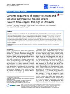
Figure 1. Dendrogram created from serological typing strains using the E. faecalis MLST database efaecalis.mlst.net. Multi-locus sequence typing (MLST) of E. faecalis isolates was based on sequences of internal gene fragments for 7 housekeeping genes. Each gene variation (for each of the seven genes) is assigned a unique allele number. The combination of the 7 allele numbers (allelic profile) for each strain defines the multi-locus sequence type, or ST. The relatedness of isolates based on sequence type is shown as an unrooted cladogram, determined by (UPGMA) analysis of the allelic profiles. Boxed isolates represent the most common serotypes found in human populations in previous studies [25,40]. doi:10.1371/journal.pone.0000582.g001
RESULTS Phylogenetic analysis of E. faecalis serotyping isolates In 1992, S. Maekawa and her colleagues [25] developed a refined serotyping test for the rapid identification of the majority of E. faecalis strains. Rabbit antisera were exhaustively cross adsorbed against a battery of E. faecalis isolates, including a smaller serotype diverse set previously created by M.E. Sharpe in 1964 [39]. Upon examining 832 strains from the U.S., Japan, and the U.K. in their study, over 78% of all isolates fell into one of 21 proposed serotypes, with serotypes 1, 2, 4, 7, and 9 representing the majority of isolates [25,40]. Because of the established diversity of this set of E. faecalis serotyping strains, it represented an ideal starting point for determining the genetic diversity of E. faecalis strains. Using MLST, we assessed the genetic relatedness of the 21 Maekawa type strains (T1-T21). A dendrogram depicting the relationships between these strains is shown in Fig. 1. These results indicate that the 21 serotypes of E. faecalis in fact span a great amount of species diversity and identify genetically distinct lineages. Only 2 of the 21 strains, strain T1 and T13, were found to be of the same sequence type.
in time. This set includes strains isolated from New Zealand to Boston, and dates ranging from the early 1900s to 2006. The collection also included clinical specimens, hospital-unrelated fecal isolates, and strains from non human sources ranging from Drosophila to swine. MLST analysis was performed on these additional isolates, listed in Table 1, and the genetic relationship of these strains to the Maekawa strains is shown in Figure 2. Within this diverse origin strain set, some sequence types (hereafter STs) occurred multiple times. The five most common STs were ST40 (n = 13), ST6 (n = 12), ST9 (n = 11), ST21 (n = 8), and ST64 (n = 7), which include 2 of the 5 serotypes identified as common by serotyping [40]. Additionally, these five most common STs had closely related single locus variants (SLVs) and/or double locus variants (DLVs) in our collection. The largest clonal clusters (CCs) in this diversity collection, defined here as a ST represented by three or more isolates, along with any SLVs and DLVs of these sequence types, include: CC21 (consisting of ST21 and ST70), CC9 (ST9 and ST106), CC2 (ST6 and ST2), CC8 (ST64, ST8, ST90, and ST112), and CC40 (ST40 and ST114). In all, the five
Test for additional species diversity The extent to which the Maekawa serotyping strains represented the diversity of the E. faecalis species, was tested by examining additionally 85 strains drawn from diverse ecologies and points PLoS ONE | www.plosone.org
7
July 2007 | Issue 7 | e582
Diversity of E. faecalis
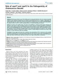
Figure 2. Dendogram of isolates aligned with capsule type, pathogenicity island segments, and antibiotic resistance traits. MLST-based dendrogram showing genetic relationship of all E. faecalis isolates in this study. Small yellow highlights indicate a serotyping type strain, while black boxes designate the five most common serotypes [40]. Arrows designate isolates used for comparative genomic microarray analysis. Abbreviations are defined as follows: ST = sequence type; CPS = capsule type; PAI = pathogenicity island fragment outlined as letter designations in Fig. S1 (A = nuc1; B = cylB; C = esp; D = hydrolase homolog similar to xylS; E = psaA homolog; F = gls-24 like); A red letter B indicates strains that readily transfer cytolysin via mating; AbR = antibiotic resistance; T = tetracycline resistance; TM = tetM+; TL = tetL+; E = ermB+; VA = vanA+; VB = vanB+; G = gentamicin resistant; C = cat+; A = blaZ+; CBH = bile salt hydrolase; GEL = gelatinase; CYL = cytolysin. More detailed strain information is listed in Table 1. doi:10.1371/journal.pone.0000582.g002
most common clonal groups encompassed 58% of the 106 isolates examined by MLST in this study. Interestingly, of the E. faecalis strains that have been used for most laboratory studies, namely strains V583 (first E. faecalis genome sequenced) [19], MMH594 (first pathogenicity island description) [10], HH22 (first b-lactamase positive isolate) [41], FA2-2 [33], and OG1RF [42,43], only one common sequence type is represented: ST6 (HH22, V583, and MMH594). We previously showed extensive sequence conservation within the 129 genes of the pathogenicity island and the 11 genes of the capsule locus of V583 and MMH594 [10], and others have observed that these strains and HH22 are closely related in genes that define the MLST assessment loci [22]. In this study, as in [24], these strains were identified as ST6. OG1RF was identified as representing the rarer sequence type ST 1, and FA2-2 is ST8. When the MLST data of the Maekawa et al. [25] serotyping strains T1-T21 (Fig. 1) are viewed on the dendrogram in Fig. 2, these isolates (highlighted in yellow) in fact span the diversity of sequence types at many grades of relatedness. This was somewhat surprising given that the molecular basis for interaction of agglutinating serotype anti-serum with the surface of the each of the sequence types is unknown, except for type 2 [44]. Of the isolates in our collection from the era preceding the widespread use of antibiotics (which we defined as having been acquired #1951), strains are found throughout the dendrogram, with only one isolate out of fifteen (SS-6) belonging to one of the most common STs of this study (ST21). No obvious clustering of older isolates is observed. PLoS ONE | www.plosone.org
Capsule locus polymorphisms and distribution among isolates All strains from Table 1 were analyzed for genes associated with virulence in E. faecalis, or homologs linked to virulence in other organisms (see Table 2 and Fig. 2). Isolates were first examined at the capsule locus (cps) to determine which of the known capsule polymorphisms occurred. The cps capsule locus of E. faecalis was discovered in part using Maekawa serotyping antiserum (serotype 2/strain T2). The T2 cps operon consists of 11 open reading frames, designated cpsA through cpsK [34,44,45]. Based on observed cps operon polymorphisms [34], primers were designed to assess these polymorphisms within our collection. Maekawa ‘type’ strains T1-T21 were verified as possessing the combinations of the 11 known genes of the cps operon previously inferred from restriction fragment length polymorphism and dot blot analysis (35, 77), and to verify the basis for the 3 previously identified polymorphisms (35, 77) using primers listed in Table S1. For all of the Maekawa serotype strains tested, the three known capsule operon polymorphisms were found: 1) that which includes all 11 genes as in strain T2 (designated CPS type 2); 2) that which includes all genes except for cpsF as in Maekawa strain T5 (CPS type 5); or only cpsA and cpsB as found in Maekawa strain T1 (CPS type 1). A limited primer set, optimized to detect the differences between these 3 capsule operon polymorphisms, was then used to determine which of the three CPS types were present in the remaining isolates of the collection. All strains tested yielded one of 8
July 2007 | Issue 7 | e582
...................................................................................................
Diversity of E. faecalis
Table 2. Bacterial virulence determinants and putative virulence factors examined. .................................................................................................................................................. Bacterial Determinant
Accession no.
Putative function
Reference
Method of detection
PAI nuc-1
EF0031
nuclease (homolog)
10
PCR, Southern hybridization
cylA,B,&M
EF0046-48
cytolysin production
35
PCR, Southern hybridization for cylB PCR, Southern hybridization
esp
EF0056
enterococcal surface protein
10
hydrolase
EF0077
glycosyl hydrolase (xylS homolog)
10
PCR, Southern hybridization
psaA
EF0095
metal binding protein (homolog)
157
PCR, Southern hybridization
gls24-like
EF0117
general stress protein
10, 158
PCR, Southern hybridization
cbh
EF0040
bile salt hydrolase
10
PCR, Southern hybridization, CBH assay
cps
EF0085-95
capsular polysaccharide
44
PCR, Southern hybridization
gelE
D85393
gelatinase
68, 159, 160
PCR, Southern hybridization, Gelatinase assay
fsrB
EF1821
accessory gene regulator
68
PCR
vanA
X56895
D-Ala-D-Lac ligase / Vancomycin resistance
52, 54
PCR, microdilution assay
vanB
L06138
D-Ala-D-Lac ligase / Vancomycin resistance
53, 54
PCR, microdilution assay
ermB
U86375
adenine methylase/ erythromycin resistance 55
PCR, microdilution assay
cat
X92945
chloramphenicol acetyl-transferase/ chloramphenicol
PCR, Southern hybridization, resistance assay FAST CAT assay
tetM
X92947
ribosomal protection / tetracycline resistance 48
PCR, Southern hybridization, resistance assay
tetL
NC_005013
efflux pump / tetracycline resistance
47
PCR, Southern hybridization, resistance assay
aac6’-aph2"
M13771
bifunctional enzyme / high-level aminoglycoside resistance
50
PCR, Southern hybridization, resistance assay
blaZ
M60253
b-lactamase / b-lactam resistance
49
PCR, nitrocefin assay, resistance assay
Other
Antibiotic resistance
51
doi:10.1371/journal.pone.0000582.t002
occurred among isolates included in this study known to be of commensal origin [46]. Of isolates tested, 31% (34/106) were sensitive to all six antibiotics. All strains isolated prior to 1954 were susceptible to all of the antibiotics tested. Less than half of CC21 isolates, which includes sequence types 21 and 70, had any antibiotic resistance, with tetracycline being the sole resistance found. Strains of CC40, which includes sequence types 40 and 114, also exhibited relatively few antibiotic resistance factors, with most isolates having only one type of resistance. Clonal clusters CC9, CC2, and CC8 contained strains that harbored many resistances, with some isolates carrying determinants for resistance to five antibiotic classes. Vancomycin resistance, conferred by the vanA or vanB genetic determinants, was found in 14% of isolates, with most occurring in CC2 and CC8 (10/15). Ampicillin resistance conferred by b-lactamase was only found in isolates from CC9 and CC2 of our collection, specifically in sequence types 9 and 6 (Fig. 2 and Table 3), and was the rarest among this collection (Table 3). The number of antibiotic resistances found in each genetic lineage is illustrated in Fig. 3. Strains were tested by PCR and dot blot for the antibiotic resistance genes most commonly conferring these resistances in E. faecalis. These included tetM and tetL for tetracycline resistance [47,48], blaZ for b-lactamase-mediated ampicillin resistance [49], aac69-aph20 for aminoglycosides [50], cat for chloramphenicol [51], vanA and vanB for glycopeptides [52–54], and ermB for macrolide resistance [55] (Table 2 and Fig. 2). Comparing antibiotic resistance phenotype and genotype, we found that all erythromycin resistant strains were positive for ermB (Fig. 4). Further, all vancomycin resistant isolates contained either vanA or vanB ligases, and all high-level ampicillin resistant (.4 mg/mL) isolates
the three CPS polymorphisms, based on characteristic PCR products. Dot blots were performed on the entire collection, using as a probe an internal fragment of cpsF, to confirm presence or absence of the cpsF gene, which is characteristic of CPS2, and to verify that negative PCR results did not derive from point changes within primer hybridization sites. The occurrence of CPS 1, 2 and 5 polymorphisms among the strains studied, as identified by this approach, is shown in Figure 2. Wherever the collection contained multiple isolates within a sequence type, CPS type was invariant among those strains. The most common CPS type among the diversity of lineages was type 1. When strains were examined by decade of isolation, CPS type 1 remained most common.
Antibiotic resistances and determinants among isolates Single-concentration broth assays were performed to test the resistance of isolates to antibiotics from six different classes: gentamicin, erythromycin, ampicillin, vancomycin, tetracycline, and chloramphenicol. For many isolates, some antibiotic resistance information was available. Reanalysis confirmed those results, with the exception of strain CH19, for which erythromycin resistance was not detected by phenotype. This strain was, however, found to harbor the ermB resistance gene. Based on the broth assay results, 58% of isolates exhibited resistance to tetracycline, 38% to erythromycin, 33% to high-level gentamicin, 14% to vancomycin, 11% to chloramphenicol, and 9% to ampicillin. An additional 6 strains exhibited low-level (#4 mg/ mL) ampicillin resistance, but were b-lactamase negative (strains F1, RMC1, SF5039, SF21520, SF21521 and Merz96). Tetracycline resistance was the only antibiotic resistance trait that PLoS ONE | www.plosone.org
9
July 2007 | Issue 7 | e582
Diversity of E. faecalis
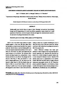
Figure 3. Dendrogram and composite virulence determinants among sequence types. MLST-based dendrogram compiling data from isolates of the 51 identified sequence type lineages. PAI = number pathogenicity island genes present per lineage; AbR = number of antibiotic resistance determinants per lineage. Brackets encompass abundant clonal isolates and their single and double locus variants where applicable. doi:10.1371/journal.pone.0000582.g003
expressed b-lactamase activity and possessed the gene, blaZ. The majority of tetracycline resistant isolates (54 of 62) carried tetM, one isolate was positive for tetL, and 12 of the 54 tetM positive isolates also carried the tetL gene. The remaining 7 tetracyclineresistant strains tested negative for either tetM or tetL by PCR, and may be resistant via one of the rarer tetracycline resistance mechanisms not examined [29]. Most gentamicin resistant isolates (32/34) were positive by PCR and dot blot for the aac69-aph20 bifunctional enzyme that confers aminoglycoside resistance [50], with the exceptions of strain JH1 and YI6-1, which had high-level gentamicin resistance and were not positive for aac69-aph20. JH1 was previously described as having a 3950-aminoglycoside phosphotransferase type III resistance gene [56]. The aminoglycoside resistance mechanism for YI6-1 was not further explored. Chloramphenicol resistance was associated with the presence of the chloramphenicol acetyl-transferase gene, cat, by PCR and dot blot, for all chloramphenicol resistant strains except RM4679, PLoS ONE | www.plosone.org
WH245, and WH571, which were phenotypically chloramphenicol resistant, but were negative for the cat gene by these tests. Because chloramphenicol acetyl transferase (CAT) is the most common mechanism for chloramphenicol resistance in enterococci [57,58], we further evaluated CAT activity in these isolates using the FAST CAT chloramphenicol acetyltransferase kit. CAT activity was confirmed for all three strains, suggesting that they harbor a divergent cat gene [57]. The number of isolates having the tested antibiotic resistance phenotypes and genotypes listed above are illustrated in Fig. 2.
Known and suspected virulence traits A number of auxiliary traits have been identified that participate in colonization or virulence in E. faecalis, or are similar to those of known activity in other intestinal pathogens [59,60]. We examined our collection of isolates for assayable activities associated with virulence, and corresponding coding sequences: cytolysin, gelati10
July 2007 | Issue 7 | e582
........................................................................................
Diversity of E. faecalis
Table 3. Distribution of putative virulence genes in five most common clonal groups. .................................................................................................................................................. clonal group
CC21
CC9
CC2
CC8
CC40
non-clustering
Total from groups
# of isolates
9
12
15
11
14
45
61
bla
0 (0/9)
58 (7/12)
20 (3/15)
0 (0/11)
0 (0/14)
0 (0/45)
16 (10/61)
cat
0 (0/9)
50 (6/12)
20 (3/15)
18 (2/11)
7 (1/14)
2 (1/45)
20 (12/61)
vanA/B
0 (0/9)
0 (0/12)
40 (6/15)
36 (4/11)
0 (0/14)
11 (5/45)
16 (10/61)
ermB
0 (0/9)
67 (8/12)
87 (13/15)
64 (7/11)
14 (2/14)
24 (11/45)
49 (30/61)
aac6’-aph2"
0 (0/9)
67 (8/12)
100 (15/15)
27 (3/11)
0 (0/14)
16 (7/45)
43 (26/61)
tetM/L
44 (4/9)
58 (7/12)
40 (6/15)
82 (9/11)
79 (11/14)
40 (18/45)
61 (37/61)
gls-24-like
0 (0/9)
0 (0/12)
67 (10/15)
9 (1/11)
0 (0/14)
18 (8/45)
18 (11/61)
cylB
22 (2/9)
33 (4/12)
47 (7/15)
36 (4/11)
29 (4/14)
31 (14/45)
34 (21/61)
nuc1*
0 (0/9)
83 (10/12)
100 (15/15)
55 (6/11)
43 (6/14)
31 (14/45)
61 (37/61)
hyd*
11 (1/9)
75 (9/12)
93 (14/15)
73 (8/11)
7 (1/14)
38 (17/45)
54 (33/61)
esp
33 (3/9)
92 (11/12)
27 (4/15)
73 (8/11)
93 (13/14)
40 (18/45)
64 (39/61)
psaA*
33 (3/9)
75 (9/12)
93 (14/15)
82 (9/11)
64 (9/14)
42 (19/45)
72 (44/61)
cbh
44 (4/9)
100 (12/12)
80 (12/15)
100 (11/11)
93 (13/14)
56 (25/45)
85 (52/61)
fsr
67 (6/9)
100 (12/12)
100 (15/15)
0 (0/11)
100 (14/14)
67 (30/45)
77 (47/61)
gelE
100 (9/9)
100 (12/12)
100 (15/15)
100 (11/11)
100 (14/14)
96 (43/45)
100 (61/61)
Groups are comprised of abundant clonal isolates (n.3) and their single and double locus variants as determined by MLST analysis (Fig 3) and eBURST [24]: CC21 = ST21&70, CC9 = ST9&106, CC2 = ST2&6, CC8 = ST8, 64, 90, &112, and CC40 = ST 40&114. Numbers in bold indicate percentage of isolates positive for the specified genotype for a given grouping as calculated from the number of positive versus total isolates in parentheses. Genes investigated above the dashed line encode antibiotic resistance determinants, while those below the dash are known to the pathogenicity island of E. faecalis and/or code for auxiliary enzymes (see Table 2). Antibiotic resistance genotypes encoded by genes other than those listed are not included in this table. * = putative nuclease and glycosyl hydrolase genes doi:10.1371/journal.pone.0000582.t003
nase, and bile salt hydrolase (Fig 2). Cytolysin and gelatinase production were characterized in E. faecalis over 40 years ago [61– 63], and many studies have linked these variable traits of the species to enterococcal virulence [64,65]. Bile salt hydrolase is expressed by some strains of E. faecalis, and also by intestinal pathogens [66,67]. It is known to contribute to a bacterium’s ability to survive in the gastrointestinal tract [15]. Analysis of
phenotypes and corresponding genotypes for cytolysin, gelatinase, and bile salt hydrolase activity, revealed more isolates that were genotypically positive than phenotypically positive in laboratory tests (Fig. 4). The greatest difference between genotype and detectable phenotype was for gelatinase activity (58 gelatinase positive of 104 gelE+), then cytolysin (26 cytolysin positive of 36 cylA/B/M+), followed by bile salt hydrolase (71 hydrolase positive
Figure 4. Virulence-associated phenotypes and corresponding genotype for all isolates.. Phenotypes were determined by microdilution assay for antibiotic resistance or enzyme-specific tests for auxiliary enzymatic traits. Genotypes were determined by PCR amplification and/or hybridization for genes known to encode each phenotype. A positive genotype is indicated by the presence of one or more genes known to produce a given phenotype (e.g. genotypically positive vancomycin strains may contain the vanA or vanB genes—a strain containing both would only be counted once). doi:10.1371/journal.pone.0000582.g004
PLoS ONE | www.plosone.org
11
July 2007 | Issue 7 | e582
Diversity of E. faecalis
of 77 cbh+). For gelatinase, the lack of phenotype in the presence of a positive genotype in many cases has been attributed to the absence of a known gelE regulator fsrB [68]. In the present study, 27 of the 46 gelE positive isolates that did not display a gelatinase positive phenotype were fsrB negative by PCR, while all gelatinase positive isolates tested positive for fsrB (Table S2). All three auxiliary trait activities assayed were present in the major MLST groups and branches of the cladogram lineages, with the exception of gelatinase activity, which was not present in any isolate of CC8. Upon further examination, it was found that all of the isolates of CC8 tested positive for the gelatinase gene, gelE, but negative for the fsrB regulator of gelatinase expression [68].
microarray information and materials and methods). To the extent that gene content reflects fitness for a particular habitat, this comparison provides a independent approach from MLST for investigating the ecological relationship of E. faecalis isolates. As shown in the dendrodram at the top of Figure 5, MMH594 and V583 (both ST6) group together as would be anticipated based on known relatedness in gene content, which includes the pathogenicity island, capsule gene and MLST genes. However, relatedness based on variable gene content did not parallel MLST (Fig. 2), indicating that as expected for mobile elements, variable traits penetrate the species independently of genome sequence drift. Each strain exhibited a minimum identity of 72% with the V583 chromosomal probe sets, and a minimum of 67% of the total E. faecalis probe sets (which includes E. faecalis genes not found in V583; Table 4). As expected, most of the differences in gene content between the strains tested and V583 appear to stem from variations in putative mobile element content, which is not measurable by MLST. Since only the genome sequence of strain V583 is known, there undoubtedly are many undiscovered variable traits in the non-V583 lineages tested, that if examined could lead to the identification of additional relationships among these strains. The core E. faecalis genome, as defined by the number of genes present in all 8 strains based on the ORFs included in the microarray, consists of 2057 of the 3091 ORFs (2129 of the 3582 total probe sets, which includes redundancies; Fig. 6). Core and variable genes were categorized by orthologous group, according to NCBI COG designation (Clusters of Orthologous Groups, http://www.ncbi.nlm.nih.gov/COG). Core and accessory genes were further analyzed and ranked by the number of strains for which a present call was made for each probe set, and for predicted cellular role (Fig. 6). All of the core genome ORFs are located on the V583 chromosome; none were associated with extrachromosomal plasmids. Of probe sets not part of the core genome but common in the majority (five or more) of the eight strains examined, 370 were identified. Of these, 236 probe sets represented poorly characterized or unassigned gene functions, and the remaining 134 spanned all of the general COG categories. Of the seven identified phage regions of the V583 genome, only one is represented in the core genome: PHAGE02 [19]. In addition, the majority (9 of 11) of predicted V583 phosphotransferase systems (PTS) [19] for the transport of sugars are present in the core genome, including those for cellobiose, fructose, glucitol, glucose, maltose, mannitol, mannose, sucrose, and trehalose. Only the galactitol and N-Acetyl galactosamine PTS systems from V583 were not present in all eight isolates. Examination of the core genome for identified twocomponent signal transduction systems from V583 [69] showed that 12 out of 17 of these systems, as well as an orphaned response regulator, are present in all strains tested. These include HK/ RR01, HK/RR02, HK/RR03, HK/RR04, HK/RR05, HK/ RR06, HK/RR07, HK/RR09, HK/RR10, HK/RR13, HK/ RR14, HK/RR17, and RR18. Furthermore, we found high conservation of genes identified as having a potential role in stress responses [19] within the core genome. Of the 56 stress-response related ORFs in V583, which represent oxidative, osmotic, and metal-ion resistance mechanisms [19], 54 are present in the core genome of E. faecalis.
Comparative genomic hybridization analysis of diverse E. faecalis isolates, and classification of core genetic elements Based on the MLST relatedness profiles of strains tested, we examined eight strains that spanned the diversity of the species by comparative genomic hybridization. The goal was to identify genes common to maximally diverse strains of E. faecalis, thereby defining the core genome. It was further of interest to assess the penetration of variable traits occurring in the V583 genome within diverse lineages of the species. Six of these strains (ARO1/DG, Com6, Fly1, HIP11704, D6, and JH1) represent diverse nodes of the MLSTbased cladogram (Fig. 2), unrelated to the known sequence strain, V583. Strain MMH594 was included as an additional positive control to verify detection of genes of the 17 kb portion of pathogenicity island known to be deleted in strain V583 [19]. The results of microarray genome hybridization analysis of the six strains, and V583 and MMH594 controls, demonstrate the relatedness between diverse strains of E. faecalis and V583 with respect to its variable gene content (Fig. 5). Probe sets for the array are ordered according to the V583 genome sequence, while additional plasmid and antibiotic resistances genes are arranged independently. Comparison of divergent E. faecalis genomes to strain V583, revealed some V583 genes that were present among all strains tested, and others that were variably present. Variable regions were found to correspond to integrated phages, plasmids, and transposable elements previously identified in silico in the V583 genome [19], as well as an additional 60 ORF phage-related variable region not previously identified (region 9). Region 9, present only in V583 and MMH594, is flanked by site-specific recombinases and contains putative metabolic and hypothetical genes, and may represent a new pathogenicity or fitness island. In strain V583, the vanB segment (region 10) is flanked by ORFs with high sequence similarity by BLASTp analysis (http://www.ncbi. nlm.nih.gov/blast) to those occuring in mobile elements. Because MMH594 isolation predated that of V583, suggesting that vanB was the more recent acquisition, the vancomycin resistance element appears to have inserted into the 60 ORF region 9, which seems to have entered the species in this lineage (ST6). Region 6, a region of the V583 genome previously identified to be atypical in GC composition [19], appears to be extended 8 additional ORFs (EF_1847-EF_1897 instead of EF_1855EF_1874) based on both the presence of these ORFs in strains identified by comparative genomic analysis, and the presence of a flanking site-specific recombinase. (see microarray data files at http://www.ebi.ac.uk/arrayexpress, accession number E-MEXP1090). This region is present in the closely related strains V583 and MMH594, and in a divergent strain, D6. Relatedness based on similarity in gene content was calculated based on the percentage of non-divergent (Present) probe sets in each strain and Pearson correlation coefficients (see supporting PLoS ONE | www.plosone.org
Strain relatedness and presence of pathogenicity island A pathogenicity island (PAI) was found to occur in E. faecalis strain MMH594, with derivatives in strains V583 and V586 [10]. This 12
July 2007 | Issue 7 | e582
Diversity of E. faecalis
...............................
Figure 5. Visualization of final absent and present calls for all probe sets across 8 distinct E. faecalis isolates. Comparative genomic hybridization was performed on DNA from isolates V583, MMH594, JH1, HIP11704, D6, ARO1/DG, Com6, and Fly1 as described in materials and methods. Probe sets are ordered according to the E. faecalis V583 gene sequence and the Affymetrix library file. Absent probe sets are in white; present probe sets are in black. Clustering of the strains was based on complete linkage using the Pearson correlation coefficient for the Absent/Present calls (A = 0, P = 1). The same clustering pattern is generated when average signal intensity values are used instead of Absent/Present calls (not shown). The numbered regions on the left correspond to the following previously identified and unidentified mobile genetic regions from strain V583: 1) EF_0125-EF_0166, 2) EF_0302-EF_0355 (PHAGE01), 3) EF_0479-EF_0628 (V583 PAI), 4) EF_1275-EF_1293 (PHAGE02), 5) EF_1416-EF_1489 (PHAGE03), 6) EF_1847EF_1897, 7) EF_1987-EF_2043 (PHAGE04), 8) EF_2084-EF_2145 (PHAGE05), 9) EF_2240-2282/EF_2335-2351, 10) EF_2284-EF_2334 (Tn/vanB), 11) EF_2512-EF_2545, 12) EF_2798-EF_2856 (PHAGE06), and 13) EF_2936-EF_2955 (PHAGE07) (see text). doi:10.1371/journal.pone.0000582.g005
Table 4. Present calls for Comparative Genomic Hybridization probe sets by isolate. .................................................................................................................................................. Strain
ARO1/DG
Com6
Fly1
JH1
HIP11704
D6
MMH594
V583
Present calls for V583 chromosome
2417
2422
2535
2404
2615
2483
3110
3256
% present calls for V583 chromosome
73.09
73.24
76.66
72.69
79.07
75.08
94.04
98.46
Present calls in all bacterial probe sets
2493
2425
2566
2407
2739
2559
3210
3409
% present calls in all bacterial probe sets
69.60
67.70
71.64
67.20
76.47
71.44
89.61
95.17
doi:10.1371/journal.pone.0000582.t004
PLoS ONE | www.plosone.org
13
July 2007 | Issue 7 | e582
Diversity of E. faecalis
Figure 6. Classification of core and dispensable sequences in 8 strains of Enterococcus faecalis. Data from comparative genomic hybridization analysis of isolates V583, MMH594, JH1, HIP11704, D6, ARO1/DG, Com6, and Fly1 are organized by the number of strains for which each probe set is detected (top to bottom). Absent probe sets are in white; present probe sets are in black. Clustering of the strains was based on complete linkage using the Pearson correlation coefficient for the Absent/Present calls (A = 0, P = 1). Genetic elements were classified as part of the core genome if present in all strains tested. COG designations were obtained from the National Center for Biotechnology Information (NCBI). Some genes/probe sets represent more than one COG category. doi:10.1371/journal.pone.0000582.g006
region encodes known and putative virulence determinants, surface adhesion factors, carbohydrate metabolism pathways, the cytolysin operon, a putative bile salt hydrolase, as well as many other genes of unknown function and homologs to virulence factors in other bacteria [10]. Many genes in this island, such as the cytolysin operon, have been identified on plasmids, transposons, or other mobile elements, and appear to have integrated into the chromosome at various points in its evolution. In order to survey which regions of the known PAI were present in the strains of our collection, we selected six functionally unrelated genes located across the pathogenicity island to serve as sampling points (Fig. S1). The pathogenicity island (PAI) associated genes we surveyed were present in many nodes of the dendrogram, and in strains from many different sources and dates of isolation. Through PCR and dot blotting for the six PAI genes from Fig. S1, we found that CC9, CC2, and CC8, possessed more of the PAI regions (and also antibiotic resistances) tested than did other clonal clusters (Fig. 3 and Table 3). Bile salt hydrolase, cytolysin, PLoS ONE | www.plosone.org
and gelatinase genes, as well as five of six PAI genes sampled (the exception being the gls24-like gene) can be found in strains isolated before 1951, including in some of the oldest strains in our collection. The exception, gene EF0117 which specifies a gls24-like gene, is located near one end of the PAI and did not appear in strains of our collection until the 1980s in isolates from ST6, of which the majority test positive. The gls24-like gene has high homology to the gls24 gene (EF0080), which has been linked to stress responses in E. faecalis [70]. This gls24-like gene appears to have penetrated other lineages (STs 11, 28, 36, and 64) in the early 1990’s. We found gls24-like in isolates of different regions of the dendrogram (Fig. 3), however, it was the rarest of the PAI genes assayed and did not appear in any CC21 or CC40 isolates surveyed. The nuclease homolog, nuc1, (EF0031) exhibits sequence similarity to staphylococcal nucleases that hydrolyze DNA and RNA [71] and is located near the opposite end of the PAI from the gls24-like gene (EF0117). nuc1 was absent in strains of our 14
July 2007 | Issue 7 | e582
....................................
Diversity of E. faecalis
Table 5. Presence of 6 representative PAI genes compared to total PAI probe sets by comparative genomic hybridization. .................................................................................................................................................. Strain
Com6
Fly1
ARO1/DG
JH1
V583
HIP11704
D6
MMH594
Origin
commensal
insect
dog
clinical
clinical
clinical
pig
clinical
Sequence type (ST)
21
101
108
40
6
4
16
6
6 gene PAI profileA
0/6
0/6
0/6
2/6
4/6
5/6
6/6
6/6
CGH PAI probe setsB
0/141
0/141
3/141
56/141
125/141
111/141
138/141
137/141
A
6 gene PAI profile includes the nuclease homolog, cylB, esp, xylS homolog, psaA homolog, and gls-24 like genes outlined in Fig S1 and Table 2. PAI probe sets were designed to the combined V583 and MMH594 PAI sequences (EF_0479-EF_0628 and EF0001-EF0129, respectively) and represent 139 ORFs. doi:10.1371/journal.pone.0000582.t005
B
collection isolated prior to 1950. It was present in every CC2 isolate and was absent from all CC21 isolates, however a pattern of nuc1 penetration the species is not evident. The other sampled PAI genes, cylB, esp, the hydrolase (EF0077), psaA, and cbh, were found in isolates of our collection dating as far back as 1926; the pattern by which these genes entered the species could not be deduced from this set. Given that the cytolysin operon is known to be on conjugative plasmids in several of the strains in our collection [11,43,72–74], we investigated whether the cytolysin determinant of other strains was highly transmissible, as is known for the cytolysin encoding plasmid, pAD1 [75], or on less mobile PAI [10] or other element [31]. Mating experiments showed that 51% (18/35) of the cytolytic strains were able to transmit cytolysin in a mixed colony at a high rate. When aligned to the position of isolates on the dendrogram (Fig. 2), the transmissibility of cytolysin corresponded to specific regions and groups. For instance, all of the cytolysin positive strains in CC21 and CC40 were able to transfer cytolysin in a mixed colony, presumably by conjugation as known for the prototype plasmid pAD1 encoded determinant [75]. Except for strain MMH594, isolates in CC2 that were positive for cylB were phenotypically not cytolytic, precluding detection of transconjugants. Transfer of the known PAI-conferred cytolysin determinant within MMH594 was not observed in the mixed colony, consistent with previous observations [32]. Within CC2, V583 and V587 which were isolated from separate patients, are known to carry vestiges of the cytolysin operon within their pathogenicity islands [18]. Part of this operon was deleted in strain V583 and a transposon insertion rendered the V587 cytolysin inactive ([10], V587 data not shown). Among the eight isolates used for comparative genomic analysis by microarray, no pathogenicity island genes were common to all of the strains, supporting the proposition that it is not part of the core E. faecalis genome. Data obtained independently by sampling for the presence of genes within the PAI, for antibiotic resistance, and for other genes detailed in this study were generally supported by the results of the microarray analysis. The sole exception was detection of a previously unknown polymorphism observed in the cps locus of strain Fly1, which had no cpsD by microarray, but otherwise fit CPS type 5. With respect to genes of the pathogenicity island, the microarray data for the 8 strains tested was consistent with the results of sampling the six PAI loci shown in Fig. S1 (Table 5), validating this set of genes as a useful tool for rapidly probing strains for the presence of various regions of the PAI. Based on the presence of the six representative PAI genes sampled in our collection, we estimate that the middle regions of the PAI between EF0056 (esp) and EF0095 (psaA) were introduced into E. faecalis by the early 1900s, if not before. Comparative hybridization analysis also indicates that the PAI has integrated into multiple MLST lineages, as previously suggested by Nallapareddy et al. [22], and is present in strains isolated from both animals and humans (Table 5). PLoS ONE | www.plosone.org
DISCUSSION This investigation was undertaken to provide new insights and a global view of the diversity of the species E. faecalis. We observed differences between phylogenetically separate groups for pathogenicity island content, antibiotic resistance determinants, and capsule operon polymorphisms, which we hypothesize are due to niche preferences within the species. Variable genetic traits confer niche specialization and virulence to bacteria, including E. faecalis. To characterize the diversity of the species, to determine what constitutes the core species characteristics, and to determine the extent of penetration of various traits into the species, we characterized a diverse collection of E. faecalis isolates by MLST, comparative genomic hybridization, and directly assessed select genotypic and phenotypic properties. As a starting point, we capitalized on the efforts of previous investigators who, over the past six decades, developed collections of serologically unique E. faecalis strains [25,39,76]. We complemented these collections with additional isolates from over the past century, and from a variety of geographic locations. Of the 51 different sequence types we identified, the majority (29 sequence types, 57%) had not been detected in previous studies [24,77]. Our studies show that the previous collections of serologically unique strains were not only antigenically diverse, but remarkably genetically diverse as well. This was somewhat surprising in that expression of a new surface protein, potentially encoded by a mobile element such as a phage or transposon, could generate a novel serotype. Only one example was identified where serological type strains from the Maekawa collection [25] were of the same MLST sequence type–namely T1 and T13, both of which were MLST sequence type 21. The remaining 19 strains of the Maekawa collection constituted 19 sequence types covering most of the unrooted dendrogram that represents the species (Fig 2). The five most common serotypes identified by Maekawa et al. [25,40] in epidemiologic studies, do not appear closely related genetically (serotypes T1, T2, T4, T7, and T9; Figs. 1 and 2; [40]). This suggests that either discrete ecologies select for the presence of these 5 lineages, or that the host mounts a transient immune response promoting a temporal rotation among these lineages. Additional isolates in the collection resolved into 31 additional STs, for a total of 51 STs. Of these 51 STs, 38 were represented only once in our collection. Of the Maekawa serotypes that were reported to be most often isolated in studies of strains from the U.K., U.S.A., and Japan [40], only T1 and T7 were members of MLST clonal clusters deriving from this collection (CC21 and CC8, respectively). Capsules are typically highly variable traits of pathogens, such as Streptococcus pneumoniae, and this variation is driven by evasion of host immune response [78–80]. The cps capsule locus of E. faecalis was discovered in part with the use of Maekawa serotyping antiserum (serotype 2/strain T2), and consists of the 11 open reading frames cpsA through cpsK [34,44,45]. The first vancomy15
July 2007 | Issue 7 | e582
Diversity of E. faecalis
Figure 7. Antibiotic determinants and pathogenicity island composition per sequence type lineage, aligned by capsule locus (cps) variation (CPS type). The percentage of sequence type lineages positive for each antibiotic resistance and pathogenicity island listed in Fig 3 were alligned by CPS capsule type and arranged in descending order. Capsule types are invariant for strains within individual sequence type lineages. Details of capsule operon (cps) arrangements are outlined in Materials and Methods. doi:10.1371/journal.pone.0000582.g007
cin-resistant isolate, V583, the first characterized pathogenicity island-containing strain MMH594, and a clinical isolate adapted for use in the laboratory FA2-2, were all shown to react with type 2 antiserum, a phenotype that is dependent on the presence of the capsule operon including, in particular, cpsF [34,45]. In studies of streptococci, where capsule variation alters serotype, an MLST type was found to correspond to one or more particular serotypes and a given serotype might be common to many clonal complexes or sequence types [81,82]. Of the 51 sequence types identified, 25/51 were found to be CPS type 1, 14/ 51 of CPS type 5, and 12/51 of CPS type 2. The vast majority of CPS type 1 lineages harbored 3 or fewer virulence traits (18/25; 72%), or 2 or fewer antibiotic resistances (19/25; 76%) (Fig. 7). Similar proportions were observed for CPS type 5 strains (79% [11/14] possess 3 or fewer virulence traits; 79% [11/14] possess 2 or fewer antibiotic resistance traits). However, half or more CPS type 2 strains (58% [7/12]) harbored 4 or more virulence traits, or 3 or more (58% [7/12]) antibiotic resistance traits (Fig. 7). This observation suggests that CPS2 strains tend to colonize sites where antibiotic resistance genes and virulence traits are advantageous. As the capsule locus, the pathogenicity island, and antibiotic resistances are known not to be physically linked in strain V583 (the only strain for which a genome sequence exists), this observation supports the hypothesis that independent selection has driven the convergence of virulence traits, antibiotic resistances and the type 2 capsule. Expression of the type 2 capsule has been associated with decreased opsonophagocytosis [44]. That ST6 isolates, which are uniformly of this capsule type and clinical in origin, had the greatest variety of both antibiotic resistances and PAI genes tested, makes the nexus of these traits of special interest in the evolution of highly virulent clones of the species (Fig. 3 and Fig. 7) Throughout our collection, the three known cps operon polymorphisms [34,83] (CPS) were consistent within individual STs, although within an ST variation in antibiotic resistance and virulence traits occurred. This suggests that although the capsule locus polymorphism is a variable trait within the species, it varies at a lower rate than other traits known to reside on mobile elements [84]. Alternatively, it suggests that a change in capsule type may give rise to a new lineage, perhaps by enabling that strain to outcompete the parental type to which the host has responded. That all three CPS variations were found in all of the major branches of the dendrogram supports the hypothesis that there is horizontal PLoS ONE | www.plosone.org
transfer. However, a lack of obvious transfer or transposition functions associated with the capsule operon leaves as an open question the mechanism. The present study revealed variation in the penetration of antibiotic resistances and genes of the pathogenicity island between major clonal clusters, as well as non-clustering isolates. The tetracycline class of antibiotics were discovered in 1945, and put into regular therapeutic use by 1950 [85,86]. Within 2– 3 years, tetracycline resistant strains of enterococci were isolated [86]. The oldest isolate with tetracycline resistance in our collection dated back to 1954. Tetracycline resistance was the only antibiotic resistance trait that occurred among isolates of known commensal origin included in this study. Its occurrence among such strains was known from previous studies of gut flora populations [46]. Tetracycline resistance in enterococci can be the result of genes encoding ribosomal protection mechanisms (TetMlike) or efflux pumps that actively transport antibiotic out of the cell (TetL-like), both of which were found in our strains [87]. Tetracycline and its derivatives are broad-spectrum antibiotics that have been commonly used for the past 67 years to treat bacterial infections, thus their regular use and spectrum of activity has exerted substantial pressure for E. faecalis strains to harbor resistance determinants. As such, tetracycline resistance is the most common resistance found in our collection (62/106 isolates) and has penetrated 25 of the 51 sequence types we identified. Vancomycin, a glycopeptide antibiotic, was discovered in 1952 and was quickly brought to market to combat infections due penicillin resistant bacteria [88]. However, vancomycin use has been limited since its introduction due to problems with toxicity, administration, and the availability of other antibiotics [88]. Resistance of group D streptococci (enterococci) to vancomycin was documented in isolates in 1968 [86], though it is unclear in which specific species this resistance occurred. The use of vancomycin increased during the 1980s, which likely contributed to the rise in resistant organisms that soon followed [88]. Vancomycin resistance in enterococci was encountered in France and the U.K. in 1986, as a result of the vanA gene cluster found in enterococcal isolates [89–91]. Vancomycin resistance mediated by vanA or vanB genes was found in only 15/106 strains in our collection. A review of our vanA-containing isolates indicates that this genotype first appeared in E. faecalis strains in ST6 (CC2) and ST28 (1990s) and was later acquired in ST108, ST104, and ST4. Vancomycin resistance due to the vanB genes was found in 1987 in 16
July 2007 | Issue 7 | e582
Diversity of E. faecalis
enzyme aac69-aph20 [50]. We found gentamicin resistance by the aac69-aph20 enzyme most frequently (33/35 gentamicin resistant isolates). Two isolates did not test postitive for the bifunctional enzyme: strain JH1 (ST 40), for which the 3950 APH gene was previously characterized [56], and strain YI6-1, for which the mechanism was not identified. Aminoglycoside resistance via aac69-aph20 first appeared in our collection in strain HH22 (ST6, CC2) in 1981, which was also the first identified b-lactamaseproducing E. faecalis [95]. Aminoglycoside resistance was previously identified in HH22, in the first publication describing highlevel gentamicin resistance in the U.S. [101]. Gentamicin resistance transferred by conjugation categorically with b-lactam resistance from HH22 [95,101], signifying that resistance to both antibiotics was acquired simultaneously in this strain. In fact, all blactamase positive isolates in our collection are also resistant to high-levels of gentamicin, though a genetic link between these determinants was not investigated. The next occurrence of aac69aph20 in our set was in ST9 and ST106 (both CC9) in the mid1980s, followed by ST64 (CC8) in 1986, ST11 (distant relative of CC9) and ST109 (CC2) in the early 1990s. The bifunctional gene was next found in ST28, a single locus variant of ST11, in the mid1990s. By 2001/2002 the aac69-aph20 had expanded to ST2 (CC2), ST4, ST103, and ST104; the gene also spread to ST16, however the date of this transfer is not known. The pervasive spread of this determinant among different lineages in the species hints that persistent use of aminoglycosides in the last several decades has permitted the expansion of this determinant between enterococcal isolates. Erythromycin, a macrolide antibiotic, came into use in 1952. Within a year, resistant isolates of group D streptococci (enterococci) were identified [86]. Many genes have been found to contribute to macrolide resistance in enterococci, however resistance to erythromycin in E. faecalis is typically mediated by the ermB rRNA methylase gene [102]. Within our collection, all 40 erythromycin resistant isolates contained the ermB gene. The first occurrence of ermB in our group was in ST40 (CC40) and ST55 from the mid 1970s. By the mid 1980s, ermB had spread to ST6 (CC2), ST9 (CC9), and ST64 (CC8). By the early 1990s, ermB could be found in five additional sequence types, and by 2002 ermB had been transferred to an additional seven sequence types. In total, 17/51 sequence type lineages were found to contain the ermB gene for macrolide resistance. In addition, all vancomycin resistant strains in our study (either vanA or vanB mediated) harbored the ermB gene. The vanA gene was previously shown to be located on the same plasmid as ermB [103], though we could find no prior work describing linkage of vanB and ermB. Antibiotic resistance genes are commonly clustered together on mobile plasmids and phages, leading to the co-selection of resistance determinants with the use of a single class of antibiotic. Moreover, antibiotic treatment of E. faecalis infections often involves the use of multiple antibiotics to achieve synergistic killing effects [104]; thus the likelihood of a strain acquiring multiple antibiotic resistances at once is great. The occurrence of PAI genes and acquired antibiotic resistances by clonal group reveals a pattern between the range of isolation dates and the average number of these genes observed: generally, the more PAI and antibiotic resistance genes found, the shorter the range of isolation dates within the clonal clusters. The five most common clonal clusters: CC21, CC9, CC2, CC8, and CC40, represented 58% of the 106 isolates examined by MLST in this study. CC2 isolates have the shortest range of isolation dates of any clonal group (1981–2003, 22+ years) and the greatest number of PAI and antibiotic resistance genes on average (7.5, 61.8), while CC21 have the widest range of isolation (1930s–2006, 75+ years)
the E. faecalis strain V583 [18], which is in ST6 (CC2). vanB next appears in ST64 (CC8) in 1991, followed by ST2 and ST103 in 2002. Our study identified vancomycin resistant strains in eight distinct STs, six of which had not been previously identified as harboring van genes [24]. The diversity of sequence types harboring van genes in our set illustrates an extensive spread of this resistance among E. faecalis lineages in less than twenty years since resistance was first discovered. Ampicillin is a b-lactam antibiotic, the first and most famous of which is penicillin [92]. Discovered in 1928, b-lactams came into widespread use in 1942 as the first class of antibiotics used to treat bacterial infections [93]. In the initial characterization of the bacteriocidal action of penicillin it was clear that the enterococci, including E. faecalis, were resistant to the antibiotic [92]. The enterococci have an intrinsic resistance to b-lactam antibiotics that is likely due to a low affinity of the penicillin-binding proteins for the antibiotic [94]. It was not until 1981 that high-level resistance to b-lactams due to penicillinase production was identified in E. faecalis in strain HH22 (CC2) [95]. Ampicillin resistance was the least encountered antibiotic resistance in our collection (10/106). The spread of ampicillin resistance conferred by b-lactamase appears to be restricted to two of the most common clonal groups in our set: CC2 and CC8. Penicillin and its derivatives have been heavily used since their inception, thus the lag time between the introduction of b-lactam antibiotics and the discovery of blactamase enzymes in E. faecalis cannot be attributed to low use. That there is likely little selective advantage for acquiring and maintaining penicillinase enzymes in a intrinsically resistant organism is the most likely reason b-lactamase has not penetrated the species more extensively. Interestingly, the first b-lactamase and vancomycin resistance determinants identified in E. faecalis were both independently identified in ST6 strains of CC2. Chloramphenicol was introduced for therapeutic use in 1949, however its use is currently restricted in the U.S. due to side effects [86]. Resistance among group D enterococci to chloramphenicol was noted immediately upon its inception [96]. In enterococci, this resistance is most commonly associated with the chloramphenicol acetyl-transferase gene, cat [57]. We evaluated our isolates for chloramphenicol resistance and found that all 12 chloramphenicol resistant strains were positive for chloramphenicol acetyl transferase. Tracing the introduction of this resistance through our collection, we found that the first isolates with cat activity belong to ST9 (CC9), and date back to 1961. Chloramphenicol resistance was then absent from our strains until the mid-1980s, when it occurred in ST9 (CC9) and ST6 (CC2). By 1991, resistance moved to ST64 (CC8), then ST28 in the mid-1990s, and finally ST40 (CC40) in 2002. It is intriguing that of the 12 chloramphenicol resistant isolates we found, only one did not belong to a common clonal cluster (strain SF21521, ST28). Though resistance to chloramphenicol was found in multiple lineages in our collection, the restricted use of chloramphenicol as a therapeutic antibiotic has probably limited the number of resistant organisms in the population by decreasing the selective pressure for maintaining the genetic determinant. Gentamicin belongs to the aminoglycoside class of therapeutic antibiotics, the first of which (streptomycin) was introduced in 1943 [97]. Enterococci are intrinsically resistant to low levels of aminoglycosides (#250 mg/ml), while high-level aminoglycoside resistance ($250–2000 mg/ml) is the result of acquired resistance [98,99]. High-level acquired resistance can be caused by three different types of aminoglycoside modifying enzymes: phosphotransferases (APHs), acetyltransferases (AACs), or nucleotidyltransferases (ANTs) [100]. The most common source of aminoglycoside resistance in E. faecalis involves the bifunctional PLoS ONE | www.plosone.org
17
July 2007 | Issue 7 | e582
Diversity of E. faecalis
However, its ubiquity within the population appears to be due to a more central role in the commensal behavior of the species. All strains isolated before the mid-1980s that were positive for cytolysin easily transmitted this activity to a non-cytolytic recipient strain by mating. Movement of the cytolysin operon to the pathogenicity island, as typified in strain MMH594 [10], appears to coincide with the emergence of the earliest ST6 lineage strains in hospital wards. Interestingly, strain MMH594 is the only isolate in CC2 of our collection that possesses an intact cytolysin operon and is phenotypically cytolytic. Strains V583 and V587 are known to have partial deletion of the cytolysin operon and IS element insertions, respectively [10]. It is possible that the other cylB+ strains in CC2 carry cytolysin genes chromosomally and that these operons have been made inactive as V583 and V587 have, however we did not test this further. The selection forces for this variation, either in vitro or in vivo, remain unknown, but may represent the cyclic generation of unusually virulent lineages, their establishment within a niche, and then their gradual attenuation. Interestingly, integration of the cytolysin operon into the PAI coincided in time with the appearance of the gls24-like gene (EF0117) in the pathogenicity island. The appearance of both regions occurred in ST6 (CC2), providing further evidence that this lineage may be particular adept at acquiring exogenous genes through recombination. Support for this proposition is found in the observation that of the 14 variable trait genes specifically analyzed in all strains in this study (nuc1, cylB, esp, hyd, psaA, gls24, blaZ, cat, vanA, vanB, ermB, aac69aph20, tetL, and tetM), all of these genes occurred at the greatest rate in lineages harboring the CPS 2 capsule locus polymorphism (which includes ST6) (Fig 7). All of this data supports a model where entry of new traits into the species mainly occurs first through a CPS 2 strain (where they are found with the greatest frequency), then radiates outward through horizontal exchange. Alternatively, entry and exit of mobile elements through the species could occur randomly, with strains of the CPS 2 polymorphism simply having a greater tendency to fix these traits through recombination; and/or strains of the CPS 2 capsule polymorphism occur in sites where virulence traits and antibiotic resistances are of greater selective advantage, i.e., infected sites, and hence become fixed in these lineages. We selected strains representing the 7 deepest nodes spanning the species dendrogram to identify those traits that are common to strains representing the maximum diversity within the E. faecalis species. This approach also allowed us to assess the extent to which all other known variable traits had penetrated into the species. It also allowed us to identify those genes that represent the core genome for E. faecalis. To the extent that variable gene content reflects niche selection, generation of a dendrogram of greatest relatedness in gene content, as revealed by comparative hybridization, would be predicted to cluster strains according to those that inhabit similar niches. This is in contrast to an MLST relatedness dendrogram, which measures genetic distance of strains. Clustering of isolates based on gene content by comparative hybridization analysis is also useful for identifying large scale insertions and deletions, which cannot be evaluated by MLST analysis. Large regions of the V583 genome were noted to be absent in other strains (represented as white in Fig. 5). Previously identified elements of presumptive extra-enterococcal origin make up the basis for most of these gaps. PHAGE02 (Fig. 5, region 4 [19]) appears to have entered the species very early in its history, is highly infectious, or includes an essential function, as all eight isolates we evaluated were positive for this element. We therefore operationally include the region previously identified as PHAGE02 as part of the core genome of E. faecalis. To our knowledge, this is the first phage associated with the core genome
and the least number of PAI and antibiotic resistance genes (1.4, 61.2). CC9 strains have a range of 31+ years and 6.8 PAI/ antibiotic genes (61.5), CC8 have a range of 60 years and 5.8 (63.5), and CC40 span 54+ years with 3.6 of these genes on average (61.4). The non-ST64 isolates of CC8 alter the age range and genetic content of the group significantly, and if left out, change the range to 13 years and the average to 7.3 (62.5). These patterns point to sudden clonal expansion of groups with greater virulence potential and mechanisms for resistance, and the persistence of clusters over time with few PAI genes and antibiotic resistances. Clusters with the most PAI genes sampled carried the most acquired antibiotic resistance traits (Fig. 3), suggesting that some lineages—particularly CC2—may be especially proficient at DNA exchange. Since the PAI is not physically linked to antibiotic resistance [10], this also indicates that there is independent selection for both antibiotic resistance and increased virulence. As has been observed in other species, the non-grouping/singleton isolates of our collection often contained fewer PAI and antibiotic resistance genes than isolates belonging to genetically similar clusters [105–107]. The frequency of occurrence of genes sampled from the pathogenicity island by lineage (51 STs) was: the psaA homolog 49% (25/51), esp, 47% (24/51), glycosyl hydrolase 41% (21/51), cylB 39% (20/51), nuc1 37% (19/51), and gls24-like 20% (10/51). 15 sequence types (29%) had none of the tested PAI genes. The majority of gls24-like positive isolates were found in CC2. Differences in the frequency of various segments within the island, even within a genetic lineage, suggests that there are modules within the island that can vary independently of the whole, as was proposed by other studies [10,32]. The nuclease homolog (nuc1) was not found in any isolate from CC21 or in any strain isolated prior to 1950. Some older strains (isolated #1950) were positive for the accessory traits bile salt hydrolase, cytolysin, and gelatinase, as well as the PAI-associated genes cylB, esp, hyd, and psaA. Two PAI genes appear to have entered the species relatively recently: nuc1, which appeared by 1951, and gls24-like (EF0117), which appeared in isolates of our collection during the mid-1980s. This gls24-like gene appeared in our collection in other sequence types in the early 1990s in genetically distinct isolates: namely STs 11, 28, 36, and 64. These data suggest the gls24-like region of the pathogenicity island is a more recent acquisition to the species, however additional samples isolated prior to the 1980s would be needed to identify the lineage and date in which this portion was attained. The PAI genes esp, hyd, psaA, and cbh, were found in isolates of our collection dating as far back as 1926. Cytolysin and gelatinase activity were present in an isolate dating from 1918 (SS-7) however, both traits were described in the 1898 report of enterococcal disease (then Micrococcus zymogenes) by McCallum and Hastings [108], indicating both entered the species before the 20th century. Because we have identified the regions of the pathogenicity island between the esp gene (EF0056) and the psaA homolog (EF0095) in isolates dating back to 1926, we propose that most of the elements of the known pathogenicity island have been present in this species for a minimum of 80 years, as have the auxiliary traits tested. Whether they occur as a single genetic element in these strains remains to be determined. As most E. faecalis do not cause infections, the high incidence of the auxiliary bile salt hydrolase and gelatinase genes in the population suggests these traits could have important roles in the commensal existence of many E. faecalis lineages. Gelatinase in particular, has been shown to affect the surface of the cell altering its behavior in biofilm formation, and increases the lethality of infections [109–111]. PLoS ONE | www.plosone.org
18
July 2007 | Issue 7 | e582
Diversity of E. faecalis
of a bacterial species. PHAGE02 has highest homology to the Lactococcus lactis bacteriophage Tuc2009 and the Streptococcus thermophilus bacteriophage Sfi11, however, based on the V583 genomic sequence, PHAGE02 appears to be less than half the size (15.3 kb) of either Tuc2009 or Sfi11 (39 and 39.8 kb, respectively) [112,113]. The major structural genes of PHAGE02 do not appear to have deteriorated to pseudogenes, as often happens when loss of function of an element occurs, yet whether PHAGE02 represents a functional phage or is merely a remnant of a once-operative bacteriophage, is not clear. The advantage or selective pressure that has allowed this region to persist is not obvious and is the subject of ongoing work. In contrast, no pathogenicity island ORFs are represented in all eight isolates examined by comparative microarray hybridization. This would be consistent with a more limited role for a pathogenicity island for an organism that is mainly a commensal, or it may reflect a more recent acquisition to the species. Because E. faecalis is capable of colonizing a wide range of hosts and environments, variants better suited for a particular niche would proliferate and fill that niche, leading to well defined genetic lineages of the species. Niche-specific phylogenetic lineages occur in other species, such as the plant pathogen Pseudomonas syringae, for which MLST analysis revealed clusters of isolates that were confined to particular plant hosts [114]. A particularly virulent and antibiotic resistant lineage of E. faecalis emerged in the mid 1980’s that harbored an antiphagocytic capsule [44], a collection of auxiliary virulence genes encoded on a pathogenicity island [10], and multiple antibiotic resistances [19]. Interestingly, we found no SLVs or DLVs of this lineage (CC2) that do not possess multiple virulence traits and antibiotic resistances. The selective forces that led to the convergence of these traits, the mechanisms involved and the origins of this strain remain the subjects of ongoing study. Additional strain references: [115–156] Additional table references: [157–160]
SUPPORTING INFORMATION Figure S1 Schematic of the pathogenicity island of E. faecalis strain MMH594. Dashed marks designate the approximate site on the island where gene products were assessed by PCR&Southern hybridization for comparative analysis of PAI components in reference strains. Each product is an amplification of the gene listed directly above and is represented in Fig. 2 by the corresponding letter designation. Found at: doi:10.1371/journal.pone.0000582.s001 (0.05 MB TIF) Table S1 Supplemental Table 1–Primers used in this study Found at: doi:10.1371/journal.pone.0000582.s002 (0.04 MB XLS) Table S2 Supplemental Table 2–Strain data chart Found at: doi:10.1371/journal.pone.0000582.s003 (0.19 MB XLS)
ACKNOWLEDGMENTS We gratefully acknowledge researchers both active and retired that kindly contributed isolates for these studies, including: Y. Ike, M. Zervos, S. Donabedian, W.G. Merz, T. Russo, M. Hayden, L.B. Jensen, L. Weigel, B. Murray, L.B. Rice, J. Patterson, R. Willems, R.R. Facklam, and anyone else we may have missed. We would like to extend a special thanks to R.R. Facklam for advice and correspondence. We also acknowledge Amy Spoering, Janet Manson, Rita Tamayo, Karen Carniol, and other members of the Gilmore lab for helpful discussions and advice on this manuscript, as well as Marcus Rauch for planning and support of the microarrays.
Author Contributions Conceived and designed the experiments: SM MG. Performed the experiments: SM. Analyzed the data: SM. Contributed reagents/ materials/analysis tools: VF DL RM. Wrote the paper: SM MG.
REFERENCES 1. Macovei L, Zurek L (2006) Ecology of antibiotic resistance genes: characterization of enterococci from houseflies collected in food settings. Appl.Environ.Microbiol 72: 4028–4035. 2. Richards MJ, Edwards JR, Culver DH, Gaynes RP (2000) Nosocomial infections in combined medical-surgical intensive care units in the United States. Infect.Control Hosp.Epidemiol. 21: 510–515. 3. Maki DG, Agger WA (1988) Enterococcal bacteremia: clinical features, the risk of endocarditis, and management. Medicine (Baltimore) 67: 248–269. 4. Jett BD, Huycke MM, Gilmore MS (1994) Virulence of enterococci. Clin.Microbiol.Rev. 7: 462–478. 5. Evans AC, Chinn AL (1947) The Enterococci: With Special Reference to Their Association with Human Disease. J.Bacteriol. 54: 495–512. 6. Facklam RR, Collins MD (1989) Identification of Enterococcus species isolated from human infections by a conventional test scheme. J.Clin.Microbiol. 27: 731–734. 7. Murray BE (1990) The life and times of the Enterococcus. Clin.Microbiol.Rev. 3: 46–65. 8. Moellering RC Jr (1992) Emergence of Enterococcus as a significant pathogen. Clin.Infect.Dis. 14: 1173–1176. 9. Ike Y, Clewell DB (1984) Genetic analysis of the pAD1 pheromone response in Streptococcus faecalis, using transposon Tn917 as an insertional mutagen. J.Bacteriol. 158: 777–783. 10. Shankar N, Baghdayan AS, Gilmore MS (2002) Modulation of virulence within a pathogenicity island in vancomycin-resistant Enterococcus faecalis. Nature 417: 746–750. 11. Dunny GM, Clewell DB (1975) Transmissible toxin (hemolysin) plasmid in Streptococcus faecalis and its mobilization of a noninfectious drug resistance plasmid. J.Bacteriol. 124: 784–790. 12. Weigel LM, Clewell DB, Gill SR, Clark NC, McDougal LK, et al. (2003) Genetic analysis of a high-level vancomycin-resistant isolate of Staphylococcus aureus. Science 302: 1569–1571. 13. Coburn PS, Gilmore MS (2003) The Enterococcus faecalis cytolysin: a novel toxin active against eukaryotic and prokaryotic cells. Cell Microbiol. 5: 661–669.
PLoS ONE | www.plosone.org
14. Chow JW, Thal LA, Perri MB, Vazquez JA, Donabedian SM, et al. (1993) Plasmid-associated hemolysin and aggregation substance production contribute to virulence in experimental enterococcal endocarditis. Antimicrob.Agents Chemother. 37: 2474–2477. 15. Dussurget O, Cabanes D, Dehoux P, Lecuit M, Buchrieser C, et al. (2002) Listeria monocytogenes bile salt hydrolase is a PrfA-regulated virulence factor involved in the intestinal and hepatic phases of listeriosis. Mol.Microbiol 45: 1095–1106. 16. Shepard BD, Gilmore MS (2002) Differential expression of virulence-related genes in Enterococcus faecalis in response to biological cues in serum and urine. Infect.Immun. 70: 4344–4352. 17. Shepard BD, Gilmore MS (2002) Antibiotic-resistant enterococci: the mechanisms and dynamics of drug introduction and resistance. Microbes.Infect. 4: 215–224. 18. Sahm DF, Kissinger J, Gilmore MS, Murray PR, Mulder R, et al. (1989) In vitro susceptibility studies of vancomycin-resistant Enterococcus faecalis. Antimicrob.Agents Chemother. 33: 1588–1591. 19. Paulsen IT, Banerjei L, Myers GS, Nelson KE, Seshadri R, et al. (2003) Role of mobile DNA in the evolution of vancomycin-resistant Enterococcus faecalis. Science 299: 2071–2074. 20. Arthur M, Reynolds PE, Depardieu F, Evers S, Dutka-Malen S, et al. (1996) Mechanisms of glycopeptide resistance in enterococci. J.Infect. 32: 11– 16. 21. Aanensen DM, Spratt BG (2005) The multilocus sequence typing network: mlst.net. Nucleic Acids Res. 33: W728–W733. 22. Nallapareddy SR, Wenxiang H, Weinstock GM, Murray BE (2005) Molecular characterization of a widespread, pathogenic, and antibiotic resistancereceptive Enterococcus faecalis lineage and dissemination of its putative pathogenicity island. J.Bacteriol. 187: 5709–5718. 23. Nallapareddy SR, Duh RW, Singh KV, Murray BE (2002) Molecular typing of selected Enterococcus faecalis isolates: pilot study using multilocus sequence typing and pulsed-field gel electrophoresis. J.Clin.Microbiol 40: 868–876.
19
July 2007 | Issue 7 | e582
Diversity of E. faecalis
51. Schwarz FV, Perreten V, Teuber M (2001) Sequence of the 50-kb conjugative multiresistance plasmid pRE25 from Enterococcus faecalis RE25. Plasmid 46: 170–187. 52. Dutka-Malen S, Molinas C, Arthur M, Courvalin P (1990) The VanA glycopeptide resistance protein is related to D-alanyl-D-alanine ligase cell wall biosynthesis enzymes. Mol.Gen.Genet. 224: 364–372. 53. Evers S, Sahm DF, Courvalin P (1993) The vanB gene of vancomycin-resistant Enterococcus faecalis V583 is structurally related to genes encoding D-Ala:D-Ala ligases and glycopeptide-resistance proteins VanA and VanC. Gene 124: 143–144. 54. Miele A, Bandera M, Goldstein BP (1995) Use of primers selective for vancomycin resistance genes to determine van genotype in enterococci and to study gene organization in VanA isolates. Antimicrob.Agents Chemother. 39: 1772–1778. 55. Oh TG, Kwon AR, Choi EC (1998) Induction of ermAMR from a clinical strain of Enterococcus faecalis by 16-membered-ring macrolide antibiotics. J.Bacteriol. 180: 5788–5791. 56. Trieu-Cuot P, Courvalin P (1983) Nucleotide sequence of the Streptococcus faecalis plasmid gene encoding the 3950-aminoglycoside phosphotransferase type III. Gene 23: 331–341. 57. Schwarz S, Kehrenberg C, Doublet B, Cloeckaert A (2004) Molecular basis of bacterial resistance to chloramphenicol and florfenicol. FEMS Microbiol Rev. 28: 519–542. 58. Trieu-Cuot P, de Cespedes G, Bentorcha F, Delbos F, Gaspar E, et al. (1993) Study of heterogeneity of chloramphenicol acetyltransferase (CAT) genes in streptococci and enterococci by polymerase chain reaction: characterization of a new CAT determinant. Antimicrob.Agents Chemother. 37: 2593–2598. 59. Kayaoglu G, Orstavik D (2004) Virulence factors of Enterococcus faecalis: relationship to endodontic disease. Crit Rev.Oral Biol.Med. 15: 308–320. 60. Begley M, Sleator RD, Gahan CG, Hill C (2005) Contribution of three bileassociated loci, bsh, pva, and btlB, to gastrointestinal persistence and bile tolerance of Listeria monocytogenes. Infect.Immun. 73: 894–904. 61. Brock TD, Davie JM (1963) Probable identity of a Group D hemolysin with a bacteriocine. J.Bacteriol. 86: 708–712. 62. Bleiweis AS, Zimmerman LN (1964) Properties of proteinase from Streptooccus faecalis var. liquefaciens. J.Bacteriol. 88: 653–659. 63. Todd EW (1934) A comparative serological study of streptolysins derived from human and from animal infections, with notes on pneumococcal h ae molysin, tetanolysin and staphylococcus toxin. J.Pathol.Bacteriol. 39: 299–321. 64. Hancock LE, Gilmore MS (2006) Pathogenicity of Enterococci. In: Fischetti VA, Novick RP, Ferretti JJ, Portnoy DA, Rood JI, eds. GramPositive Pathogens, 2nd edition. pp 299–311. 65. Mundy LM, Sahm DF, Gilmore M (2000) Relationships between enterococcal virulence and antimicrobial resistance. Clin. Microbiol Rev. 13: 513–522. 66. Begley M, Gahan CG, Hill C (2005) The interaction between bacteria and bile. FEMS Microbiol Rev. 29: 625–651. 67. Rince A, Le Breton Y, Verneuil N, Giard JC, Hartke A, et al. (2003) Physiological and molecular aspects of bile salt response in Enterococcus faecalis. Int.J.Food Microbiol 88: 207–213. 68. Qin X, Singh KV, Weinstock GM, Murray BE (2000) Effects of Enterococcus faecalis fsr genes on production of gelatinase and a serine protease and virulence. Infect.Immun. 68: 2579–2586. 69. Hancock L, Perego M (2002) Two-component signal transduction in Enterococcus faecalis. J Bacteriol. 184: 5819–5825. 70. Giard JC, Verneuil N, Auffray Y, Hartke A (2002) Characterization of genes homologous to the general stress-inducible gene gls24 in Enterococcus faecalis and Lactococcus lactis. FEMS Microbiol Lett. 206: 235–239. 71. Anfinsen CB, Cuatrecasas P, Taniuchi H (1971) Staphylococcal nuclease: chemical properties and catalysis. In: Boyer P, ed. The Enzymes, Vol. 4. New York: Academic Press. pp 177–204. 72. Oliver DR, Brown BL, Clewell DB (1977) Characterization of plasmids determining hemolysin and bacteriocin production in Streptococcus faecalis 5952. J.Bacteriol. 130: 948–950. 73. Jacob AE, Douglas GJ, Hobbs SJ (1975) Self-transferable plasmids determining the hemolysin and bacteriocin of Streptococcus faecalis var. zymogenes. J.Bacteriol. 121: 863–872. 74. Tomich PK, An FY, Damle SP, Clewell DB (1979) Plasmid-related transmissibility and multiple drug resistance in Streptococcus faecalis subsp. zymogenes strain DS16. Antimicrob.Agents Chemother. 15: 828–830. 75. Dunny GM, Craig RA, Carron RL, Clewell DB (1979) Plasmid transfer in Streptococcus faecalis: production of multiple sex pheromones by recipients. Plasmid 2: 454–465. 76. Sharpe ME, Shattock PMF (1952) The Serological Typing of Group-D Streptococci Associated with Outbreaks of Neonatal Diarrhoea. J.Gen.Microbiol. 6: 150–165. 77. Shankar N, Baghdayan AS, Willems R, Hammerum AM, Jensen LB (2006) Presence of pathogenicity island genes in Enterococcus faecalis isolates from pigs in Denmark. J Clin.Microbiol 44: 4200–4203. 78. Bentley SD, Aanensen DM, Mavroidi A, Saunders D, Rabbinowitsch E, et al. (2006) Genetic Analysis of the Capsular Biosynthetic Locus from All 90 Pneumococcal Serotypes. PLoS.Genet. 2: e31. 79. Roberts IS (1995) Bacterial polysaccharides in sickness and in health. The 1995 Fleming Lecture. Microbiology 141 ( Pt 9): 2023–2031.
24. Ruiz-Garbajosa P, Bonten MJ, Robinson DA, Top J, Nallapareddy SR, et al. (2006) Multilocus sequence typing scheme for Enterococcus faecalis reveals hospital-adapted genetic complexes in a background of high rates of recombination. J Clin.Microbiol 44: 2220–2228. 25. Maekawa S, Yoshioka M, Kumamoto Y (1992) Proposal of a new scheme for the serological typing of Enterococcus faecalis strains. Microbiol.Immunol. 36: 671–681. 26. Manson JM, Keis S, Smith JM, Cook GM (2003) A clonal lineage of VanAtype Enterococcus faecalis predominates in vancomycin-resistant Enterococci isolated in New Zealand. Antimicrob.Agents Chemother. 47: 204–210. 27. SambrookJoseph, RussellDavid W (2001) Molecular Cloning. Cold Spring Harbor, NY: Cold Spring Harbor Press. pp 999. 28. Dutka-Malen S, Evers S, Courvalin P (1995) Detection of glycopeptide resistance genotypes and identification to the species level of clinically relevant enterococci by PCR. J.Clin.Microbiol 33: 1434. 29. Roberts MC (2005) Update on acquired tetracycline resistance genes. FEMS Microbiol.Lett. 245: 195–203. 30. LeBlanc DJ, Lee LN, Clewell DB, Behnke D (1983) Broad geographical distribution of a cytotoxin gene mediating beta-hemolysis and bacteriocin activity among Streptococcus faecalis strains. Infect.Immun. 40: 1015–1022. 31. Ike Y, Clewell DB (1992) Evidence that the hemolysin/bacteriocin phenotype of Enterococcus faecalis subsp. zymogenes can be determined by plasmids in different incompatibility groups as well as by the chromosome. J.Bacteriol. 174: 8172–8177. 32. Coburn PS, Baghdayan AS, Dolan G, Shankar N (12-5-2006) Horizontal transfer of virulence genes encoded on the Enterococcus faecalis pathogenicity island. Mol.Microbiol. 33. Jacob AE, Hobbs SJ (1974) Conjugal transfer of plasmid-borne multiple antibiotic resistance in Streptococcus faecalis var. zymogenes. J.Bacteriol. 117: 360–372. 34. Hancock LE (2001) Identification and Characterization of an Enterococcus faecalis capsular polysaccharide genetic determinant and the contribution of the capsular polysaccharide to the biology of the organism. 35. Gilmore MS, Segarra RA, Booth MC, Bogie CP, Hall LR, et al. (1994) Genetic structure of the Enterococcus faecalis plasmid pAD1-encoded cytolytic toxin system and its relationship to lantibiotic determinants. J.Bacteriol. 176: 7335–7344. 36. Dashkevicz MP, Feighner SD (1989) Development of a differential medium for bile salt hydrolase-active Lactobacillus spp. Appl.Environ.Microbiol 55: 11–16. 37. Waar K, Muscholl-Silberhorn AB, Willems RJ, Slooff MJ, Harmsen HJ, et al. (2002) Genogrouping and incidence of virulence factors of Enterococcus faecalis in liver transplant patients differ from blood culture and fecal isolates. J Infect.Dis. 185: 1121–1127. 38. Zervos MJ, Patterson JE, Edberg S, Pierson C, Kauffman CA, et al. (1987) Single-concentration broth microdilution test for detection of high-level aminoglycoside resistance in enterococci. J.Clin.Microbiol. 25: 2443–2444. 39. Sharpe ME (1964) Serological types of Streptococcus faecalis and its varieties and their cell wall type antigen. J.Gen.Microbiol 36: 151–160. 40. Yoshioka M, Kumamoto Y, Hirose T, Shimizu T, Maekawa S (1995) [Study of distribution of serovars of Enterococcus faecalis clinical isolates]. Kansenshogaku Zasshi 69: 189–198. 41. Murray BE, Mederski-Samaroj B (1983) Transferable beta-lactamase. A new mechanism for in vitro penicillin resistance in Streptococcus faecalis. J Clin.Invest 72: 1168–1171. 42. Gold OG, Jordan HV, van Houte J (1975) The prevalence of enterococci in the human mouth and their pathogenicity in animal models. Arch.Oral Biol. 20: 473–477. 43. Dunny GM, Brown BL, Clewell DB (1978) Induced cell aggregation and mating in Streptococcus faecalis: evidence for a bacterial sex pheromone. Proc.Natl.Acad.Sci.U.S.A 75: 3479–3483. 44. Hancock LE, Gilmore MS (2002) The capsular polysaccharide of Enterococcus faecalis and its relationship to other polysaccharides in the cell wall. Proc.Natl.Acad.Sci.U.S.A 99: 1574–1579. 45. Hancock LE, Shepard BD, Gilmore MS (2003) Molecular analysis of the Enterococcus faecalis serotype 2 polysaccharide determinant. J.Bacteriol. 185: 4393–4401. 46. Mathur S, Singh R (2005) Antibiotic resistance in food lactic acid bacteria– a review. Int.J.Food Microbiol 105: 281–295. 47. Francia MV, Clewell DB (2002) Amplification of the tetracycline resistance determinant of pAMalpha1 in Enterococcus faecalis requires a site-specific recombination event involving relaxase. J.Bacteriol. 184: 5187–5193. 48. Perreten V, Kolloffel B, Teuber M (1997) Conjugal transfer of the Tn916-like transposon TnFO1 from Enterococcus faecalis isolated from cheese to other Gram-positive bacteria. Syst.Appl.Microbiol. 20: 27–38. 49. Zscheck KK, Murray BE (1991) Nucleotide sequence of the beta-lactamase gene from Enterococcus faecalis HH22 and its similarity to staphylococcal betalactamase genes. Antimicrob.Agents Chemother. 35: 1736–1740. 50. Ferretti JJ, Gilmore KS, Courvalin P (1986) Nucleotide sequence analysis of the gene specifying the bifunctional 69-aminoglycoside acetyltransferase 20aminoglycoside phosphotransferase enzyme in Streptococcus faecalis and identification and cloning of gene regions specifying the two activities. J.Bacteriol. 167: 631–638.
PLoS ONE | www.plosone.org
20
July 2007 | Issue 7 | e582
Diversity of E. faecalis
109. Hancock LE, Perego M (2004) The Enterococcus faecalis fsr two-component system controls biofilm development through production of gelatinase. J Bacteriol. 186: 5629–5639. 110. Mylonakis E, Engelbert M, Qin X, Sifri CD, Murray BE, et al. (2002) The Enterococcus faecalis fsrB gene, a key component of the fsr quorum-sensing system, is associated with virulence in the rabbit endophthalmitis model. Infect.Immun. 70: 4678–4681. 111. Engelbert M, Mylonakis E, Ausubel FM, Calderwood SB, Gilmore MS (2004) Contribution of gelatinase, serine protease, and fsr to the pathogenesis of Enterococcus faecalis endophthalmitis. Infect.Immun. 72: 3628–3633. 112. Arendt EK, Daly C, Fitzgerald GF, van de Guchte M (1994) Molecular characterization of lactococcal bacteriophage Tuc2009 and identification and analysis of genes encoding lysin, a putative holin, and two structural proteins. Appl.Environ.Microbiol 60: 1875–1883. 113. Lucchini S, Desiere F, Brussow H (1999) Comparative genomics of Streptococcus thermophilus phage species supports a modular evolution theory. J Virol. 73: 8647–8656. 114. Hwang MS, Morgan RL, Sarkar SF, Wang PW, Guttman DS (2005) Phylogenetic characterization of virulence and resistance phenotypes of Pseudomonas syringae. Appl.Environ.Microbiol 71: 5182–5191. 115. Avery OT, Cullen GE (1919) The use of the final Hydrogen ion concentration in differentiation of Streptococcus haemolyticus of human and bovine types. J.Exp.Med. 29: 215–234. 116. Yoshioka M, Kumamoto Y, Hirose T, Maekawa S (1995) [Study of pathogenicity of various serovars of Enterococcus faecalis]. Kansenshogaku Zasshi 69: 181–188. 117. Maekawa S, Habadera S (1996) Comparative distribution of the serotypes of Enterococcus faecalis isolated from the urine of patients with urinary tract infections and the feces of healthy persons as determined by the slide agglutination reaction. Kansenshogaku Zasshi 70: 168–174. 118. Rice LB, Eliopoulos GM, Wennersten C, Goldmann D, Jacoby GA, et al. (1991) Chromosomally mediated beta-lactamase production and gentamicin resistance in Enterococcus faecalis. Antimicrob.Agents Chemother. 35: 272–276. 119. Birkhaug K (1927) Bacteriologic Studies in Acute Rheumatic Fever with Reference to Soluble Toxin Production. Proc.Soc.Exp.Biol.Med. 24: 541–545. 120. Birkhaug K (1927) Rheumatic Fever. J.Infect.Dis. 40: 549–569. 121. Wheeler SM, Foley GE (1945) Studies on the Streptococci (Enterococci) of Lancefield Group-D. 2. Recovery of Lancefield Group D Streptococci from Antemortem and Postmortem Cultures from Infants and Young Children. Amer.J.Dis.Child. 70: 207–213. 122. Patterson JE, Zervos MJ (1989) Susceptibility and Bactericidal Activity Studies of 4 Beta-Lactamase-Producing Enterococci. Antimicrob.Agents Chemother. 33: 251–253. 123. Patterson JE, Wanger A, Zscheck KK, Zervos MJ, Murray BE (1990) Molecular Epidemiology of Beta-Lactamase-Producing Enterococci. Antimicrob.Agents Chemother. 34: 302–305. 124. Patterson JE, Colodny SM, Zervos MJ (1988) Serious Infection Due to BetaLactamase Producing Streptococcus faecalis with High-Level Resistance to Gentamicin. J.Infect.Dis. 158: 1144–1145. 125. Patterson JE, Masecar BL, Zervos MJ (1988) Characterization and Comparison of 2 Penicillinase-Producing Strains of Streptococcus (Enterococcus) faecalis. Antimicrob.Agents Chemother. 32: 122–124. 126. Huycke MM, Spiegel CA, Gilmore MS (1991) Bacteremia caused by hemolytic, high-level gentamicin-resistant Enterococcus faecalis. Antimicrob.Agents Chemother. 35: 1626–1634. 127. Luginbuhl LM, Rotbart HA, Facklam RR, Roe MH, Elliot JA (1987) Neonatal Enterococcal Sepsis - Case-Control Study and Description of An Outbreak. Pediatric Infectious Disease Journal 6: 1022–1030. 128. Shattock PMF (1949) The Streptococci of Group D - the Serological Grouping of Streptococcus bovis and Observations on Serologically Refractory Group-D Strains. J.Gen. Microbiol. 3: 80–92. 129. McDonald JS, McDonald TJ (1975) Differentiation of streptococci recovered from bovine intramammary infections. Proc.Am.Assoc.Vet.Lab.Diagnosticians 18: 251–256. 130. Shattock PMF, Mattick ATR (1943) The serological grouping of Streptococcus lactis (group N) and its relationship to Streptococcus faecalis. Journal of Hygiene 43: 173–188. 131. Thal LA, Chow JW, Patterson JE, Perri MB, Donabedian, et al. (1993) Molecular characterization of highly gentamicin-resistant Enterococcus faecalis isolates lacking high-level streptomycin resistance. Antimicrob.Agents Chemother. 37: 134–137. 132. Thal LA, Chow JW, Clewell DB, Zervos MJ (1994) Tn924, a chromosomeborne transposon encoding high-level gentamicin resistance in Enterococcus faecalis. Antimicrob.Agents Chemother. 38: 1152–1156. 133. Patterson JE, Masecar BL, Kauffman CA, Schaberg DR, Hierholzer WJ Jr, et al. (1988) Gentamicin resistance plasmids of enterococci from diverse geographic areas are heterogeneous. J Infect.Dis. 158: 212–216. 134. Kornberg HA, Langdon RS, Cheldelin VH (1948) Microbiological Assay for Riboflavin. Anal.Chem. 20: 81–83. 135. Ackermann HW, Caprioli T, Kasatiya SS (1975) Large New Streptococcus Bacteriophage. Can.J.Microbiol. 21: 571–574. 136. Clewell DB, Tomich PK, Gawron-Burke MC, Franke AE, Yagi Y, et al. (1982) Mapping of Streptococcus faecalis plasmids pAD1 and pAD2 and studies relating to transposition of Tn917. J.Bacteriol. 152: 1220–1230.
80. Roberts IS (1996) The biochemistry and genetics of capsular polysaccharide production in bacteria. Annu.Rev.Microbiol 50: 285–315. 81. Luan SL, Granlund M, Sellin M, Lagergard T, Spratt BG, et al. (2005) Multilocus sequence typing of Swedish invasive group B streptococcus isolates indicates a neonatally associated genetic lineage and capsule switching. J.Clin.Microbiol 43: 3727–3733. 82. Jefferies JM, Smith A, Clarke SC, Dowson C, Mitchell TJ (2004) Genetic analysis of diverse disease-causing pneumococci indicates high levels of diversity within serotypes and capsule switching. J.Clin.Microbiol 42: 5681–5688. 83. Hufnagel M, Hancock LE, Koch S, Theilacker C, Gilmore MS, et al. (2004) Serological and genetic diversity of capsular polysaccharides in Enterococcus faecalis. J.Clin.Microbiol. 42: 2548–2557. 84. Kozlowicz BK, Dworkin M, Dunny GM (2006) Pheromone-inducible conjugation in Enterococcus faecalis: A model for the evolution of biological complexity? Int.J.Med.Microbiol. 296: 141–147. 85. Walker JC (1958) Benjamin Minge Duggar. In: Biographical Memoirs of the National Academy of Sciences. Washington, D.C.: The National Academies Press. pp 113–131. 86. Toala P, McDonald A, Wilcox C, Finland M (1969) Susceptibility of group D streptococcus (enterococcus) to 21 antibiotics in vitro, with special reference to species differences. Am.J.Med.Sci. 258: 416–430. 87. Speer BS, Shoemaker NB, Salyers AA (1992) Bacterial resistance to tetracycline: mechanisms, transfer, and clinical significance. Clin.Microbiol Rev. 5: 387–399. 88. Moellering RC Jr (2006) Vancomycin: a 50-year reassessment. Clin.Infect.Dis. 42 Suppl 1: S3–S4. 89. Leclercq R, Derlot E, Duval J, Courvalin P (1988) Plasmid-mediated resistance to vancomycin and teicoplanin in Enterococcus faecium. N.Engl.J Med 319: 157–161. 90. Uttley AH, Collins CH, Naidoo J, George RC (1988) Vancomycin-resistant enterococci. Lancet 1: 57–58. 91. Dutka-Malen S, Leclercq R, Coutant V, Duval J, Courvalin P (1990) Phenotypic and genotypic heterogeneity of glycopeptide resistance determinants in gram-positive bacteria. Antimicrob.Agents Chemother. 34: 1875–1879. 92. Fleming A (1929) On the antibacterial action of cultures of a Penicillum, with special reference to their use in the isolation of B. influenzae. Brit.J.Exp.Path. 10: 226–236. 93. Fletcher C (1984) 1st Clinical Use of Penicillin. Brit.Med.J. 289: 1721–1723. 94. Fontana R, Amalfitano G, Rossi L, Satta G (1992) Mechanisms of resistance to growth inhibition and killing by beta-lactam antibiotics in enterococci. Clin.Infect.Dis. 15(3): 486–489. 95. Murray BE, Mederski-Samaroj B (1983) Transferable beta-lactamase. A new mechanism for in vitro penicillin resistance in Streptococcus faecalis. J Clin.Invest 72: 1168–1171. 96. Finland M, Wilcox C, Frank PF (1950) Invitro Sensitivity of Human Pathogenic Strains of Streptococci to 7 Antibiotics-(Penicillin, Streptomycin, Bacitracin, Polymyxin, Aerosporin, Aureomycin and Chloromycetin). Amer. J.Clin.Pathol. 20: 208–217. 97. Schatz A, Bugie E, Waksman SA (1944) Streptomycin, a substance exhibiting antibiotic activity against gram positive and gram-negative bacteria. Proc.Soc.Exp.Biol.Med. 55: 66–69. 98. Moellering RC Jr, Wennersten C, Medrek T, Weinberg AN (1970) Prevalence of high-level resistance to aminoglycosides in clinical isolates of enterococci. Antimicrobial Agents Chemother.(Bethesda.) 10: 335–340. 99. Moellering RC, Korzeniowski OM, Sande MA, Wennersten CB (1979) Species-Specific Resistance to Anti-Microbial Synergism in Streptococcus faecium and Streptococcus faecalis. J.Infect.Dis. 140: 203–208. 100. Benvenis R, Davies J (1973) Mechanisms of Antibiotic-Resistance in Bacteria. Ann.Rev.Biochem. 42: 471–506. 101. Mederskisamoraj BD, Murray BE (1983) High-Level Resistance to Gentamicin in Clinical Isolates of Enterococci. J.Infect.Dis. 147: 751–757. 102. Roberts MC, Sutcliffe J, Courvalin P, Jensen LB, Rood J, et al. (1999) Nomenclature for macrolide and macrolide-lincosamide-streptogramin B resistance determinants. Antimicrob.Agents Chemother. 43: 2823–2830. 103. Uttley AH, George RC, Naidoo J, Woodford N, Johnson AP, et al. (1989) High-level vancomycin-resistant enterococci causing hospital infections. Epidemiol.Infect. 103: 173–181. 104. Moellering RC Jr (1983) Rationale for use of antimicrobial combinations. Am.J Med 75: 4–8. 105. Peacock SJ, Moore CE, Justice A, Kantzanou M, Story L, et al. (2002) Virulent combinations of adhesin and toxin genes in natural populations of Staphylococcus aureus. Infect.Immun. 70: 4987–4996. 106. Melles DC, Gorkink RF, Boelens HA, Snijders SV, Peeters JK, et al. (2004) Natural population dynamics and expansion of pathogenic clones of Staphylococcus aureus. J.Clin.Invest 114: 1732–1740. 107. Booth MC, Pence LM, Mahasreshti P, Callegan MC, Gilmore MS (2001) Clonal associations among Staphylococcus aureus isolates from various sites of infection. Infect.Immun. 69: 345–352. 108. MacCallum WG, Hastings TW (1899) A Case of Acute Endocarditis Caused by Micrococcus zymogenes (Nov. Spec.), with a Description of the Microorganism. J.Exp.Med. 4: 421–534.
PLoS ONE | www.plosone.org
21
July 2007 | Issue 7 | e582
Diversity of E. faecalis
150. Zervos MJ, Mikesell TS, Schaberg DR (1986) Heterogeneity of plasmids determining high-level resistance to gentamicin in clinical isolates of Streptococcus faecalis. Antimicrob.Agents Chemother. 30: 78–81. 151. Peset V, Tallon P, Sola C, Sanchez E, Sarrion A, et al. (2000) Epidemiological, microbiological, clinical, and prognostic factors of bacteremia caused by highlevel vancomycin-resistant Enterococcus species. Eur.J.Clin.Microbiol.Infect.Dis. 19: 742–749. 152. Huebner J, Wang Y, Krueger WA, Madoff LC, Martirosian G, et al. (1999) Isolation and chemical characterization of a capsular polysaccharide antigen shared by clinical isolates of Enterococcus faecalis and vancomycin-resistant Enterococcus faecium. Infect.Immun. 67: 1213–1219. 153. Hufnagel M, Kropec A, Theilacker C, Huebner J (2005) Naturally acquired antibodies against four Enterococcus faecalis capsular polysaccharides in healthy human sera. Clin.Diagn.Lab.Immunol. 12: 930–934. 154. Manson JM, Keis S, Smith JM, Cook GM (2003) Characterization of a vancomycin-resistant Enterococcus faecalis (VREF) isolate from a dog with mastitis: further evidence of a clonal lineage of VREF in New Zealand. J Clin.Microbiol 41: 3331–3333. 155. Oprea SF, Zaidi N, Donabedian SM, Balasubramaniam M, Hershberger E, et al. (2004) Molecular and clinical epidemiology of vancomycin-resistant Enterococcus faecalis. J.Antimicrob.Chemother. 53: 626–630. 156. Harrington SM, Ross TL, Gebo KA, Merz WG (2004) Vancomycin resistance, esp, and strain relatedness: a 1-year study of enterococcal bacteremia. J Clin.Microbiol 42: 5895–5898. 157. Johnston JW, Myers LE, Ochs MM, Benjamin WH Jr, Briles DE, et al. (2004) Lipoprotein PsaA in virulence of Streptococcus pneumoniae: surface accessibility and role in protection from superoxide. Infect.Immun. 72: 5858–5867. 158. Giard JC, Rince A, Capiaux H, Auffray Y, Hartke A (2000) Inactivation of the stress- and starvation-inducible gls24 operon has a pleiotrophic effect on cell morphology, stress sensitivity, and gene expression in Enterococcus faecalis. J Bacteriol. 182: 4512–4520. 159. Su YA, Sulavik MC, He P, Makinen KK, Makinen PL, et al. (1991) Nucleotide sequence of the gelatinase gene (gelE) from Enterococcus faecalis subsp. liquefaciens. Infect.Immun. 59: 415–420. 160. Singh KV, Qin X, Weinstock GM, Murray BE (1998) Generation and testing of mutants of Enterococcus faecalis in a mouse peritonitis model. J.Infect.Dis. 178: 1416–1420.
137. LeBlanc DJ, Lee LN (1982) Characterization of two tetracycline resistance determinants in Streptococcus faecalis JH1. J Bacteriol. 150: 835–843. 138. Brock TD, Peacher B, Pierson D (1963) Survey of the Bacteriocines of Enterococci. J.Bacteriol. 86: 702–707. 139. Yagi Y, Kessler RE, Shaw JH, Lopatin DE, An F, et al. (1983) Plasmid content of Streptococcus faecalis strain 39-5 and identification of a pheromone (cPD1)induced surface antigen. J Gen.Microbiol 129: 1207–1215. 140. Clewell DB, Yagi Y, Dunny GM, Schultz SK (1974) Characterization of three plasmid deoxyribonucleic acid molecules in a strain of Streptococcus faecalis: identification of a plasmid determining erythromycin resistance. J.Bacteriol. 117: 283–289. 141. Banai M, LeBlanc DJ (1983) Genetic, molecular, and functional analysis of Streptococcus faecalis R plasmid pJH1. J Bacteriol. 155: 1094–1104. 142. Moellering RC Jr, Weinberg AN (1971) Studies on antibiotic syngerism against enterococci. II. Effect of various antibiotics on the uptake of 14 C-labeled streptomycin by enterococci. J.Clin.Invest. 50: 2580–2584. 143. Moellering RC Jr, Wennersten C, Weinberg AN (1971) Studies on antibiotic synergism against enterococci. I. Bacteriologic studies. J.Lab.Clin.Med. 77: 821–828. 144. Gardner P, Smith DH, Beer H, Moellering RC Jr (1969) Recovery of resistance (R) factors from a drug-free community. Lancet 2: 774–776. 145. Rosan B, Williams NB (1964) Hyaluronidase production by oral enterococci. Arch.Oral Biol 11: 291–298. 146. Tomich PK, An FY, Clewell DB (1980) Properties of erythromycin-inducible transposon Tn917 in Streptococcus faecalis. J.Bacteriol. 141: 1366–1374. 147. Bridge PD, Sneath PH (1982) Streptococcus gallinarum sp. nov. and Streptococcus oralis sp. nov. Int.J.Syst.Bacteriol. 32: 410–415. 148. Collins MD, Jones D, Farrow JAE, Kilpper-Baelz R, Schleifer KH (1984) Enterococcus avium nom. rev., comb. nov.; E. casseliflavus nom. rev., comb. nov.; E. durans nom. rev., comb. nov.; E. gallinarum comb. nov.; and E. malodoratus sp. nov. Int.J.Syst.Bacteriol. 34: 220–223. 149. Murray BE, Singh KV, Markowitz SM, Lopardo HA, Patterson JE, et al. (1991) Evidence for clonal spread of a single strain of beta-lactamase-producing Enterococcus (Streptococcus) faecalis to six hospitals in five states. J Infect.Dis. 163: 780–785.
PLoS ONE | www.plosone.org
22
July 2007 | Issue 7 | e582