Abstract. The genetic diversity and relationships among 154 Bacillus cereus/B. thuringiensis isolates recovered from soil samples from five geographic areas in ...
CURRENT MICROBIOLOGY Vol. 37 (1998), pp. 80–87
An International Journal
R Springer-Verlag New York Inc. 1998
Genetic Diversity of Bacillus cereus/B. thuringiensis Isolates from Natural Sources Erlendur Helgason,1,2 Dominique A. Caugant,2 Marguerite-M. Lecadet,3 Yahua Chen,4 Jacques Mahillon,4 Ann Lo¨vgren,5 Ida Hegna,1 Kirsti Kvaløy,1 Anne-Brit Kolstø1 1Biotechnology
Centre of Oslo and Institute of Pharmacy, University of Oslo, PB1125, 0316 Oslo 3, Norway of Bacteriology, National Institute of Public Health, N-0403 Oslo, Norway 3Bacte ´ ries Entomopathoge´nes, Institut Pasteur, 75724 Paris Cedex 15, France 4Laboratoire de Ge ´ ne´tique Microbienne, Universite´ catholique de Louvain, B-1348 Louvain-la-Neuve, Belgium 5Department of Microbiology, Stockholm University, S-106 91 Stockholm, Sweden 2Department
Received: 15 December 1997 / Accepted: 26 January 1998
Abstract. The genetic diversity and relationships among 154 Bacillus cereus /B. thuringiensis isolates recovered from soil samples from five geographic areas in Norway were investigated with multilocus enzyme electrophoresis (MEE). Cluster analysis revealed two major groups (designated cluster I and cluster II) separated at genetic distance greater than 0.55. Cluster I included 62 electrophoretic types (ETs) originating from all five locations, whereas, in cluster II, all but one isolate were from the same location. The isolates were also serotyped with B. thuringiensis flagellar antisera, and 28 distinct serotypes were identified. In general, serotyping did not show correlation to the genetic diversity of the isolates. The presence of IS231- and IS240-like transposable elements was detected in 14% of the strains of cluster II only. Parasporal crystals were observed in three strains; ten other strains were toxic to Trichoplusia ni. We conclude that B. cereus/B. thuringiensis from soil exhibit a high degree of recombination.
Bacillus cereus and B. thuringiensis are Gram-positive, spore-forming bacteria commonly found in soil [8]. B. thuringiensis is phenotypically distinguished from B. cereus only by the presence of intracellular protein crystals during sporulation. We have recently shown, using a limited number of laboratory strains, that B. thuringiensis and B. cereus are essentially identical [5–7]. The present study was undertaken to analyze the genetic diversity of B. cereus / B. thuringiensis in their natural environment, the soil. In order to evaluate the genetic diversity within and between soil samples, bacterial isolates were collected from a few selected areas in Norway, 400 to 1200 km apart. For this analysis, multilocus enzyme electrophoresis (MEE) was used as a convenient method to assess allelic diversity in natural populations of bacteria [28]. In contrast to serotyping, where variation in only one or a few antigenic determinants is monitored, MEE gives information about allelic variation of randomly selected genes coding for
Correspondence to: A.-B. Kolstø
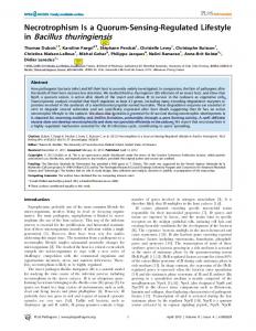
metabolic enzymes. However, since serotyping is a well-established method for B. thuringiensis strain identification, the isolated strains were also serotyped. We also included toxicity assays against larvae of the lepidoptera Trichoplusia ni, known to be sensitive to Cry toxins such as CryIA [31]. Finally, since the mobile elements IS231, IS232, and IS240 were found associated with various cry genes [22], their distribution among the natural isolates was also investigated. Materials and Methods Strain isolation. In total, 154 strains were isolated from soil samples in five locations in Norway (Fig. 1): Moss (Østfold), 77 strains from four sample sites, all sample sites were on the Southern part of Jeløy island in Moss; Singsås (Sør-Trøndelag), 24 strains; Brekkvasselv (NordTrøndelag), 15 strains; Steigen (Nordland), 14 strains; and Tromsø (Troms), 24 strains, the sample site was on the Tromsø island. Samples from Moss were taken in September, the others about one month earlier. Strains isolated in Tromsø, Singsås, and Steigen were from heather soil, and in Brekkvasselv from a woodland at riverside. The four soil sampling sites from Moss were taken from a leaf and grass compost (sample 1, 6 strains), a strawberry field (sample 2, 25 strains), a
81
E. Helgason et al.: Genetic Diversity of Bacillus cereus
Table 1. Genetic diversity in Bacillus cereus / B. thuringiensis isolates from soil samples in Norway Locality/ sample Moss (total) Sample 1 Sample 2 Sample 3 Sample 4 Steigen Brekkvasselv Singsås Tromsø Total/mean
Fig. 1. Geographic map of Norway with the locations where the 154 B. cereus / B. thuringiensis strains were isolated.
cabbage field (sample 3, 15 strains) and a beech grove (sample 4, 31 strains). Isolation of B. cereus / B. thuringiensis from soil was done according to the method of Travers et al. [32]. Colonies with morphological appearance of B. cereus / B. thuringiensis were randomly picked and subcultured on T3 agar medium. Cultures were then grown for 1 and 2 days on Oxoid B. cereus selective agar base (CH617) with supplement SR99, and sterile Egg Yolk Emulsion (SR 47) for confirmation of B. cereus / B. thuringiensis. Serotyping. H-serotyping based on B. thuringiensis flagellar antigens was performed by the agglutination method, as described by de Barjac [9]. Parasporal crystal observation. The bacteria were grown on Schaeffer Sporulation Medium [27] plates for 5–7 days at 28°C. The samples were stained with carbolfuchsin and observed under a microscope (Nikon, Labophot-2). Insect toxicity assay. Insect toxicity was tested by feeding the cabbage looper Trichoplusia ni as previously described [1]. Bacterial cultures were mixed with the normal T. ni diet to a final concentration of 20% (vol/vol). Mortality was analyzed after 1, 3, and 5 days. Electrophoresis of enzymes. Protein extracts were prepared as described previously [6]. Starch-gel electrophoresis and selective enzyme staining were performed as described by Selander et al. [28]. The 13 enzymes assayed were: nucleoside phosphorylase (NSP); three leucylleucyl-glycine peptidases (P3T), (P3M), (P3B); alanine dehydrogenase (ALD); phosphoglucose isomerase (PGI); adenylate kinase (ADK); malic enzyme (ME); malate dehydrogenase (MDH); a-naphthylpropionate esterase (EST); aconitase (ACO); indophenol oxidase (IPO); and catalase (CAT). Electromorphs of each enzyme, numbered in order of decreasing anodal mobility, were equated with alleles at the corresponding structural gene locus. Each strain was characterized by its combination of alleles at the 13 enzyme loci, and distinctive
No. isolates
No. ETs
Mean no. of alleles per locus
Isolate diversity
ET diversity
77 6 25 15 31 14 15 24 24 154
57 6 17 14 26 12 10 18 22 112
4.6 2.3 3.2 3.0 3.2 1.9 1.7 2.9 3.2 5.2
0.453 0.410 0.391 0.501 0.424 0.242 0.180 0.379 0.377 0.440
0.485 0.410 0.437 0.511 0.429 0.252 0.231 0.399 0.388 0.526
multilocus genotypes were designated as electrophoretic types (ETs) (data on allelic profiles are available on request). Alleles were not cognate with those previously recorded by Carlson et al. [6]. Statistical analyses of the genetic diversity were performed as described by Selander et al. [28], clustering was performed from a matrix of genetic distance by the average-linkage method [30], and variance in allele frequencies within and among groups was estimated by F-statistic [29]. PCR amplification for IS detection. Primers corresponding to the conserved repeats at the end of IS elements were designed for PCR amplification [21]. Bacterial DNA samples used as PCR templates were obtained from bacterial colonies by a boiling method [3], and PCR was performed as described previously [21]. For each independent experiment, each IS’s original B. thuringiensis strain was included in the PCR reaction as positive control.
Results Overall genetic diversity. In the collection of the 154 strains from five distinct geographical locations in Norway (Fig. 1), all the 13 enzyme loci were polymorphic. The number of alleles ranged from 2 to 8 with an average of 5.2 alleles per locus. In total, 112 ETs were distinguished. Among all ETs, the mean genetic diversity (H) was 0.526 (Table 1), with NSP (0.053) and P3T (0.767) being the least and the most diverse loci, respectively (data not shown). At a genetic distance of 0.55, there were two large clusters of ETs, designated cluster I (ET 1 to ET 62) and cluster II (ET 63 to 112), including the genotypes of 84 and 70 isolates, respectively. Genetic diversity within and between locations. Each soil sample harbored B. cereus / B. thuringiensis strains of various multilocus genotypes. The genetic diversity was highest in Moss, where all four samples were highly diverse, with mean ET diversity ranging from 0.410 to 0.511 (Table 1). There was little sharing of ETs between the four sample sites from Moss, i.e., few strains having the same ET in more than one sample site. However, the coefficient of genetic differentiation between the Moss
82
CURRENT MICROBIOLOGY Vol. 37 (1998)
sample sites, FST, indicated no significant difference in the genetic composition of strains between the different samples (data not shown), owing to the fact that the same alleles were represented in all four samples. Ninety percent of the isolates from Moss had genotypes that belong to cluster II, and cluster II included only one strain from another location (Fig. 2). In contrast, in cluster I, ETs from all five locations sampled in Norway were found. However, cluster I could be differentiated into smaller clusters, where the genotypes of isolates from the same location were represented (Fig. 2, Table 2). Thirteen of the 59 alleles (20%) were unique for isolates from Moss and not found in isolates from other locations. Alleles specific to isolates from other geographic areas were rare. This specificity of Moss is also confirmed by measuring the degree of genetic differentiation between locations. Comparing genetic differences between all locations gives an FST of 0.173 (data not shown), but if isolates from Moss are not included, FST falls to 0.039. Taking Moss as a specific location vs. the others joined together as one location, then FST becomes 0.105. The extent of differentiation of the ETs of isolates from Moss vs. those from other locations is, therefore, obvious. Genetic variation in relation to serotype. Among the 150 isolates serotyped for B. thuringiensis H-antigen, 90 were typeable, and 28 serotypes were identified (Table 2). Thus, more than 55% of the B. cereus / B. thuringiensis isolates were typeable. A few strains (3%) were autoagglutinable and thus non-typeable, while the remaining isolates (42%) did not cross-react with any of the known B. thuringiensis antigens. The number of isolates assigned to each serotype varied from 1 to 17. The most common serotype was H34. Eighteen serotypes were represented by isolates in cluster I, while 15 different serotypes were present in cluster II. A larger proportion of strains in cluster II were non-typeable (57% in cluster II versus 30% of the strains in cluster I). Strains of the same ET often harbor different serotype antigens: for example, the 3 ET 86 strains were H35, H24, and non-typeable serotype. Within each of the most frequent serotypes (H34, H10, H11, H8, and H3a3b3c), genetic diversity ranged from 0.318 to 0.505 with a mean of 0.403 (data not shown). Thus, no close association could be found between the serotypes and multilocus genotypes of the soil isolates. = Fig. 2. Genetic relationships between 112 ETs of B. cereus / B. thuringiensis strains. The dendrogram was generated by the averagelinkage method of clustering from a matrix of coefficient of genetic distance based on 13 enzyme loci. ETs are numbered sequentially. ET differing solely by null alleles are on the same branch of the dendrogram. Symbols indicate the isolates from the four sample sites of Moss: k, sample 1; r, sample 2; l, sample 3; j, sample 4.
E. Helgason et al.: Genetic Diversity of Bacillus cereus
Insecticidal activity, parasporal crystals, and IS elements. Since strains were originally isolated regardless of the presence of parasporal crystals in the sporangia, a systematic observation of sporulated culture of each strain was undertaken. Only four strains (3%) produced obvious parasporal inclusions, three of which having the inclusions apparently attached to the spore, whereas the last strain had a free, small, irregular inclusion. The strains with appendix inclusions, from two sample sites in Moss, were indistinguishable by MEE (ET 86) and also their IS element pattern (Table 2). The strain with the free, small, irregular inclusion (ET 104) recovered from Moss had no detectable IS element (Table 2). Only 10 of the 154 isolates, all of them with genotypes in cluster I, were significantly toxic to Trichoplusia ni (average mortality rate of 80% or more after five days), but none were as active as B. thuringiensis subsp. kurstaki (H3a3b3c), the reference strain for lepidoptera (Table 3). Only two out of seven of the kurstaki (serotype H3a3b3c) strains in this study were highly toxic against the lepidoptera (Tables 2 and 3). It is interesting to note, however, that none of the kurstaki strains isolated in this study produced any detectable crystals during sporulation, none of the crystal-producing strains were toxic to T. ni, and none of the strains toxic against T. ni had any detectable crystals. Of the 154 isolates, 28 strains (14%) gave positive results to IS-elements, 20 amplified by primer P231A, 2 by primer P231V, and 8 by primer P240 (Table 2). None of the 154 strains gave any reliable PCR product of IS232. IS231- and IS240-related elements occurred in strains of various serotypes and with multilocus genotypes in both clusters. However, 26 of the 29 IS-positive strains were from Moss.
Discussion An understanding of the genetic structure in B. cereus / B. thuringiensis population is of interest since these bacteria, which are so common in nature, are known to be potentially pathogenic both in humans and insects. The crystal proteins are toxic to some insects, and B. thuringiensis has been used to control agricultural pests for more than 30 years [16]. B. cereus is frequently isolated as a contaminant of milk, cereals, and various other foods and has been the cause of isolated cases and outbreaks of food poisoning, which may be caused by enterotoxins [10]. Occasionally, B. cereus strains are responsible for wound and eye infections in humans, as well as systemic infections [10, 33]. It has been reported that B. thuringiensis strains produce enterotoxins and have homologous DNA sequence to the enterotoxin gene of B. cereus [2, 4]. Recently, B. thuringiensis was identified as the cause of a
83 diarrhea outbreak in Canada [18]. To analyze the genetic diversity of soil isolates of B. cereus / B. thuringiensis, we have used MEE. The advantage of this method is that the characters tested are minimally subject to evolutionary convergence, because the electrophoretic variants (allozyme) are essentially selectively neutral or nearly so [11, 12, 15, 34]. This is the first time B. cereus and B. thuringiensis in the soil have been thoroughly analyzed. The total genetic diversity of the soil strains examined in this study (0.526) was similar to the diversity found among B. cereus / B. thuringiensis from various culture collections (0.556) and, thus, from much more diverse origins than presented here [6]. The genetic diversity in soil population of B. cereus / B. thuringiensis was higher than that reported for B. subtilis from soil by Istock et al. [17]. The high diversity in multilocus genotypes supports the observation that B. thuringiensis strains displayed greater diversity in rRNA gene restriction patterns than all other species examined by that technique [26]. The soil isolates from four locations in Norway fall into two main clusters (Fig. 2), one containing isolates from all locations and the other comprising almost exclusively isolates from one location. At a genetic distance of 0.1–0.2, clusters of isolates from the same sample were observed. The occurrence of closely related ETs among isolates from the same sample site suggests that recombination may generate the observed genotypic diversity. This finding is similar to what was observed in the analysis of B. subtilis strains from soil, where recombination appeared to be frequent, giving rise to high local genotypic diversity [17]. As in B. subtilis populations, transformation is probably the mechanism responsible for recombination in natural population of B. cereus / B. thuringiensis, though other forms of chromosomal gene transfer may occur [14]. In B. cereus / B. thuringiensis, transfer of plasmids containing the d-endotoxin genes is known to occur [14], and it is thus likely that other plasmids and genes are transferred as well. All the B. cereus /B. thuringiensis natural isolates investigated so far have plasmids, usually larger that 50 kb [Kolstø and Helgason, unpublished results], which in some cases may carry housekeeping genes [5] and contribute to a high genetic diversity in the species. Several strains also produce bacteriocins [23; P. Rønning and A.-B. Kolstø, unpublished results], which may contribute to the availability of closely related DNA for transformation. The fact that some B. thuringiensis and B. cereus strains have a very efficient conjugation system [14, 19] facilitates the exchange of genetic material between strains. In spite of this evidence of extensive local recombination, two observations suggests that B.
84
CURRENT MICROBIOLOGY Vol. 37 (1998)
Table 2. Characteristics of 154 B. cereus / B. thuringiensis isolates from five soil locations in Norway
ET
Isolate
1 2 3 4 5 6 7 8 9 10 11 12 13 14 15 16 17 18 19 20 21 22 23 24 25 26 27 28 29 30 31 32 33 34 35 36 37
AH 618 AH 636‡ AH 671 AH 628 AH 688 AH 689 AH 632 AH 624, 635 AH 663 AH 656 AH 658 AH 642 AH 621 AH 694 AH 691 AH 673–675 AH 692 AH 693 AH 554‡ AH 552‡ AH 521 AH 690 AH 679 AH 678‡ AH 676‡ AH 677 AH 547 AH 550 AH 525 AH 686‡ AH 654 AH 630 AH 685 AH 683 AH 652 AH 655 AH 659, 660, 665, 668 AH 661 AH 664 AH 670 AH 637, 641 AH 640 AH 646 AH 649 AH 633 AH 669 AH 634 AH 650 AH 687 AH 530 AH 662 AH 684 AH 647 AH 648, 653 AH 651 AH 681‡
38 39
40 41 42 43 44 45 46 47 48
Serotype a
IS231/ 240
Singsås Singsås Tromsø Singsås Tromsø Tromsø Singsås Singsås Brekkvasselv Brekkvasselv Brekkvasselv Steigen Singsås Tromsø Tromsø Tromsø Tromsø Tromsø Moss 3 Moss 3 Moss 2 Tromsø Tromsø Tromsø Tromsø Tromsø Moss 3 Moss 3 Moss 2 Tromsø Steigen Singsås Tromsø Tromsø Steigen Steigen Brekkvasselv
H31 H11 H34 H34 H-b HH34 H34 (2) H34 H34 H34 H34 H34 H34 H3a3b3c H34 (3) H15 H3a3b3c HH5a5b H29 H16 H35 H10 H11 H34 HH29 H15 H3a3b3c H25 HH3a3b3c H11 H19 H19 H11 (4)
0 0 0 0 0 0 0 0 0 0 0 0 0 0 0 0 0 0 0 0 1650 e 0 0 0 0 0 1650 e 0 0 0 0 0 0 0 0 0 0
Brekkvasselv Brekkvasselv Brekkvasselv Singsås Singsås Steigen Steigen Singsås Brekkvasselv Singsås Steigen Tromsø Moss 2 Brekkvasselv Tromsø Steigen Steigen Steigen Tromsø
H43 nd c H11 H6, AA d H6 HAA HH5 HHH35 H25 H3a3d3e H3a3b3c H3a3b3c H24 (2) H34 H3a3b3c
0 0 0 0 0 0 0 0 850 f 0 0 0 0 0 0 0 0 0 0
Site/ sample
Table 2 (Continued)
ET
Isolate
49
AH 657 AH 682‡ AH 644 AH 643 AH 622‡ AH 645 AH 619‡ AH 620 AH 666 AH 667 AH 535 AH 680 AH 638 AH 625, 626 AH 627 AH 631 AH 639 AH 623 AH 629 AH 520 AH 518 AH 536 AH 537, 538 AH 513, 540, 541, 565 AH 531 AH 572 AH 574 AH 542 AH 560 AH 576 AH 519 AH 526 AH 527 AH 532 AH 551 AH 557 AH 543, 544 AH 516 AH 571 AH 564 AH 579 AH 567, 568 AH 580 AH 569 AH 570 AH 517● AH 563● AH 581● AH 558 AH 548 AH 559 AH 524 AH 587 AH 588 AH 515, 523 AH 528, 529, 534 AH 533 AH 522
50 51 52 53 54 55 56 57 58 59 60
61 62 63 64 65 66 67
68 69 70 71 72 73 74 75 76 77 78 79 80 81 82 83 84 85 86
87 88 89 90 91
Site/ sample
Serotype a
Brekkvasselv Tromsø Steigen Steigen Singsås Steigen Singsås Singsås Brekkvasselv Brekkvasselv Moss 2 Tromsø Singsås Singsås Singsås Singsås Singsås Singsås Singsås Moss 2 Moss 2 Moss 2 Moss 2 Moss 1, 2, 2, 4
H14 HHHH5 HHH44 H10 nd AA AA HAA (2) nd H34 H4a4c AA HHHH14 H- (2) H- (4)
Moss 2 Moss 4 Moss 4 Moss 2 Moss 4 Moss 4 Moss 2 Moss 2 Moss 2 Moss 2 Moss 3 Moss 3 Moss 3 Moss 1 Moss 4 Moss 4 Moss 4 Moss 4 Moss 4 Moss 4 Moss 4 Moss 1 Moss 4 Moss 4 Moss 4 Moss 3 Moss 4 Moss 2 Moss 4 Moss 4 Moss 1, 2 Moss 2 Moss 2 Moss 2
H14 H10 HH19 HH10 HH19 HHHHH- (2) nd HHHH- (2) HHHH35 HH24 H8a8b H10 H10a10b H10 HH10 H- (2) H8 (3) H8a8b H-
IS231/ 240 0 0 0 0 850 f 0 0 0 0 0 1950 e 0 0 0 0 0 0 0 0 0 1950 e 0 0 0 0 1950 e 850 f 0 0 0 0 0 0 1950 e 0 0 0 0 1950 e 0 0 0 0 1650 e 1650 e 0 1650 e 1650 e 1650 e 0 1950 e 1950 e 1950 e 0 0 0 0 0 1950/1650 e
85
E. Helgason et al.: Genetic Diversity of Bacillus cereus Table 2 (Continued)
ET 92 93 94 95 96 97 98 99 100 101 102 103 104 105 106 107 108 109 110 111 112
Isolate AH 575 AH 562 AH 583 AH 555 AH 566 AH 512 AH 514 AH 578 AH 577 AH 585 AH 586 AH 539 AH 672 AH 582* AH 584 AH 561 AH 573 AH 556 AH 545 AH 546 AH 553 AH 549
Site/ sample Moss 4 Moss 4 Moss 4 Moss 3 Moss 4 Moss 1 Moss 1 Moss 4 Moss 4 Moss 4 Moss 4 Moss 2 Tromsø Moss 4 Moss 4 Moss 4 Moss 4 Moss 3 Moss 3 Moss 3 Moss 3 Moss 3
Serotype a H22 HH11 HHH8 H7 H7 H17 H3 H7 HH17 H5 H10 H16 HHHH8a8b HH-
IS231/ 240
Table 3. Toxicity of soil isolates against larvae of Trichoplusia ni. Strains that showed mortality rates of 80% or more for both overnight and sporulated cultures of all isolates are listed Strain
ET
Serotype
Mortality a
AH 636 AH 554 AH 552 AH 678 AH 676 AH 686 AH 681 AH 682 AH 622 AH 619 Control, no bacteria added B. thuringiensis 213
2 19 20 24 25 30 48 49 52 54
H11 HH5a5b H10 H11 H3a3b3c H3a3b3c HH5 H-
100 92 87 87 80 87 92 95 92 97
H3a3b
0 100
1650 e 0 0 0 0 850 f 0 850 f 850 f 1650 e 850 f 1850 g 0 0 0 0 1650 g 850 f 0 1950 e 1650 e 0 0
a Numbers in parentheses indicate number of strains with the same serotype found in that location. b H-, non typeable. c nd, not done. d AA, auto-agglutinable. e Length (bp) of PCR product obtained with the P231A primer. f Length (bp) of PCR product obtained with the P240 primer. g Length (bp) of PCR product obtained with the P231V primer. ● Crystal inclusion apparently attached to the spore. * Irregular crystal inclusion. ‡ Toxic against the larvae Trichoplusia ni.
cereus / B. thuringiensis populations in soil do not possess a fully panmictic population structure. First, some isolates displayed identical ET although they originated from very different locations, such as ET 58 strains isolated from both Moss and Tromsø, a geographic distance of about 1200 km (Table 2). This conservation of the electrophoretic pattern could reflect an increased genetic stability in a subset of strains, as has been suggested for B. thuringiensis subsp. israelensis [26]. Second, the strains from Moss presented a significant amount of geographic differentation, with 22% of the alleles identified in this study being seen solely in isolates from Moss. This significant geographic differentation compared with the B. cereus / B. thuringiensis populations from other localities was shown by F-statistic, which quantifies degrees of genetic differentiation among groups by partitioning the total genetic diversity between groups [25]. On the other hand, isolates from the four sample sites from Moss (Fig. 2) did not show significant
a Calculated from number of surviving larvae out of total larvae after 5 days of feeding.
genetic heterogeneity between samples, as measured by F-statistic, although the samples were from different types of soil (Table 2). The reason for the geographic differentiation of the Moss population is unclear: it may, at least in part, be due to a rather great difference in the climate, since the summers in Moss are longer and warmer than in most other areas of Norway, and particularly compared with the Northern areas where the other samples were located (Fig. 1); The flora in Moss is very rich, and several plants have their northern growth limit on the island of Jeløy, which is a moraine and lava island. It is thus possible that, because of special ecology, the B. cereus / B. thuringiensis strains from Moss have evolved some degree of a subspecific structure. Thus, as to B. subtilis strains from soil [17], the results from our study indicate a mixed population structure of B. cereus / B. thuringiensis, with evidence of both extensive recombination and a low degree of clonality. B. thuringiensis subsp. kurstaki, serotype H3a3b3c, is frequently used in various commercially available biopesticide formulations against lepidopteran insects [16]. In this study, seven strains were serotyped as kurstaki, although only two turned out to be toxic to the lepidoptera T. ni, and none of the strains produced any detectable crystal inclusions. The H3a3b3c-positive strains in this study were all genetically different, as judged by MEE, suggesting that the protein(s) specifying the serotype is expressed in strains with different genetic background, supporting the evidence of extensive genetic recombination in soil. Analysis of these strains by pulsed field gel electrophoresis confirmed their genomic differences and clearly differentiates them from the ATCC
86 reference kurstaki strains [Helgason and Kolstø, unpublished results]. This is in contrast to the conserved ribotype patterns of six kurstaki strains reported by Priest and co-workers [26] and the conserved DNA of kurstaki strains shown by hybridization [24]. Thus, it is possible that certain strains containing plasmids with insecticidal genes are more stable and more similar to each other, as suggested by Priest et al. [26]. None of the strains active on T. ni had parasporal crystal, and none of them gave IS-PCR signals. This observation could certainly be set into perspective with the possibility of other, non-crystal-forming d-endotoxins, entomopathogenic virulence factors. The recent discoveries of soluble d-endotoxins [20] and vegetative insecticidal Vip3A/B toxins [13] in some B. thuringiensis strains might be relevant to these issues. In conclusion, this study indicated high genetic diversity among B. cereus /B. thuringiensis isolates from soil. In spite of a high percentage of strains serotyped with B. thuringiensis antisera, IS-elements, crystal inclusion, and toxicity against T. ni were detected in only few strains. The population structure of B. cereus /B. thuringiensis in our study appears to be mixed, with both extensive recombination, resulting in closely related ETs among isolates from the same sample, and a low degree of clonality, since few identical ETs were observed in different locations. ACKNOWLEDGMENTS This work was supported by grants from the Norwegian Research Council and from the Norwegian Directorate for Nature Management (A.-B. Kolstø). We are very grateful to Mrs. V. Cosmao-Dumanoir and Ms. H. Ripoutean for serotyping the strains. Y. Chen received fellowships from the State Education Commission of P.R. China and from the Catholic University of Louvain (UCL, Belgium). J. Mahillon is Research Associate at the National Fund for Scientific Research (FNRS, Belgium). Part of this work (J. Mahillon) has been sponsored by grant FRSM # 3.4578.90 from FNRS and a FDS grant from UCL.
Literature Cited 1. Abdel-Hameed A, Lande´n R (1994) Studies on Bacillus thuringiensis strains isolated from Swedish soils: insect toxicity and production of B. cereus-diarrhoeal-type enterotoxin. World J Microbiol Biotechnol 10:406–409 2. Asano S-I, Nukumizu Y, Bando H, Iizuka T, Yamamoto T (1997) Cloning of novel enterotoxin genes from Bacillus cereus and Bacillus thuringiensis. Appl Environ Microbiol 63:1054–1057 3. Brousseau R, Saint-Onge A, Pre´fontaine G, Mason L, Cabana J (1993) Arbitrary primer polymerase chain reaction, a powerful method to identify Bacillus thuringiensis serovars and strains. Appl Environ Microbiol 59:114–119 4. Carlson CR, Kolstø A-B (1993) A complete map of a Bacillus thuringiensis chromosome. J Bacteriol 175:1053–1060 5. Carlson CR, Kolstø A-B (1994) A small (2.4 Mb) Bacillus cereus chromosome corresponds to a conserved region of a larger (5.3 Mb) Bacillus cereus chromosome. Mol Microbiol 13:161–169
CURRENT MICROBIOLOGY Vol. 37 (1998) 6. Carlson CR, Caugant DA, Kolstø A-B (1994) Genotypic diversity among Bacillus cereus and Bacillus thuringiensis strains. Appl Environ Microbiol 60:1719–1725 7. Carlson CR, Johansen T, Lecadet M-M, Kolstø A-B (1996) Genomic organization of the entomopathogenic bacterium Bacillus thuringiensis subsp. berliner 1715. Microbiol 142:1625–1634 8. Claus D, Berkley RCW (1986) Genus Bacillus Cohn 1872 In: Smith PHA (ed) Bergey’s manual of systematic bacteriology, vol 2. Baltimore: The Williams & Wilkins Co, pp 1105–1139 9. de Barjac H (1981) Identification of H-serotypes of Bacillus thuringiensis. In: Burges HD (ed) Microbial control of pest and plant diseases 1970–1980. London: Academic Press, Inc (London), Ltd, pp 35–43 10. Drobniewski FA (1993) Bacillus cereus and related species. Clin Microbiol Rev 6:324–338 11. Dykhuizen DE, Hartl DL (1980) Selective neutrality of 6PGD in Escherichia coli and the effects of genetic background. Genetics 96:801–817 12. Dykhuizen DE, Hartl DL (1983) Functional effects of PGI allozymes in Escherichia coli. Genetics 105:1–18 13. Estruch JJ, Warren GW, Mullins MA, Nye GJ, Craig JA, Koziel MG (1996) Vip3A, a novel Bacillus thuringiensis vegetative insecticidal protein with a wide spectrum of activities against lepidopteran insects. Proc Natl Acad Sci USA 93:5389–5394 14. Gonzales JM Jr, Brown BJ, Carlton BC (1982) Transfer of Bacillus thuringiensis plasmids coding for d-endotoxin among strains of B. thuringiensis and B. cereus. Proc Natl Acad Sci USA 79:6951– 6955 15. Hartl DL, Dykhuizen DE (1981) Potential for selection among nearly neutral allozymes of 6-phospogluconate dehydrogenase in Escherichia coli. Proc Natl Acad Sci USA 78:6344–6348 16. Ho¨fte H, Whiteley HR (1989) Insecticidal crystal proteins of Bacillus thuringiensis. Microbiol Rev 53:242–255 17. Istock CA, Duncan KE, Ferguson N, Zhou X (1992) Sexuality in natural populatio n of bacteria; Bacillus subtilis challenges the clonal paradigm. Mol Ecol 1:95–103 18. Jackson SG, Goodbrand RB, Ahmed R, Kasatiya S (1995) Bacillus cereus and Bacillus thuringiensis isolated in a gastroenteritis outbreak investigation. Appl Microbiol 21:103–105 19. Jensen GB, Andrup L, Wilcks A, Smidt L, Poulsen OM (1996) The aggregation-mediated conjugation system of Bacillus thuringiensis subsp. israeliensis: host range and kinetics of transfer. Curr Microbiol 33:1–10 20. Kostichka K, Warren GW, Mullins MA, Mullins AD, Craig JA, Koziel MG, Estruch JJ (1996) Cloning of a cryV-type insecticidal protein gene from Bacillus thuringiensis: the cryV-encoded protein is expressed early in stationary phase. J Bacteriol 178:2141–2144 21. Le´onard C, Chen Y, Mahillon J (1997) Diversity and differential distribution of IS231, IS232 and IS240 among B. cereus, B. thuringiensis and B. mycoides. Microbiology 143:2537–2547 22. Mahillon J, Rezso¨hazy R, Hallet B, Delcour J (1994) IS231 and other Bacillus thuringiensis transposable elements: a review. Genetica 93:13–26 23. Naclerio G, Ricca E, Sacco M, De Felice M (1993) Antimicrobial activity of a newly identified bacteriocin of Bacillus cereus. Appl Environ Microbiol 59:4313–4316 24. Nakamura LK (1994) DNA Relatedness among Bacillus thuringiensis serovars. Int J Syst Bacteriol 44:125–129 25. Nei M (1977) F-statistics and analysis of gene diversity in subdivided populations. Ann Hum Genet 41:225–233 26. Priest FG, Kaji DA, Rosato YB, Canhos VP (1994) Characterization of Bacillus thuringiensis and related bacteria by ribosomal
E. Helgason et al.: Genetic Diversity of Bacillus cereus RNA gene restriction fragment length polymorphisms. Microbiology 140:1015–1022 27. Schaeffer P, Millet JA, Aubert J-P (1965) Catabolic repression of bacterial sporulation. Proc Natl Acad Sci USA 54:704–711 28. Selander RK, Caugant DA, Ochman H, Musser JM, Gilmour MN, Whittam TS (1986) Methods of multilocus enzyme electrophoresis for bacterial population genetics and systematics. Appl Environ Microbiol 51:873–884 29. Selander RK, Caugant DA, Whittam TS (1987) Genetic structure and variation in natural population of Escherichia coli. In: Neidhardt FC, Ingraham JL, Low KB, Magasanik B, Umbarger HE (eds) Escherichia coli and Salmonella typhimurium: cellular and molecular biology. Washington DC: American Society of Microbiology, pp 1625–1648
87 30. Sneath PHA, Sokal RR (1973) Numerical taxonomy: the principles of numerical classification. San Francisco: W.H. Freeman & Co 31. Travers RS, Martin PAW (1989) Worldwide abundance and distribution of Bacillus thuringiensis isolates. Appl Environ Microbiol 55:2437–2442 32. Travers RS, Martin PAW, Reichelderfer CF (1987) Selective process for efficient isolation of soil Bacillus spp. Appl Environ Microbiol 53:1263–1266 33. Turnbull PCB, Kramer J, Melling J (1990) Bacillus. In: Parker MT, Duerden BI (eds) Principples of bacteriology, virology and immunity, Vol 2. London: Systematic Edward Arnold, pp 187–210 34. Whittam TS, Ochman H, Selander RK (1983) Multilocus genetic structure in natural populations of Escherichia coli. Proc Natl Acad Sci USA 80:1751–1755