Mar 3, 2005 - Commonwealth Ave., St. Paul, Minnesota 55108,1 and Anti-Infective Research, ...... Ziebandt, A. K., H. Weber, J. Rudolph, R. Schmid, D. Hoper, ...
JOURNAL OF BACTERIOLOGY, Aug. 2005, p. 5486–5492 0021-9193/05/$08.00⫹0 doi:10.1128/JB.187.15.5486–5492.2005 Copyright © 2005, American Society for Microbiology. All Rights Reserved.
Vol. 187, No. 15
Global Regulation of Gene Expression by ArlRS, a Two-Component Signal Transduction Regulatory System of Staphylococcus aureus Xudong Liang,1 Li Zheng,1 Christina Landwehr,1 Dwayne Lunsford,2 David Holmes,2 and Yinduo Ji1* Department of Veterinary and Biomedical Sciences, College of Veterinary Medicine, University of Minnesota, 1971 Commonwealth Ave., St. Paul, Minnesota 55108,1 and Anti-Infective Research, GlaxoSmithKline Research and Development, 1250 S. Collegeville Rd., Collegeville, Pennsylvania 194262 Received 3 March 2005/Accepted 26 April 2005
Staphylococcus aureus expresses various cell wall-associated and extracellular virulence factors, coordinately controlled by different two-component signal transduction systems and transcriptional regulators. In this study, we used microarray technology to identify the genes regulated by ArlR. The microarray data indicate that ArlR functions as a positive regulator and also as a negative repressor to directly and/or indirectly mediate the expression of at least 114 genes involved in different functions, including autolysis, cell division, growth, and pathogenesis.
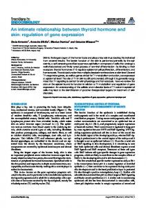
effect of the arlR mutation on the bacterial growth rate, CFU, phenotype, and stress responses to different antibacterial agents, including cell wall synthesis inhibitors such as bacitracin, phosphomycin, and vancomycin. No significant effect of the arlR mutation on stress response to antibacterial agents was observed (data not shown). However, Sa316ko grew slightly slower than WCUH29 between early log and stationary phases (Fig. 1). Growth curves as measured by optical density (OD) were confirmed by determining the effect of arlR mutation on colony size and viable cell counts at intervals during growth. The ArlR mutant strain, Sa316ko, displayed similarsized colonies on a tryptic soy agar plate and similar morphological features of individual cells under microscopic observation (data not shown) but decreased approximately one log CFU compared to wild-type strain between early log and stationary phases (data not shown). These results suggest that the arlRS regulon may be involved in the modulation of expression of genes associated with growth and cell division. Although it is unlikely that the arlR allelic gene replacement mutation has a polar effect on arlRS downstream gene expression (since arlR and arlS loci are located in a single operon), we examined the transcription level of arlRS downstream gene odhA, encoding 2-oxoglutarate dehydrogenase E1. Our real-time reverse transcriptase (RT)-PCR analyses showed that the arlR mutation had no significant influence on the expression of odhA (see Table 5). Identification of the arlRS regulon using microarray assay. To better define the arlRS regulon, gene expression profiles of the arlR mutant and parent cells were analyzed by using Affymetrix S. aureus arrays as described previously (11). The S. aureus array (Affymetrix) contains probe sets to over 3,300 S. aureus open reading frames (ORFs) based on the updated S. aureus genomic sequences of N315, Mu50, NCTC 8325, and COL. Total RNA was extracted from S. aureus cells grown to mid-log phase (OD at 600 nm, 0.4) by using the RNAPrep kit (Promega, MI) and treated with a DNA-free kit (Ambion). The RNA (10 g) was reverse transcribed to cDNA by using Superscript II reverse transcriptase and random primers (In-
Staphylococcus aureus is an important human and animal pathogen that causes a wide range of infections, including life-threatening endocarditis and toxic shock syndrome (22, 26). The ability of this organism to cause a variety of diseases is partly due to the expression of different cell wall-associated and secreted virulence factors which enable the bacteria to adhere to and colonize host cells (13) or cause toxic shock syndrome (26). The expression of virulence factors is coordinately controlled by two-component signal transduction systems, such as agr (1), sae (16), arl (14), and srrAB (30), and global regulators, including sar (8, 9), sigB (3, 31), rot (2, 24), and mgr (23). Therefore, the elucidation of the regulons of these regulatory systems is important for better understanding molecular mechanisms of pathogenesis. Recently, S. aureus regulons of agr, sar, sigB, and rot have been revealed by using a microarray-based approach (4, 11, 27). In our studies, we identified target genes controlled by ArlR by a comparison of the transcriptional profile between an arlR mutant and the wild-type strain during the mid-exponential phase of growth by using Affymetrix S. aureus arrays. Construction of the arlR deletion mutant. The arlR deletion mutant (Sa316ko) was constructed by bacteriophage 11-mediated transduction of a cassette containing the tetA gene, flanked by chromosomal fragments upstream and downstream of the alrR, from strain RN4220 into a clinical human isolate strain WCUH29 as described previously (12). Selection for tetracycline resistance and screening for the loss of the erythromycin resistance marker carried by the vector indicated that allelic replacement had occurred and resulted in the arlR mutant strain Sa316ko. The mutation in arlR was verified by PCR and Southern blot analysis (data not shown). To characterize the arlR mutant strain, we examined the
* Corresponding author. Mailing address: Department of Veterinary and Biomedical Sciences, College of Veterinary Medicine, University of Minnesota, 1971 Commonwealth Ave., St. Paul, MN 55108. Phone: (612) 624-2757. Fax: (612) 625-5203. E-mail: jixxx002@umn .edu. 5486
VOL. 187, 2005
NOTES
5487
FIG. 1. Growth curve of the arlR mutant. The arlR mutant strain Sa316ko and parent strain WCUH29 were incubated in TSB at 37°C overnight with shaking; the cultures were inoculated in fresh TSB. The cell growth was monitored at 37°C by a measuring of the OD at 600 nm every 15 min with 1 min of mixing before each reading. These curves represent one of three reproducible experiments.
vitrogen). The cDNA was treated with NaOH, purified by using the QIAquick PCR purification kit (QIAGEN), and digested with DNase I. The fragmented cDNAs were then directly labeled with biotin by using a biotin-ddUTP kit (Affymetrix). Biotinylated cDNA (3 g) was hybridized to the GeneChips. The GeneChips were then washed and subjected to a series of staining procedures as described in the manual for the Affymetrix array. Each GeneChip was washed and scanned at a 570-nm wavelength and a 3-m resolution in an Affymetrix GeneChip scanner. The Affymetrix Microarray Suite 4.0 algorithms calculated the signal intensities (average differences) and the present or absent determinations for each open reading frame. The GeneChips were then normalized, and their backgrounds were defined by using GeneSpring 4.0 (Silicon Genetics). The GeneSpring software was used to further analyze the transcription patterns of genes. To identify genes with significantly altered expression levels, a series of statistical analyses (filtering) were performed; cutoff values for ratio of expression levels of 1.80 and 0.55 were used to filter genes with expression level changes (n-fold) greater than ⫾1.8 in all three independent biological samples. Genes with variations (n-fold) of ⬎1.5 across the three samples were excluded. Furthermore, a statistical group comparison using the Student t test/analysis of variance was conducted to compare the mean expression levels of the control and the arlR mutant samples. The genes with significant differential expression levels (P value, ⬍0.05) were selected. The results of three independent experiments demonstrated that on average, transcripts for 73% of all genes on the arrays were detected by the Affymetrix arrays in the mid-log phase of bacterial cells of WCUH29. A comparison analysis of gene expression levels between wild-type WCUH29 and the arlR mutant revealed that the expression levels of 114 genes were significantly altered in the arlR mutant. Of these, 37 genes
showed a decrease (Table 1) and 77 genes showed an increase (Table 2) in expression level after the mutation of arlR. The array data indicated that ArlR positively regulates a two-component system, lytR-lytS, which encodes a response regulator and a sensor histidine kinase and is involved in autolysis (6, 14). The positive regulation of lytS expression by ArlR was confirmed by real-time RT-PCR analysis using the Stratagene Mx3000P real-time PCR system. Gene-specific primers were designed to yield ⬃100 bp of specific products (Table 3), and the housekeeping gene 16S rRNA was used as an endogenous control (29). All samples were analyzed in triplicate and normalized against 16S rRNA gene expression. The results were statistically analyzed for correlation to the microarray results. Compared to the wild type, the arlR mutant possessed low levels of lytS mRNA in the early log, mid-log, and stationary phases of growth (Tables 4 and 5). We also found that the mutation in arlR significantly down-regulates the expression of lrgA and lrgB, encoding different holin-like proteins involved in murein hydrolase transport and inhibition of murein hydrolase activity (7, 17). These enzymes are involved in the cleavage of specific cell wall components, which are important for cell division and growth (7, 17). To determine whether the growth defect of the mutant is attributable to increased susceptibility to cell lysis, we examined the effect of ArlR on autolysis induced by Triton X-100 and detected the cell wall murein hydrolase activity by using a zymographic assay as described previously (19). Consistent with a previous report (14), the arlR mutant cells displayed increased lysis in the presence of 0.01% Triton and showed enhanced peptidoglycan hydrolase activity compared to the parent control (unpublished data). These results indicated that increased autolysis in the arlR mutant may result from the significant down-regulation of the lytSR and lrgAB operons and, in turn, may partially affect bacterial growth. Although it
5488
NOTES
J. BACTERIOL. TABLE 1. S. aureus genes up-regulated by ArlR
N315 ORF
SA1844 SA1842 SA1843 SAS066 SA1248 SAS065 SA0250 SA0251 SA0144 SA0147 SA0211 SA0212 SA0220 SA0252 SA0253 SA0269 SA0270 SA0271 SA0272 SA0275 SA0276 SA0417 SA0519 SA0520 SA0521 SA0746 SA0893 SA1269 SA1270 SA1271 SA1272 SA1305 SA1583 SA2222 SA2303 SA2486
N315 gene
N315 description
Change in expression level (n-fold)a
agrA agrB agrC agrD arlR hld lytS lytR cap5A cap5D
Response regulator Accessory gene regulator B Sensor histidine kinase AgrD protein Response regulator Delta-hemolysin Two-component sensor histidine kinase Two-component response regulator Capsular polysaccharide synthesis enzyme Cap5A Capsular polysaccharide synthesis enzyme Cap5D Putative oxidoreductase Hypothetical protein Hypothetical protein Holin-like protein LrgA Holin-like protein LrgB Hypothetical protein Secretory antigen precursor SsaA Hypothetical protein Hypothetical protein Hypothetical protein Similar to diarrheal toxin Hypothetical protein Ser-Asp-rich fibrinogen-binding protein SdrC Ser-Asp-rich fibrinogen-binding protein SdrD Ser-Asp-rich fibrinogen-binding protein SdrE Staphylococcal nuclease Hypothetical protein Blt-like protein Similar to amino acid pearmease IlvA threonine deaminase Alanine dehydrogenase Cell-dividion initiation protein Repressor of toxin Bicyclomycin-resistant protein TcaB Hypothetical protein 2-Oxoglutarate/malate translocator
⫺2.8 ⫺2.2 ⫺2.4 ⫺1.6 ⫺602.2 ⫺2.2 ⫺3.1 ⫺2.2 ⫺1.8 ⫺1.5 ⫺2.1 ⫺1.9 ⫺2.8 ⫺16.5 ⫺19.7 ⫺2.3 ⫺2 ⫺5.9 ⫺7.4 ⫺4.2 ⫺4.4 ⫺2.5 ⫺2.3 ⫺3.7 ⫺2.1 ⫺1.9 ⫺1.9 ⫺3.1 ⫺1.8 ⫺3 ⫺2.4 ⫺4.9 ⫺1.9 ⫺2 ⫺11.2 ⫺1.8
lrgA lrgB ssaA
sdrC sdrD sdrE
rot
agr, sar, rot, or sigB effectb
agr ⫹ rot ⫹ B ⫹ B ⫹ rot ⫹ sar ⫹, sigB ⫺ sar ⫹, sigB ⫺ sigB agr ⫹, rot ⫹, sigB ⫺ sigB ⫺ sar ⫹, rot ⫹
rot ⫹
a
Normalized values in the wild-type strain over values in the arlR mutant. agr and sar effects are as described by Dunman et al. (13), rot effect is as described by Said-Salim et al. (41), and sigB effect is as described by Bischoff et al. (4). ⫹, up-regulated; ⫺, down-regulated. b
has been reported that the mutation in rat, another autolysis regulator, exhibited a growth defect and enhanced autolysis partly due to increased murein hydrolase activity (19), the reason why the mutation in arlR led to a slight impact on growth remains undefined, since the lrgAB mutation did not show significant impact on the cell shape and growth rate (17). Also, our microarray data showed that ArlR positively regulates virulence factor genes, such as sdrC, sdrD, and sdrE, encoding different Ser-Asp-rich bone sialoprotein-binding proteins (Table 1) (28). The result for sdrD was confirmed by using real-time RT-PCR and demonstrated that the level of sdrD mRNA in the arlR mutant strain is significantly decreased compared with that in the wild-type strain (Table 4). The up-regulation of sdrC by ArlR may indirectly function via upregulated rot expression, since Rot positively regulates the expression of sdrC (11). In addition, ArlR positively regulates the tcaB gene (which encodes a bicyclomycin-resistant protein), the secretory antigen precursor ssaA, and toxin genes, such as hld and SA0276, encoding delta-hemolysin and diarrheal toxin, respectively (Table 1). Moreover, the array data showed that ArlR positively regulates the accessory gene regulator (agr) (Table 1). A real-time
RT-PCR was employed to validate this result and demonstrated that the mutation in arlR significantly decreased the level of agrA expression (Table 4). The expression of agrA was constantly up-regulated by ArlR at different times of growth (Table 5). These results are inconsistent with the finding that the mutation in arlS leads to the overexpression of Agr (15). This controversy may be due to different sensitivities, culture conditions, and time points of sampling between different assays. In addition, the array results indicated that ArlR also positively regulates the expression of the repressor of toxins (rot). This result was confirmed by real-time RT-PCR and demonstrated that the mutation in arlR causes a decrease in rot expression (Table 4). Our findings are consistent with previous reports that the expression of certain secreted enzymes, toxins, and ureases are repressed by Rot (9, 27). Most virulence factors negatively regulated by ArlR were also repressed by Rot but were up-regulated by Agr (5, 25). Therefore, the downregulation of these toxins, proteases, and adhesins may be mediated directly by ArlR or indirectly controlled via Rot or Agr (Fig. 2). To determine the role of the ArlRS regulatory system in pathogenesis, we examined the effect of the arlR mutation on
VOL. 187, 2005
NOTES
5489
TABLE 2. S. aureus genes repressed by ArlR N315 ORF
SA0104 SA0123 SA0124 SA0125 SA0126 SA0127 SA0135 SA0136 SA0137 SA0138 SA0165 SA0299 SA0304 SA0318 SA0319 SA0320 SA0321 SA0331 SA0332 SA0333 SA0395 SA0710 SA0850 SA0851 SA0899 SA0904 SA0956 SA0978 SA1090 SA1091 SA1145 SA1154 SA1266 SA1267 SA1268 SA1552 SA1577 SA1630 SA1637 SA1638 SA1752 SA1848 SA1882 SA1883 SA1991 SA1992 SA1993 SA1994 SA1995 SA1996 SA1997 SA2006 SA2007 SA2008 SA2081 SA2082 SA2083 SA2084 SA2085 SA2086 SA2087 SA2088 SA2091 SA2092
N315 gene
cpsM
nanA
sspC isdC lytN fmhC
ebhA ebhB splB lukD lukE hlb nrgA kdpD kdpE lacG lacF lacD lacC lacB lacA alsS ureA ureB ureC ureE ureF ureG ureD
N315 description
Hypothetical protein Hypothetical protein Hypothetical protein Hypothetical protein Hypothetical protein Capsular polysaccharide repeat, Unit transporter cpsM Hypothetical protein Hypothetical protein Hypothetical protein Hypothetical protein Hypothetical protein PfkB family carbohydrate kinase N-Acetylneuraminate lyase Putative membrane protein Hypothetical protein Hypothetical protein Putative PTS multidomain regulator Hypothetical protein Hypothetical protein Conserved hypothetical protein Hypothetical protein Conserved hypothetical protein Hypothetical protein Oligopeptide ABC transporter, ATP-binding protein Cysteine protease Hypothetical protein Hypothetical protein Conserved hypothetical protein LytN protein FmhC protein Host factor-1 protein Conserved hypothetical protein Hypothetical protein Hypothetical protein Hypothetical protein Hypothetical protein Hypothetical protein Serine protease, V8 protease Leukotoxin, LukD Leukotoxin, LukE Truncated beta-hemolysin Probable ammonium transporter Two-component sensor kinase KdpD Two-component response regulator KdpE 6-Phospho-beta-galactosidase PTS system, lactose-specific IIBC component PTS system, lactose-specific IIA component Tagatose 1,6-diphosphate aldolase Tagatose-6-phosphate kinase Galactose-6-phosphate isomerase LacB subunit Galactose-6-phosphate isomerase LacA subunit Hypothetical protein Hypothetical protein Alpha-acetolactate synthase Urea transporter Urease gamma subunit Urease beta subunit Urease alpha subunit Urease accessory protein UreE Urease accessory protein UreF Urease accessory protein UreG Urease accessory protein UreD Hypothetical protein Hypothetical protein
Change in expression level (n-fold)a
1.9 22.1 35 39.6 20.4 4 1.6 2.7 3.6 5.6 1.8 2.5 2.5 2.5 4.7 5.1 5.5 14.2 8 10.6 2.2 7.4 10.7 2.2 3.1 10.7 2.9 1.9 10.4 3.9 3 9.1 6.4 15.1 28.6 3.7 17.2 4.3 1.9 1.6 2.5 1.9 2.9 2 4.1 3.5 6.5 4.4 4.9 7.2 5.1 5.5 2.8 2.4 3.1 9.3 12.1 7 5.2 4.6 3.6 4.5 6.7 4.5
agr, sar, rot, or sigB effectb
rot⫺
rot ⫺
rot ⫺
rot ⫺ rot ⫺ agr ⫹, rot ⫺ rot ⫺ agr ⫹, sar ⫹, rot ⫺, sigB ⫺ rot ⫺
rot ⫺ agr ⫹, rot ⫺ agr ⫹, sar ⫹, rot ⫺, sigB ⫺
rot rot rot rot rot rot rot
⫺ ⫺ ⫺ ⫺ ⫺ ⫺ ⫺
Continued on following page
5490
NOTES
J. BACTERIOL. TABLE 2—Continued
N315 ORF
N315 gene
SA2209 SA2287 SA2315 SA2319 SA2320 SA2329 SA2337 SA2338 SA2382 SA2455
hlgC
SA2456
cap8B
SA2457 SA2482 SAV0397
capA arcA
feoB cap8C
Change in expression level (n-fold)a
N315 description
Gamma-hemolysin component C precursor Staphylococcal regulator SarH2 Putative membrane protein Putative L-serine dehydratase Putative membrane protein Murine hydrose exporter Ferrous iron transporter protein Hypothetical protein Hypothetical protein Capsular polysaccharide synthesis enzyme Cap8C Capsular polysaccharide synthesis enzyme Cap8B Capsular polysaccharide biosynthesis CapA Arginine deiminase Hypothetical protein
agr, sar, rot, or sigB effectb
3.2 7.9 2.6 3.8 4.7 3.8 4 7.3 5.4 12.7
agr ⫹, rot ⫺
18.1
rot ⫺, sigB ⫹
31.2 2 12.1
rot ⫺, sigB ⫹
rot ⫺, sigB ⫹
a
Normalized values in the arlR mutant over values in the wild-type strain. agr and sar effects are as described by Dunman et al. (13), rot effect is as described by Said-Salim et al. (41), and sigB effect is as described by Bischoff et al. (4). ⫹, up-regulated; ⫺, down-regulated. b
virulence by using a murine hematogenous pyelonephritis model as described previously (20). The virulence of the arlR mutant was significantly attenuated compared to that of the parent control (unpublished data). Collectively, these findings suggest that ArlRS is a global two-component virulence regulatory system which can interact with other regulators to modulate the expression of virulence factors. On the other hand, ArlR functions as a repressor of virulence factors. Our microarray data showed that ArlR negatively regulates some toxin genes, lukD, lukE, phlC (hlb), and hlgC, which encode leukotoxin D, leukotoxin E, beta-hemolysin component C, and gamma-hemolysin component C, respectively (Table 2). Gamma-hemolysin is an S. aureus virulence factor that has been shown to play a role in S. aureus
endophthalmitis and corneal pathogenesis (10, 21). The microarray result for lukD was confirmed by real-time RT-PCR. As shown in Table 4, the lukD transcript was present at higher levels in the arlR mutant strain than in the wild-type strain. The array results also demonstrated that ArlR negatively regulates splB (encoding V8 protease), sspC (encoding cysteine protease), and ebhA and ebhB (encoding adhesins) (Table 2). The negative regulation of ebhB expression by ArlR was confirmed by a real-time RT-PCR (Table 4). Furthermore, the array results showed that ArlR negatively regulates the expression of genes involved in different PTS systems, such as the lacG, lacF, lacD, lacC, lacB, and lacA operons, as well as hypothetical proteins. To validate these results, real-time RT-PCR was performed, and it demonstrated that the mutation of arlR increases the expression of hypothetical proteins (SA0123 and SA0319) and constantly
TABLE 3. Primers used in real-time RT-PCR Primer
Sequence (5⬘-3⬘)
SA0123for ...........................ATATTACGGCGAACGGACGAC SA0123rev ...........................TGGCTTGTTATGCTCAAATGAATCG SA0250for ...........................GCATGGTTCTATCGTCGGTACATTG SA0250rev ...........................ACTTACTTTGCGTTTCGGCTTCAC SA0252lrgAfor....................TGAAACAACAAAAAGACGCATCAAAACCAG SA0252lrgArev....................ACTTCGCCTAACTTAACAGCACCAG SA0270ssaAfor ...................GGCATCCAAGTCAATTAAACCAAGATAATG SA0270ssaArev ...................CAGTACGGTAGCTGTTTGTGTTGTAAC SA0319for ...........................GCACCATCTGATATCGAAGTTGAAC SA0319rev ...........................TAGGCGTTCGGCATTTTCAGC SA1248for ...........................TGACAAAGTTGCTGGGCTTGATTAC SA1248rev ...........................TGTGGCTGACGACGTAAAATTGC SA1583for ...........................TCAGCGAGATTGAAAGCGAATAC SA1583rev ...........................CTGTCCATTTCTTTAAGCGTCATAG SA1637for ...........................TGGGGCGGTAAGTATAATGTTTCG SA1637rev ...........................GATCCATTCAATCCACCTGATAAGC SA1882for ...........................GAAAGACAAGCTGGTGCAACAAC SA1882rev ...........................AACGGCGAGAGAAAGTTCATTTAAC SA1844agrAfor ...................GTGAAATTCGTAAGCATGACCCAGTTG SA1844agrArev...................TGTAAGCGTGTATGTGCAGTTTCTAAAC SA1993for ...........................GAAGGAAACAATTGCATTGCTGAAG SA1993rev ...........................ATATCATCACCTTGCGCTTCTTTAG SA1245odhA1665for..........GCAATGAACCACCCGTAGAATAGC SA1245odhA1833rev..........ACGAGAGCAGCACAAGATGATACAC 16S rRNAfor ......................CTGTGCACATCTTGACGGTA 16S rRNArev ......................TCAGCGTCAGTTACAGACCA
TABLE 4. Real-time RT-PCR analysis of expression of genes regulated by ArlR N315 ORF
N315 gene
N315 description
Change in expression level (n-fold)a RT-PCR
SA0123 SA0319 SA1637 SA1882
lukD kdpD
SA1993
lacF
SA1844
agrA
SA0250
lytS
SA0252 SA0270
lrgA ssaA
SA1583
rot
a
Hypothetical protein Hypothetical protein Leukotoxin LukD Two-component sensor kinase PTS system, lactose-specific IIA component Two-component response regulator Two-component sensor kinase Holin-like protein LrgA Secretory antigen precursor SsaA Repressor of toxin
42.8 5.1 2 3.1 17
Microarray
22.1 4.7 1.9 2.9 6.5
⫺5.7
⫺2.8
⫺2.8
⫺3.1
⫺64.8 ⫺1.7
⫺16.5 ⫺2
⫺4
⫺1.9
Normalized values in the arlR mutant over values in the wild-type strain. Negative numbers denote up-regulation in the wild-type strain.
VOL. 187, 2005
NOTES
5491
TABLE 5. Real-time RT-PCR analysis of expression of genes regulated by ArlR in different growth phases N315 ORF
SA0250 SA1844 SA1882 SA1993 SA1245b a b
N315 gene
lytS agrA kdpD lacF odhA
Change in expression level (n-fold)a in RT-PCR by phase
N315 description
Two-component sensor kinase Two-component response regulator Two-component sensor kinase PTS system, lactose specific IIA component 2-Oxoglutarate dehydrogenase E1
Early log
Mid-log
Stationary
⫺4.4 ⫺2.7 ⫺2.6 6.3 NC
⫺7 ⫺2.3 13.8 4 NC
⫺6.7 ⫺3.9 1.2 13 NC
Normalized values in the arlR mutant over values in the wild-type strain. Negative numbers denote up-regulation in the wild-type strain. Downstream gene of arlRS. NC, no detectable change.
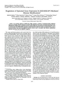
up-regulates lacF expression at different phases of growth (Tables 4 and 5). Also, we found that in the arlR mutant, the levels of kdpD and kdpE transcripts (which encode a two-component sensor kinase and response regulator and involve K⫹ transport [18]) were obviously higher than those in the wild-type strain. To confirm this result, real-time RT-PCR was performed, and the results demonstrated that the mutation of arlR increases kdpD expression in the mid-log phase of growth but decreases kdpD expression in the early log phase of growth (Tables 4 and 5). These results suggest that ArlR differentially regulates kdpD expression at different times of growth. Conclusion. The regulon of ArlRS has been identified by employing transcriptome technology using Affymetrix S. aureus arrays. The results demonstrate that ArlRS is a global transcriptional regulator which directly and/or indirectly interacts with other regulators in regulatory networks and modulates the expression of genes involved in autolysis, cell growth, and pathogenesis (Fig. 2). Some genes/operons mediated by ArlRS may be missed, due to their lack of stability and/or kinetic regulation as well as low detection sensitivity. Further studies to investigate which genes identified using microarray assays are directly regulated by ArlR and are involved in autolysis and/or pathogenesis are in progress.
FIG. 2. Schematic figure showing how the staphylococcal two-component signal transduction regulatory system, ArlRS, directly and/or indirectly modulates gene expression. 3, positive regulation; ⬜, negative regulation.
We thank Junsong Sun for his assistance in the real-time RT-PCR analysis, Aaron Becker for his assistance in the microarray analysis, and Doug Weiss for his critical reading of the manuscript and for his helpful suggestions. This work was supported by grant O3-O2 from the Academic Health Center at the University of Minnesota. This research was also supported in part by grant AI057451 from the National Institute of Allergy and Infectious Diseases. REFERENCES 1. Abdelnour, A., S. Arvidson, T. Bremell, C. Ryden, and A. Tarkowski. 1993. The accessory gene regulator (agr) controls Staphylococcus aureus virulence in a murine arthritis model. Infect. Immun. 61:3879–3885. 2. Arvidson, S., and K. Tegmark. 2001. Regulation of virulence determinants in Staphylococcus aureus. Int. J. Med. Microbiol. 291:159–170. 3. Bischoff, M., J. M. Entenza, and P. Giachino. 2001. Influence of a functional sigB operon on the global regulators sar and agr in Staphylococcus aureus. J. Bacteriol. 183:5171–5179. 4. Bischoff, M., P. Dunman, J. Kormanec, D. Macapagal, E. Murphy, W. Mounts, B. Berger-Ba ¨chi, and S. Projan. 2004. Microarray-based analysis of the Staphylococcus aureus B regulon. J. Bacteriol. 186:4085–4099. 5. Bronner, S., H. Monteil, and G. Prevost. 2004. Regulation of virulence determinants in Staphylococcus aureus: complexity and applications. FEMS Microbiol. Rev. 28:183–200. 6. Brunskill, E. W., and K. W. Bayles. 1996. Identification and molecular characterization of a putative regulatory locus that affects autolysis in Staphylococcus aureus. J. Bacteriol. 178:611–618. 7. Brunskill, E. W., and K. W. Bayles. 1996. Identification of LytSR-regulated genes from Staphylococcus aureus. J. Bacteriol. 178:5810–5812. 8. Cheung, A. L., J. M. Koomey, C. A. Butler, S. J. Projan, and V. A. Fischetti. 1992. Regulation of exoprotein expression in Staphylococcus aureus by a locus (sar) distinct from agr. Proc. Natl. Acad. Sci. USA 89:6462–6466. 9. Cheung, A. L., K. Schmidt, B. Bateman, and A. C. Manna. 2001. SarS, a SarA homolog repressible by agr, is an activator of protein A synthesis in Staphylococcus aureus. Infect. Immun. 69:2448–2455. 10. Dajcs, J. J., M. S. Austin, G. D. Sloop, J. M. Moreau, E. B. Hume, H. W. Thompson, F. M. McAleese, T. J. Foster, and R. J. O’Callaghan. 2002. Corneal pathogenesis of Staphylococcus aureus strain Newman. Investig. Ophthalmol. Vis. Sci. 43:1109–1115. 11. Dunman, P. M., E. Murphy, S. Haney, D. Palacios, G. Tucker-Kellogg, S. Wu, E. L. Brown, R. J. Zagursky, D. Shlaes, and S. J. Projan. 2001. Transcription profiling-based identification of Staphylococcus aureus genes regulated by the agr and/or sarA loci. J. Bacteriol. 183:7341–7353. 12. Fan, F., R. D. Lunsford, D. Sylvester, J. Fan, H. Celesnik, S. Iordanescu, M. Rosenberg, and D. McDevitt. 2001. Regulated ectopic expression and allelicreplacement mutagenesis as a method for gene essentiality testing in Staphylococcus aureus. Plasmid 46:71–75. 13. Foster, T. J, and M. Ho ¨o ¨k. 1998. Surface protein adhesins of Staphylococcus aureus. Trends Microbiol. 6:484–488. 14. Fournier, B., and D. C. Hooper. 2000. A new two-component regulatory system involved in adhesion, autolysis, and extracellular proteolytic activity of Staphylococcus aureus. J. Bacteriol. 182:3955–3964. 15. Fournier, B., A. Klier, and G. Rapoport. 2001. The two-component system ArlS-ArlR is a regulator of virulence gene expression in Staphylococcus aureus. Mol. Microbiol. 41:247–261. 16. Giraudo, A. T., A. L. Cheung, and R. Nagel. 1997. The sae locus of Staphylococcus aureus controls exoprotein synthesis at the transcriptional level. Arch. Microbiol. 168:53–58. 17. Groicher, K. H., B. A. Firek, D. F. Fujimoto, and K. W. Bayles. 2000. The Staphylococcus aureus lrgAB operon modulates murein hydrolase activity and penicillin tolerance. J. Bacteriol. 182:1794–1801. 18. Heermann, R., K. Altendorf, and K. Jung. 2003. The N-terminal input
5492
19. 20. 21.
22. 23. 24.
NOTES
domain of the sensor kinase KdpD of Escherichia coli stabilizes the interaction between the cognate response regulator KdpE and the corresponding DNA-binding site. J. Biol. Chem. 278:51277–51284. Ingavale, S. S., W. Van Wamel, and A. L. Cheung. 2003. Characterization of Rat, an autolysis regulator in Staphylococcus aureus. Mol. Microbiol. 48: 1451–1466. Ji, Y., A. Marra, M. Rosenberg, and G. Woodnutt. 1999. Regulated antisense RNA eliminates alpha-toxin virulence in Staphylococcus aureus infection. J. Bacteriol. 181:6585–6590. Karlsson, A., P. Saravia-Otten, K. Tegmark, E. Morfeldt, and S. Arvidson. 2001. Decreased amounts of cell wall-associated protein A and fibronectinbinding proteins in Staphylococcus aureus sarA mutants due to up-regulation of extracellular proteases. Infect. Immun. 69:4742–4748. Lowy, F. D. 1998. Staphylococcus aureus infections. N. Engl. J. Med. 339: 520–532. Luong, T. T., S. W. Newell, and C. Y. Lee. 2003. mgr, a novel global regulator in Staphylococcus aureus. J. Bacteriol. 185:3703–3710. McNamara, P. J., K. C. Milligan-Monroe, S. Khalili, and R. A. Proctor. 2000. Identification, cloning, and initial characterization of rot, a locus encoding a regulator of virulence factor expression in Staphylococcus aureus. J. Bacteriol. 182:3197–3203.
J. BACTERIOL. 25. Novick, R. P. 2003. Autoinduction and signal transduction in the regulation of staphylococcal virulence. Mol. Microbiol. 48:1429–1449. 26. Projan, S., and R. Novick. 1997. The molecular basis of pathogenicity, p. 55–81. In G. Archer and K. Crossley (ed.), Staphylococci in human diseases. Churchill Livingstone, New York, N.Y. 27. Saı¨d-Salim, B., P. M. Dunman, F. M. McAleese, D. Macapagal, E. Murphy, P. J. McNamara, S. Arvidson, T. J. Foster, S. J. Projan, and B. N. Kreiswirth. 2003. Global regulation of Staphylococcus aureus genes by Rot. J. Bacteriol. 185:610–619. 28. Tung, H., B. Guss, U. Hellman, L. Persson, K. Rubin, and C. Ryden. 2000. A bone sialoprotein-binding protein from Staphylococcus aureus: a member of staphylococcal Sdr family. Biochem. J. 345:611–619. 29. Yarwood, J. M., J. K. McCormick, M. L. Paustian, V. Kapur, and P. M. Schlievert. 2002. Repression of the Staphylococcus aureus accessory gene regulator in serum and in vivo. J. Bacteriol. 184:1095–1101. 30. Yarwood, J. M., J. K. McCormick, and P. M. Schlievert. 2001. Identification of a novel two-component regulatory system that acts in global regulation of virulence factors of Staphylococcus aureus. J. Bacteriol. 183:1113–1123. 31. Ziebandt, A. K., H. Weber, J. Rudolph, R. Schmid, D. Hoper, S. Engelmann, and M. Hecker. 2001. Extracellular proteins of Staphylococcus aureus and the role of SarA and sigma B. Proteomics 1:480–493.