BIOLOGY OF REPRODUCTION 65, 1364–1370 (2001)
Glucose Transporter Expression in Rat Embryo and Uterus During Decidualization, Implantation, and Early Postimplantation1 Emin Tu¨rkay Korgun,3,4 Ramazan Demir,4 Astrid Hammer,3 Gottfried Dohr,3 Gernot Desoye,5 Gerhard Skofitsch,6 and Tom Hahn2,3 Institute of Histology and Embryology,3 University of Graz, A-8010 Graz, Austria Institute of Histology and Embryology,4 Akdeniz University, 07070 Antalya, Turkey Department of Obstetrics and Gynecology,5 University of Graz, A-8036 Graz, Austria Institute of Zoology,6 University of Graz, A-8010 Graz, Austria ABSTRACT Efficient transfer of glucose from the mother to the embryonic compartment is crucial to sustain the survival and normal development of the embryo in utero, because the embryo’s production of this primary substrate for oxidative metabolism is minimal. In the present study, the temporal sequence of expression of the sodium-independent facilitative glucose transporter isoforms GLUTs 1, 3, 4, and 5 was investigated in the developing rat uteroembryonic unit between conception and Gestational Day 8 using immunohistochemistry. The GLUTs 1, 3, and 4 were expressed in the embryonic tissues after the start of implantation, being colocalized in the parietal endoderm, visceral endoderm, primary ectoderm, extraembryonic ectoderm, and the ectoplacental cone. In the uterus, a faint GLUT1 labeling emerged, but not until Gestational Day 3, in the luminal epithelium, endometrial stroma, and decidual cells. The intensity of GLUT1 staining increased in the latter population with progressing decidualization. Endometrial glands and myometrial smooth muscle cells stained neither for GLUT1 nor for GLUT3 until postimplantation. During all developmental stages examined, GLUT4 was visualized throughout the pregnant rat uterus, as was GLUT3 (with the above-mentioned exceptions). The density of GLUT5 was generally less than the sensitivity of the immunohistochemical detection method in all tissues investigated. In conclusion, the data point to a significant expression of the high-affinity glucose transporters GLUTs 1, 3, and 4 in the rat uteroembryonic unit, providing supportive evidence for an important role of facilitative glucose diffusion during peri-implantation development.
decidua, early development, implantation, placental transport, uterus
INTRODUCTION
Early mammalian embryos are not able to utilize glucose as an energy source until compactation. The situation changes radically with the beginning of blastocyst formation, when the embryonic fuel metabolism switches its preferred substrate from the more oxidized pyruvate to glucose, an alternation that is signaled by glucose transport Supported by grants from the Austrian Science Foundation FWF (P13721MED) and the Jubilaeumsfonds of the Oesterreichische Nationalbank (7361). 2 Correspondence: Tom Hahn, Institute of Histology and Embryology, University of Graz, Harrachgasse 21, A-8010 Graz, Austria. FAX: 43 316 380 9625; e-mail:
[email protected] 1
Received: 13 February 2001. First decision: 19 March 2001. Accepted: 13 June 2001. Q 2001 by the Society for the Study of Reproduction, Inc. ISSN: 0006-3363. http://www.biolreprod.org
proteins [1]. From this stage onward, adequate transfer of glucose from the mother to the embryonic compartment is decisive to the survival and normal development of the embryo in utero [2]. In general, glucose transport can be brought about by a sodium-coupled mechanism or by facilitated diffusion along a concentration gradient. The latter process is mediated by carrier proteins rendering substrate entry approximately 10 000-fold faster than that calculated for diffusion across the lipid membrane layer. The transport facilitators are approximately 500 amino acids in length and belong to a growing superfamily of integral membrane glycoproteins with 12 membrane-spanning domains. The genes of these glucose transporters have been designated as GLUT1 through GLUT9, in the order in which they were identified. Very recently, GLUT8 and GLUT9 have both been described [3, 4], but their similarity with the ‘‘classical’’ GLUT isoforms is not higher than that with bacterial inositol, arabinose, or xylose transporters [5]. Therefore, they constitute a separate branch within the family of hexose transporters. The clone termed GLUT7 has turned out to be an artifact and does not, as suggested previously, encode a liver endoplasmic reticulum transporter. Additionally, GLUT6 encodes a pseudogene that is not translated into protein, and GLUT2 has, alternatively to its glucose transport function, also been considered to serve as a glucose sensor and/or a fructose transporter. It is somewhat unusual, in that it operates with a significantly lower affinity for glucose than any of the other isoforms and can, therefore, not function efficiently in the low-glucose environment prevailing in the uterus. In contrast, the remaining isoforms (GLUTs 1, 3, 4, and 5) represent high-affinity transport facilitators. Because of their low Michaelis constant (Km), these transporters function at rates close to maximal velocity. Thus, their level of cell surface expression greatly influences the rate of glucose uptake into the cells. Because considerable agreement now exists that the significance of sodium-dependent transport in supplying preimplantation embryos with glucose is not worth mentioning (for review, see [1]), it seems reasonable to assume that facilitative glucose transporters might play a pivotal role in meeting the energy requirements of the embryo during the critical period between blastocyst formation and implantation. For obvious ethical reasons, no information is generally available regarding glucose transporter expression in the human uterus or developing (i.e., nonpolyploid) embryo during the first weeks of pregnancy. However, in the commonly used animal models, the spatiotemporal distribution pattern of GLUT proteins also has not been investigated in the uteroembryonic unit from conception until postimplantation. To close this gap, we have performed experiments to study, in parallel, expression of the high-affinity sugar
1364
GLUCOSE TRANSPORTERS IN RAT EMBRYO AND UTERUS
1365
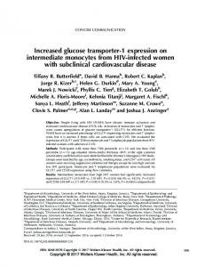
FIG. 1. Distribution of glucose transporters GLUT1 (yellow), GLUT3 (blue), and GLUT4 (pink) in the developing rat uteroembryonic unit between GD 1 and GD 8. Empty cells in the table indicate that the concerned tissue is either not yet differentiated or has not been investigated at the respective GD. 1, Weak staining; 11, moderate staining; 111, strong staining.
transporters GLUTs 1, 3, 4, and 5 during decidualization, implantation, and early postimplantation (Gestational Days [GDs] 1–8) in the rat conceptus and uterus. MATERIALS AND METHODS Immunohistochemistry was performed on paraffin-embedded sections of the developing rat uteroembryonic unit between conception and GD 8. Briefly, 1 ml of 1% (v/v) Evans blue in 0.9% (v/v) NaCl was injected into the posterior right femoral vein of 32 impregnated white Wistar rats under deep ether anesthesia to identify the implantation areas within the uterus. Ten minutes after the injection, the anterior abdominal wall was opened, and tissue samples were taken from parts of the uterus containing the dye. These samples were fixed in Holland fixative for 4 h, embedded, and then serial sections (6 mm) were collected on SuperFrost Plus slides (Novoglas, Berne, Switzerland). After rehydration, samples were transferred to 0.01 M citrate buffer (pH 6) and subsequently heated twice in a microwave oven for 5 min each time at 750 W for antigen retrieval. After cooling for 20 min at room temperature, the sections were washed with PBS. To remove endogenous peroxidase activity, sections were kept in 3% H2O2 for 20 min and afterward washed with PBS. Sections were incubated for 60 min at room temperature in a moist chamber with rabbit antisera against the C-terminal sequences of GLUT1 (CGLFHPLGADSQV), GLUT3 (NSMQPVKEPGNA), GLUT4 (CTELEYLGPEND), and GLUT5 (ELKELPPVTSEQ) (all from Chemicon, Temecula, CA). Antisera were diluted 1:1000 (v/v) (GLUT1), 1:500 (GLUT3), 1:100 (GLUT4), and 1:500 (GLUT5) with Antibody Diluent (Dako, Carpinteria, CA). Labeling was visualized using the Universal LSAB kit (Dako) according to the manufacturer’s instructions. The sections were counterstained with Mayer hemalum (Merck, Darmstadt, Germany) and mounted with Kaiser glycerol gelatin (Merck). For controls, sections were incubated with antisera preadsorbed with corresponding oligopeptides (10 mg/ml; Pichem, Graz, Austria) based on the sequences used for the immunization of the antibodygenerating rabbits. Pictures were taken with an Axiophot microscope (Zeiss, Oberkochen, Germany). Investigations were conducted in accordance with the Guide for the Care and Use of Laboratory Animals (Institute for Laboratory Animal Research of the National Academy of Science, Bethesda, MD; 1996).
RESULTS
The density of GLUT5 in rat uterus and developing embryo was generally less than the sensitivity of the immunohistochemical detection method between GD 1 and GD 8. The distribution of the GLUTs 1, 3, and 4 in the rat uteroembryonic unit is detailed in Figure 1. Major findings are presented below. Gestational Day 1
At the first day of gestation, GLUT3 was localized in distinct populations of endometrial stromal cells and in the uterine epithelium (Fig. 2A). The GLUT4 antiserum immunoreacted with the uterine epithelium, endometrial stromal cells, epithelium of the endometrial glands, and myometrial smooth muscle cells (Fig. 2B). The significant GLUT4 labeling in uterine glands as well as in endometrial stroma and myometrial smooth muscle cells was observed throughout the whole gestational period investigated, with no (or negligible) decrease in intensity (Fig. 1). In contrast to the homogeneously distributed GLUT4 signal within the cells of the luminal uterine epithelial layer (Fig. 2B), GLUT3 staining was limited to plasma membranes, being more pronounced in the apical membranes of the uterine epithelium than in the basolateral areas (Fig. 2A). However, GLUT1 could not be detected in the uterus of the newly impregnated rats. Gestational Day 2
The localization of GLUTs 1, 3, and 4 was similar to that observed at GD 1.
1366
KORGUN ET AL.
GLUCOSE TRANSPORTERS IN RAT EMBRYO AND UTERUS
Gestational Day 3
A faint GLUT1 reaction emerged in the luminal uterine epithelium, endometrial stroma, and primary endometrial decidual cells with progressing decidualization at GD 3. Endometrial glands and myometrial smooth muscle cells neither stained for GLUT1 nor for GLUT3, and the respective cells remained negative for both transporters until postimplantation (Fig. 1). In contrast, GLUT3 staining was even increased in the apical region of uterine epithelial cells compared with GDs 1 and 2. Endometrial stromal cells and primary endometrial decidual cells also reacted with the GLUT3 antiserum, but the latter stained more intensely. Gestational Day 4
The GLUT1 staining became more apparent in the cell populations where it was first recognized the day before. The GLUT4 immunoreactivity was visualized throughout the uterus, as was GLUT3 immunoreactivity (with the above-mentioned exceptions). Both transport facilitators were most abundant in the primary decidual cells. Gestational Day 5
Implantation starts at this developmental stage, and the rat blastocyst that became visible in these sections was immunopositive for GLUTs 1, 3, and 4 (Fig. 2, C–E). This reaction pattern remained constant until GD 8, with only slight changes in intensity (Fig. 1). The glucose transporter expression of the uterus roughly resembled that observed on GD 4, apart from a drastically reduced GLUT1 and GLUT4 staining in the epithelium, which was found exclusively in the antimesometrial region (Fig. 2, C and E). Gestational Day 6
Parietal endodermal, visceral endodermal, primary ectodermal, extraembryonic ectodermal cells, and the ectoplacental cone immunoreacted with antisera to GLUTs 1, 3, and 4 (Fig. 2, F–H). The newly emerging secondary decidual cells were weakly or moderately labeled for b FIG. 2. Transverse sections of the developing rat uteroembryonic unit from GD 1 (A and B), GD 5 (C–E), and GD 6 (F–H). For every developmental stage and GLUT isoform, control sections were incubated with antisera preadsorbed with corresponding oligopeptides; examples are shown as insets in A and C. A) GLUT3 at GD 1. Arrowheads indicate uterine luminal epithelium; the arrow points to an immunonegative endometrial gland. B) GLUT4 at GD 1. Arrowheads indicate uterine luminal epithelium; the arrow points to an immunopositive endometrial gland. C) GLUT1 at GD 5. Arrowheads indicate uterine luminal epithelium. The embryo (arrow) is located antimesometrially in the uterus lumen. D) GLUT3 at GD 5. Arrowheads indicate uterine luminal epithelium; the arrow points to the embryo. E) GLUT4 at GD 5. Arrowheads indicate uterine luminal epithelium; the arrow points to the embryo. F) GLUT1 at GD 6. The black arrow points to ectoplacental cone, the red arrow to parietal endoderm, the black arrowhead to visceral endoderm, the red arrowhead to extraembryonic ectoderm, and the yellow arrowhead to primary ectoderm. G) GLUT3 at GD 6. The red arrow indicates parietal endoderm. The black arrowhead points to visceral endoderm, the red arrowhead to extraembryonic ectoderm, and the yellow arrowhead to primary ectoderm. H) GLUT4 at GD 6. The black arrow points to ectoplacental cone, the red arrow to parietal endoderm, the black arrowhead to visceral endoderm, the red arrowhead to extraembryonic ectoderm, and the yellow arrowhead to primary ectoderm. ESC, Endometrial stromal cells; MSMC, myometrial smooth muscle cells; PDZ, primary decidual zone; SDZ, secondary decidual zone; UL, uterus lumen. Magnification 3150 for A–E, 3300 for F and H, 3600 for G, and 350 for the insets in A and C.
1367
GLUT1 (Fig. 2F), GLUT3, and GLUT4; otherwise, no considerable changes were observed in uterine glucose transporter distribution compared to that found at GD 5. Gestational Day 7
Embryonic tissues stained more intensively for GLUT1 at GD 7 than at GD 6. Simultaneously, a striking increase of this isoform was observed in the secondary decidual zone of the uterus (Fig. 3A). No significant changes were observed in embryonic GLUT3 and GLUT4 expression between GD 6 and GD 7 (Fig. 3, B and C). Generally, together with the capsule and the glycogenic area, the secondary decidual zone represented the major site of GLUT expression in the uterus. Gestational Day 8
Except for the primary decidual region, where GLUT1 was no longer detectable and GLUT3 and GLUT4 staining was markedly reduced (Fig. 3, D–F), the antisera labeled the same uterine structures as they did at GD 7. Conspicuous GLUT3 expression was visualized in the glycogenic area (Fig. 3H) accompanied by immunoreactivity for GLUT1 (Fig. 3G) and GLUT4 (Fig. 3F). Trophectoderm, inner cell mass, and the ectoplacental cone expressed GLUTs 1, 3, and 4 (see insets in Fig. 3, D–F). DISCUSSION
In the present study, data concerning the expression of facilitative glucose transporter proteins are provided, to our knowledge for the first time, from conception until an early postimplantation stage of the rat. The heterogeneously glucosylated GLUT1 isoform was originally considered to represent the specific transport machinery for glucose in epithelial cells of blood-tissue barriers, such as the placental trophoblast [6, 7]. The data presented here for the rat implantation period demonstrated GLUT1 to be expressed in all embryonic cell populations examined, which is in good agreement with the preceding GLUT1 expression in mouse morulae [8] as well as an intense labeling of the respective transcript throughout in the early postimplantation rat embryo [9]. During subsequent developmental stages of the rat conceptus, GLUT1 was even reported to represent the predominant glucose transporter isoform [10]. Collectively, these results support the concept of a more ubiquitous occurrence of GLUT1, which might play a kind of ‘‘housekeeping’’ role [11], thus covering the cellular glucose requirement for ATP production and biosynthesis of sugar-containing macromolecules, at least during embryonic development. In the uterus, decidual cells above all were found to contain appreciable amounts of GLUT1 protein, also coinciding with the earlier, significant GLUT1 mRNA levels detected in this tissue [9]. The transporter isoform characteristic for cells with highglucose requirements, such as neurons or tumor cells [12], is GLUT3, because it has a particularly high affinity for glucose. In this study, GLUT3 protein was most abundant in embryonic cell populations, in accordance with noticeable mRNA staining demonstrated previously for rats at GD 8 [9]. Rat embryos were also shown to express significant amounts of GLUT3 during organogenesis [13]. Within the rat uterus, the highest GLUT3 levels were found in decidual cells, which is in contrast to the relatively little GLUT3 mRNA detected there by in situ hybridization [9]. These divergent data may reflect a significant posttranscrip-
1368
KORGUN ET AL.
GLUCOSE TRANSPORTERS IN RAT EMBRYO AND UTERUS
tional GLUT3 regulation, such as a reduced turnover rate of the transporter protein, and/or a stabilization of the GLUT3 mRNA. In the nonpregnant rat uterus, GLUT3 has not been detectable at all [12, 14], which suggests that uterine expression of this isoform is only induced in the wake of specific differentiation programs associated with gestation. Expression of GLUT4 is considered to be limited to insulin-responsive tissues. Welch and Gorski [14] have demonstrated GLUT4 mRNA and protein in the nonpregnant rat uterus, and the immunohistochemical data presented here clearly identified GLUT4 as a glucose carrier protein expressed in this organ between GD 1 and GD 8. Endometrial glands and myometrial smooth muscle cells even contained GLUT4 exclusively. In uterine epithelial cells coexpressing GLUT4 with GLUT1 and/or GLUT3, a difference in the intracellular staining pattern of these molecules became apparent at the light microscopic level. The GLUT4 signal was homogeneously distributed throughout the cells, whereas the GLUT1 and GLUT3 signals were more restricted to the plasma membranes (Fig. 2, A and B). The homogeneous GLUT4 staining might be ascribed to intracellular reserve pools of this molecule. Reserve pools could not be detected for the other isoforms examined, although a growing body of evidence suggests that the level of functional plasma membrane GLUT1 transporters is also determined via subcellular trafficking of the protein [15]. In contrast to the pregnant rat uterus, GLUT4 reactivity in embryo-derived tissues and in the embryo proper was rather moderately, but clearly, detectable. In general, GLUT4 expression in embryonic cell populations is a controversial issue. Because we are the first, to our knowledge, to investigate rat embryos around implantation in this context, our positive results for GLUT4 are unique so far. Until now, only the transcripts were identified in the developing rat brain as early as GD 14 [16] and in the rat placenta between GD 14 and GD 21 [17]. Indeed, recent reports concerning other species support our findings. The b FIG. 3. Transverse sections of the rat uteroembryonic unit from GD 7 (A–C) and GD 8 (D–H). For every developmental stage and GLUT isoform, control sections were incubated with antisera preadsorbed with corresponding oligopeptides; an example is shown as an inset in C. A) GLUT1 at GD 7. The black arrow points to ectoplacental cone, the red arrow to parietal endoderm, the black arrowhead to visceral endoderm, the red arrowhead to extraembryonic ectoderm, and the yellow arrowhead to primary ectoderm. B) GLUT3 at GD 7. The red arrow indicates parietal endoderm. The black arrowhead points to visceral endoderm, the red arrowhead to extraembryonic ectoderm, and the yellow arrowhead to primary ectoderm. C) GLUT4 at GD 7. The black arrow points to ectoplacental cone, the red arrow to parietal endoderm, the black arrowhead to visceral endoderm, the red arrowhead to extraembryonic ectoderm, and the yellow arrowhead to primary ectoderm. D) GLUT1 at GD 8. The arrow points to the embryo. Inset: The red arrow indicates parietal endoderm, and the black arrowhead points to visceral endoderm, the red arrowhead to extraembryonic ectoderm, and the yellow arrowhead to primary ectoderm. E) GLUT3 at GD 8. The arrow points to the embryo. Inset: The red arrow indicates parietal endoderm, and the black arrowhead points to visceral endoderm, the red arrowhead to extraembryonic ectoderm, and the yellow arrowhead to primary ectoderm. F) GLUT4 at GD 8. The arrow points to the embryo. Inset: The red arrow indicates parietal endoderm, and the black arrowhead points to visceral endoderm, the red arrowhead to extraembryonic ectoderm, and the yellow arrowhead to primary ectoderm. G) GLUT1 at GD 8 in the glycogenic area. Arrows point to blood vessels. H) GLUT3 at GD 8 in the glycogenic area. Arrows point to blood vessels. C, Capsule; GA, glycogenic area; PDZ, primary decidual zone; SDZ, secondary decidual zone. Magnification 3150 for A–C, 375 for D–F, 3600 for G and H, 3100 for the inset in C, and 3200 for the insets in D–F.
1369
GLUT4 mRNA was visualized in bovine blastocysts [18], and both transcript and protein were found in murine neuroepithelium as early as GD 9 [19]. However, several earlier attempts failed to detect GLUT4 mRNA [9, 10, 13] and protein [20] in the rat conceptus between GD 9 and GD 14, and a number of older investigations concerning mice provided evidence against GLUT4 expression during periimplantation development [21–24]. Interestingly, the tissues staining for GLUT4 in this study are also richly endowed with the insulin receptor (unpublished results). This finding coincides with the presence of insulin receptor mRNA in preimplantation rat embryos [25]. In addition, embryonic and uterine insulin receptor expression has also been demonstrated in various mammalian species different from rat [26–30]. During the period of time investigated here, the uterus undergoes extensive remodeling to prepare for the invasion of the embryo. These changes impose acute metabolic demands on the cells, which can largely be met by utilization of glucose. Thus, having the uterine GLUT staining pattern in mind, it is conceivable that insulin not only promotes cellular proliferation and differentiation events in the uterus but also stimulates uterine glucose uptake mediated by the insulinsensitive GLUT4. In addition, later during gestation, GLUT4 would be ideally suited for covering the increasing glucose requirements of the gravid uterus, because its activity could increase in response to the elevated plasma insulin levels that can be observed with continuing pregnancy [31]. The appearance of insulin receptors on embryonic cells is delayed until the compacted eight-cell stage [23] and parallels the switch of the conceptus to a glucose-based metabolism. Accordingly, one may expect that insulin, apart from acting mitogenically, exerts its effect on glucose transport in the embryo as well. Increasing evidence suggests that diabetes-like conditions retard embryonic development via down-regulation of GLUTs and the triggering of apoptosis [6, 32, 33], but the most important outcome of the rare studies dealing with GLUT regulation by insulin during early pregnancy is that these deleterious effects of hyperglycemia on reproductive performance can be prevented by treatment with insulin [2, 34, 35]. In this context, reports showing that the overall expression levels of GLUTs 1, 3, and 4 remain unaffected by the hormone in various rat embryonic cells [10, 36] seem rather perplexing. However, the supposed discrepancy no longer appears as such when insulin is considered to bring about the translocation of GLUT4 from intracellular vesicles to the cell membrane. This mechanism, which could compensate for impaired GLUT expression, is well known from other insulin-dependent tissues (for review, see [37]) and has also been demonstrated in rat embryonic myoblasts [36]. The activity of the previously unidentified GLUT8 glucose carrier, which was among other sites detected in the mouse blastocyst, is regulated similarly by insulin [3]. Therefore, GLUT8 seems to be a candidate for supplementing, or even overriding, the action of GLUT4 in the embryo, because its Km is closer to that of GLUT3 than to those of the other transporters. In summary, the results demonstrate the paramount importance of high-affinity glucose transport facilitators for rat peri-implantation embryos, and they suggest a different functional significance for the individual isoforms in the developing uteroembryonic unit. ACKNOWLEDGMENT Our sincere thanks go to Rudolf Schmied for excellent technical assistance.
1370
KORGUN ET AL.
REFERENCES 1. Pantaleon M, Kaye PL. Glucose transporters in preimplantation development. Rev Reprod 1998; 3:77–81. 2. Moley KH, Chi MM, Knudson CM, Korsmeyer SJ, Mueckler MM. Hyperglycemia induces apoptosis in preimplantation embryos through cell death effector pathways. Nat Med 1998; 4:1421–1424. 3. Carayannopoulos MO, Chi MM, Cui Y, Pingsterhaus JM, McKnight RA, Mueckler M, Devaskar SU, Moley KH. GLUT8 is a glucose transporter responsible for insulin-stimulated glucose uptake in the blastocyst. Proc Natl Acad Sci U S A 2000; 97:7313–7318. 4. Phay JE, Hussain HB, Moley JF. Cloning and expression analysis of a novel member of the facilitative glucose transporter family, SLC2A9 (GLUT9). Genomics 2000; 66:217–220. 5. Doege H, Schurmann A, Bahrenberg G, Brauers A, Joost HG. GLUT8, a novel member of the sugar transport facilitator family with glucose transport activity. J Biol Chem 2000; 275:16275–16280. 6. Hahn T, Barth S, Weiss U, Mosgoeller W, Desoye G. Sustained hyperglycemia in vitro down-regulates the GLUT1 glucose transport system of cultured human term placental trophoblast: a mechanism to protect fetal development. FASEB J 1998; 12:1221–1231. 7. Hahn T, Barth S, Graf R, Engelmann M, Beslagic D, Reul JM, Holsboer F, Dohr G, Desoye G. Placental glucose transporter expression is regulated by glucocorticoids. J Clin Endocrinol Metab 1999; 84: 1445–1452. 8. Sasaki R, Nakayama T, Kato T. Microelectrophoretic analysis of changes in protein expression patterns in mouse oocytes and preimplantation embryos. Biol Reprod 1999; 60:1410–1418. 9. Zhou J, Bondy CA. Placental glucose transporter gene expression and metabolism in the rat. J Clin Invest 1993; 91:845–852. 10. Maeda Y, Akazawa S, Akazawa M, Takao Y, Trocino RA, Takino H, Kawasaki E, Yokota A, Okuno S, Nagataki S. Glucose transporter gene expression in rat conceptus during early organogenesis and exposure to insulin-induced hypoglycemic serum. Acta Diabetol 1993; 30:73–78. 11. Mueckler M. Family of glucose-transporter genes. Implications for glucose homeostasis and diabetes. Diabetes 1990; 39:6–11. 12. Hahn T, Barth S, Hofmann W, Reich O, Lang I, Desoye G. Hyperglycemia regulates the glucose transport system of clonal choriocarcinoma cells in vitro. A potential molecular mechanism contributing to the adjunct effect of glucose in tumor therapy. Int J Cancer 1998; 78:353–360. 13. Takao Y, Akazawa S, Matsumoto K, Takino H, Akazawa M, Trocino RA, Maeda Y, Okuno S, Kawasaki E, Uotani S. Glucose transporter gene expression in rat conceptus during high glucose culture. Diabetologia 1993; 36:696–706. 14. Welch RD, Gorski J. Regulation of glucose transporters by estradiol in the immature rat uterus. Endocrinology 1999; 140:3602–3608. 15. Hahn T, Hahn D, Blaschitz A, Korgun ET, Desoye G, Dohr G. Hyperglycemia-induced subcellular redistribution of GLUT1 glucose transporters in cultured human term placental trophoblast cells. Diabetologia 2000; 43:173–180. 16. Royer C, Lachuer J, Crouzoulon G, Roux J, Peyronnet J, Mamet J, Pequignot J, Dalmaz Y. Effects of gestational hypoxia on mRNA levels of Glut3 and Glut4 transporters, hypoxia inducible factor-1 and thyroid hormone receptors in developing rat brain. Brain Res 2000; 856:119–128. 17. Hauguel-de Mouzon S, Boileau P, Cau¨zac M, Girard J. Expression of genes involved in placental glucose transport and phosphorylation. Placenta 1995; 16:A25. 18. Navarrete Santos A, Augustin R, Lazzari G, Galli C, Sreenan JM, Fischer B. The insulin-dependent glucose transporter isoform 4 is expressed in bovine blastocysts. Biochem Biophys Res Commun 2000; 271:753–760.
19. Vannucci SJ, Rutherford T, Wilkie MB, Simpson IA, Lauder JM. Prenatal expression of the GLUT4 glucose transporter in the mouse. Dev Neurosci 2000; 22:274–282. 20. Trocino RA, Akazawa S, Takino H, Takao Y, Matsumoto K, Maeda Y, Okuno S, Nagataki S. Cellular-tissue localization and regulation of the GLUT-1 protein in both the embryo and the visceral yolk sac from normal and experimental diabetic rats during the early postimplantation period. Endocrinology 1994; 134:869–878. 21. Hogan A, Heyner S, Charron MJ, Copeland NG, Gilbert DJ, Jenkins NA, Thorens B, Schultz GA. Glucose transporter gene expression in early mouse embryos. Development 1991; 113:363–372. 22. Aghayan M, Rao LV, Smith RM, Jarett L, Charron MJ, Thorens B, Heyner S. Developmental expression and cellular localization of glucose transporter molecules during mouse preimplantation development. Development 1992; 115:305–312. 23. Schultz GA, Hogan A, Watson AJ, Smith RM, Heyner S. Insulin, insulin-like growth factors and glucose transporters: temporal patterns of gene expression in early murine and bovine embryos. Reprod Fertil Dev 1992; 4:361–371. 24. Smith DE, Gridley T. Differential screening of a PCR-generated mouse embryo cDNA library: glucose transporters are differentially expressed in early postimplantation mouse embryos. Development 1992; 116:555–561. 25. Zhang X, Kidder GM, Watson AJ, Schultz GA, Armstrong DT. Possible roles of insulin and insulin-like growth factors in rat preimplantation development: investigation of gene expression by reverse transcription-polymerase chain reaction. J Reprod Fertil 1994; 100:375– 380. 26. Heyner S, Smith RM. Schultz GA. Temporally regulated expression of insulin and insulin-like growth factors and their receptors in early mammalian development. Bioessays 1989; 11:171–176. 27. Nagamani M, Stuart CA. Specific binding sites for insulin in the human myometrium and leiomyomas of the uterus. Fertil Steril 1992; 58:120–123. 28. Glatstein IZ, Choi YM, Osathanondh R, Yeh J. Human fetal uterine cells: culture, characterization, and analysis of growth factor receptor gene expression. J Clin Endocrinol Metab 1994; 79:126–133. 29. Kaye PL, Harvey MB. The role of growth factors in preimplantation development. Prog Growth Factor Res 1995; 6:1–24. 30. Puscheck EE, Pergament E, Patel Y, Dreschler J, Rappolee DA. Insulin receptor substrate-1 is expressed at high levels in all cells of the peri-implantation mouse embryo. Mol Reprod Dev 1998; 49:386–393. 31. Leturque A, Hauguel S, Ferre P, Girard J. Glucose metabolism in pregnancy. Biol Neonate 1987; 51:64–59. 32. Moley KH, Chi MM, Manchester JK, McDougal DB, Lowry OH. Alterations of intraembryonic metabolites in preimplantation mouse embryos exposed to elevated concentrations of glucose: a metabolic explanation for the developmental retardation seen in preimplantation embryos from diabetic animals. Biol Reprod 1996; 54:1209–1216. 33. Moley KH. Diabetes and preimplantation events of embryogenesis. Semin Reprod Endocrinol 1999; 17:137–151. 34. Diamond MP, Moley KH, Pellicer A, Vaughn WK, DeCherney AH. Effects of streptozotocin- and alloxan-induced diabetes mellitus on mouse follicular and early embryo development. J Reprod Fertil 1989; 86:1–10. 35. Moley KH, Vaughn WK, DeCherney AH, Diamond MP. Effect of diabetes mellitus on mouse preimplantation embryo development. J Reprod Fertil 1991; 93:325–332. 36. Guillet-Deniau I, Leturque A, Girard J. Expression and cellular localization of glucose transporters (GLUT1, GLUT3, GLUT4) during differentiation of myogenic cells isolated from rat fetuses. J Cell Sci 1994; 107:487–496. 37. Rea S, James DE. Moving GLUT4: the biogenesis and trafficking of GLUT4 storage vesicles. Diabetes 1997; 46:1667–1677.