Plant Physiology Preview. Published on November 3, 2014, as DOI:10.1104/pp.114.242602
1
Running head: glutamate receptor 3.5 and mitochondria
Corresponding author: Ildiko Szabo University of Padova, Department of Biology, Viale G. Colombo 3. 35131 Padova Tel: 00390498276324 e-mail: ildi @civ.bio.unipd.it
Membranes, Transport and Biophysics
Downloaded from www.plantphysiol.org on December 11, 2014 - Published by www.plant.org Copyright © 2014 American Society of Plant Biologists. All rights reserved.
Copyright 2014 by the American Society of Plant Biologists
2
Alternative splicing-mediated targeting of the plant glutamate receptor AtGLR3.5 to mitochondria affects organelle morphology Enrico Teardo1, Luca Carraretto1, Sara De Bortoli1 , Alex Costa2, Smrutisanjita Behera2, Richard Wagner3, Fiorella Lo Schiavo1, Elide Formentin1, Ildiko Szabo1 1 2 3
Department of Biology, viale G. Colombo 3. 35121, Padova, Italy
Department of Biosciences, University of Milan, 20133 Milan, Italy.
Biophysics, Department of Biology/Chemistry, University of Osnabrueck, Osnabrueck, Germany.
E.F. and I.S. share last authorship
Summary: A novel mitochondrial ion channel affects organelle physiology and its lack is associated with anticipated senescence in the Arabidopsis model plant.
Downloaded from www.plantphysiol.org on December 11, 2014 - Published by www.plant.org Copyright © 2014 American Society of Plant Biologists. All rights reserved.
3 Footnotes: Financial source: This work was supported by the PRIN grant 2010CSJX4F_005 and Progetto di Ateneo 2009 to I.S. and by Ministero dell’Istruzione, dell’Università e della Ricerca (FIRB) 2010 (RBFR10S1LJ_001) grant to A.C. E.T. is recipient of a senior post-doc fellowship of the University of Padova.
Corresponding author: Ildiko Szabo e-mail:
[email protected],
[email protected]
Downloaded from www.plantphysiol.org on December 11, 2014 - Published by www.plant.org Copyright © 2014 American Society of Plant Biologists. All rights reserved.
4
ABSTRACT
Since the discovery of 20 genes encoding for putative ionotropic glutamate receptors (iGLRs) in the Arabidopsis genome, there has been considerable interest in uncovering their physiological functions. For many of these receptors neither their channel formation and/or physiological roles nor their localization within the plant cells is known. Here, we provide new information about in vivo protein localization and give insight into the biological roles of the so far uncharacterized AtGLR3.5, a member of subfamily 3 of plant glutamate receptors. Using the pGREAT vector designed for expression of fusion proteins in plants, we show that a splicing variant of AtGLR3.5 targets the inner mitochondrial membrane, while the other variant localizes to chloroplasts. Mitochondria of knockout or silenced plants showed a strikingly altered ultrastructure, lack of cristae and swelling. Furthermore, using a genetically encoded mitochondria-targeted calcium probe, we measured a slightly reduced mitochondrial calcium uptake capacity in the KO mutant. These observations indicate a functional expression of AtGLR3.5 in this organelle. Furthermore, AtGLR3.5-less mutant plants undergo anticipated senescence.Our data thus represent the first evidence of splicingregulated, organellar targeting of a plant ion channel and they identify the first cation channel in plant mitochondria from a molecular point of view.
INTRODUCTION
Downloaded from www.plantphysiol.org on December 11, 2014 - Published by www.plant.org Copyright © 2014 American Society of Plant Biologists. All rights reserved.
5 In vertebrates, ionotropic glutamate receptors (iGluRs) are ligand-gated cation channels that mediate the majority of the excitatory neurotransmission in the central nervous system. (1). In the model plant Arabidopsis thaliana, 20 genes encoding homologues of animal iGluRs have been identified (2). According to phylogenetic analyses, the A. thaliana glutamate receptor homologues can be subdivided into three separate subgroups (3, 4). Some evidence for the channel-forming ability by plant iGLRs has been obtained only recently, and only for AtGLR3.4 and AtGLR1.4 expressed in heterologous systems (5, 6). Studies with transgenic plants suggested roles of members of the plant GLR family in Ca2+ fluxes (AtGLR2) (7), coordination of mitotic activity in the root apical meristem (8), regulation of abscisic acid biosynthesis and water balance (AtGLR1.1) (9, 10), carbon/nitrogen sensing (AtGLR1.1)(9), resistance against fungal infection (11), leaf-to-leaf wound signaling (12) and lateral root initiation (13). Application of antagonists and agonists of animal iGluRs revealed that plant GLRs might be involved in regulation of root growth and branching (14), in light signal transduction (2), and the response to aluminum (15). In various plant cell types, the agonists glutamate and glycine induced plasma membrane depolarization and a rise in intracellular Ca2+ concentration that were inhibited by blockers of non-selective cation channels (NSCCs) and by antagonists of animal iGluRs (16-21). Furthermore, glutamate-activated cation currents in patchclamped root protoplasts were inhibited by NSCC blockers such as La3+ and Gd3+ (22). Therefore, it was proposed that plant iGLRs can form Ca2+-permeable NSCCs, are inhibited by animal iGLR antagonists, and might contribute to the shaping of plant Ca2+ signaling (23).
Studies using
AtGLR3.3 mutant plants showed that intracellular Ca2+ rise and membrane depolarization induced by glutamate in Arabidopsis hypocotyls and root cells are correlated with the presence of AtGLR3.3 (24, 25). However, most plant iGLRs, when expressed in heterologous systems, do not give rise to any current (e.g., in Xenopus oocytes) or are toxic to host cells (e.g., in mammalian cells) (26). Recently, to examine whether AtGLR homologues possess functional ion channel domains, Tapken and Hollmann (27) transplanted the pore loop together with two adjacent intracellular loops of 17 AtGLR subunits into two rat iGluR subunits and tested the resulting chimeric receptors for ion channel activity in the heterologous expression system Xenopus oocyte. The authors showed that AtGLR1.1 and AtGLR1.4 have functional ion pore domains. The AtGLR1.1 pores are permeable to Na+, K+, and Ca2+ and are blocked by the non-specific cation channel blocker La3+ (27). Recent work has demonstrated that the expression of full-length AtGLR1.4 in oocytes gives rise to an amino acid-activated, non-selective calcium permeable channel that was found to be inhibited by animal iGluR modulators DNQX and CNQX (6).
Downloaded from www.plantphysiol.org on December 11, 2014 - Published by www.plant.org Copyright © 2014 American Society of Plant Biologists. All rights reserved.
6 The study of these channels has so far been restricted to those members that are located in the plasma membrane and were proved to be functional in the expression systems used. Instead, various localization prediction tools suggest that some of the plant GLRs might have chloroplast and mitochondrial targeting. In general, determining the subcellular localization of a protein is an important step toward understanding its function. We have recently reported localization of GLR3.4 to the inner chloroplast membrane (28), which was also shown to harbor a DNQX-sensitive, calcium-permeable channel activity (29). No other studies have addressed eventual subcellular localization of other putative glutamate receptors. In the present work, we show that an isoform of GLR3.5 is efficiently targeted to the mitochondria. Functional expression of the channel in this organelle is indicated by the fact that its absence in knockout plants leads to a dramatically altered ultrastructure of mitochondria that impacts the plant physiology, ultimately leading to an anticipated senescence.
RESULTS Cloning of the two splicing variants of AtGLR3.5 The Arabidopsis glutamate receptor AtGLR3.5 is encoded by the At2g32390 gene that is transcribed in two splicing variants, NM_128798 (isoform 1) and NM_001036387 (isoform 2), corresponding to the gene models At2g32390.1 and At2g32390.2, respectively. The 5’ sequence is affected by the splicing, , with the consequent modification of the putative targeting peptide between long (isoform 1) and short (isoform 2) translated proteins (see Fig. 1 A). Although a third gene model has been generated in the TAIR10 version of the Arabidopsis genome database, only two isoforms have been demonstrated to be expressed so far. Their mRNA sequences correspond to accession numbers AF170494 and AY495449. At the protein level, the isoform 1 (NP_565743.1) shows a putative signal peptide for the localization to the mitochondria that is missing in the isoform 2 (NP_001031464.1) (Aramemnon database: http://aramemnon.uni-koeln.de and ChloroP (30)). To confirm the predicted localization of the two isoforms to the respective organelles, we isolated and cloned the cDNAs corresponding to transcripts NM_128798 and NM_001036387 from leaf RNA by RT-PCR using primers listed in Table I. As the sequence recognized by the primers, corresponding to the beginning of the coding sequences (CDS) of isoform 1, is also present at the 5’ UTR (untranslated region) of isoform 2 transcript , the PCR product comprised both cDNAs. We designed a primer spanning the 8 nucleotides in position 150-158 of the isoform 2 that are missing in the isoform 1 to discriminate between the two isoforms. Thus, the E. coli clones harboring the two different isoforms have been separated by PCR.
Downloaded from www.plantphysiol.org on December 11, 2014 - Published by www.plant.org Copyright © 2014 American Society of Plant Biologists. All rights reserved.
7 AtGLR3.5 isoform 1 is located in the mitochondria and isoform 2 targets chloroplasts The coding sequences of the two isoforms have been cloned into binary vectors developed in our laboratory (31) (pGREAT-2x35S-EGFP and pGREAT-2x35S-DsRed2) and transformed into A. tumefaciens strain GV3101 for subsequent Arabidopsis leaf agro-infiltration. Fig. 1B shows the targeting of the isoform 1 (GLR3.5v1) to highly motile structures in the cytoplasm resembling mitochondria visualized using mitochondria-targeted mCherry (32), both for size and motility (Fig. 1B and video (Videos 1 and 2)). The fluorescence of the GFP-fused short isoform (GLR3.5v2) instead co-localizes with the autofluorescence of chlorophyll in the chloroplasts (with a Pearson coefficient greater than 0.8 at the chloroplast level) (Fig. 1B). The relative expression of the mitochondrial isoform over the plastidial one was assessed in several tissues (Fig.2A), showing an overall lower expression level for the mitochondrial GLR3.5. Knocking out and silencing of the At2g32390 gene alters the mitochondria ultrastructure and dimension Two knockout homozygous mutant lines have been obtained from the NASC stock center: N656359 (SALK_035264C, atglr3.5-1) and N661846 (SALK_023880C, atglr3.5-2). Both lines have been tested for kanamycin resistance, T-DNA insertion and absence of At2g3290 gene expression. Using different primer combinations to discriminate between the two isoforms, we observed that only the atglr3.5-1 mutant lacks the transcripts (Fig. 2B). Given that isoform 1 is the first GLR located in the mitochondria, we focused our attention on elucidating the physiological role of AtGLR3.5 in this organelle. First, we selected a homozygous KO line (N656359, SALK_035264C, atglr3.5-1) as the unique line not expressing the gene (Fig. 2B). We then checked the presence or absence of the protein in the mitochondria isolated from WT or KO plants using an antibody, which is able to specifically recognize the recombinant AtGLR3.5 (Fig. 2C) with an apparent molecular weight (MW) of approximately 90 kDa which is close to the predicted MW. We clearly detected an approximately 88 kDa band in the isolated mitochondria (enriched in cytochrome c (Fig. S1)) from WT Col-0 plants, while the protein, as expected, was completely missing in the mutant mitochondria (Fig. 2D). In order to assess the localization of GLR3.5 within the mitochondria and its membrane topology, we used GLR3.5v1 in fusion with a pH-sensitive ratiometric probe, SypHer. The mitochondria-targeted version of this probe has been reported to report dynamic changes in the mitochondrial pH gradient in HeLa cells (33). In mitochondria, the proton motive force comprises an electrical component, and a chemical component, the transmembrane pH gradient (ΔpHm approximately 0.8, alkaline inside). Thus, the observation that SypHer fused to the C-terminus of GLR3.5v1 was able to sense matrix acidification induced by H+ ionophores (Fig. 3A), indicated that
Downloaded from www.plantphysiol.org on December 11, 2014 - Published by www.plant.org Copyright © 2014 American Society of Plant Biologists. All rights reserved.
8 the C-terminal part of the protein is facing the matrix, meaning also that GLR3.5 is inserted into the inner mitochondrial membrane. Next, the number, dimension and motility of mitochondria were analyzed using the cationic membrane-potential sensitive dye TMRM (tetramethylrhodamine methyl ester) in roots of 10 dayold plants. No significant differences were seen in either the number or motility of these organelles when comparing WT (Fig 3B and Video 3) and KO (Fig. 3C and Video 4) or silenced (Fig. 3D-E) plants. Post-transcriptional silencing was performed as described in (31) (Fig. S2). Moreover, the clear TMRM detection in WT and KO or silenced plants suggests that in both cases the mitochondria in these young plants were sufficiently energized to take up the potentiometric dye. Interestingly, in all lines lacking GLR3.5, we observed a significantly higher percentage of mitochondria with an increased size, which was found by measuring the area of each single organelle. In particular, mutant plants display a higher percentage of organelles with a size larger than 4 μm2 (Fig. 3F). When we investigated the ultrastructure of these linesat the cellular level by TEM, we observed that in 3-week-old KO plants, part of the mitochondria displays enlargement and partial loss of cristae (Fig. 4A). This result is in agreement with the larger size of part of the mitochondria observed with TMRM in 10-day-old root cells. At 6 weeks, this phenotype became even more pronounced: most mitochondria are less electron-dense, swollen and lack cristae (Fig. 4B and Fig. S3). Altered mitochondrial ultrastructure was evident also in the silenced plants (Fig. 4C and S3). In contrast to mitochondria, no dramatic alterations are noticeable with respect to WT for chloroplasts, where the isoform 2 is located (Fig S3), possibly due to the presence in these organelles of other members of iGLRs as AtGLR3.4 (28, 29), which might compensate for the loss of function of AtGLR3.5.
AtGLR3.5 mutants show accelerated senescence An altered mitochondrial size/structure is one of the characteristics of plants subjected to stress and may lead to cell death and senescence (34,35). Therefore, we investigated whether this aspect is affected in KO plants. Mutant plants grow similarly to the WT plants up to the bolting stage, undergoing accelerated senescence in the subsequent phases. In fact, 5 to 6 weeks after germination, the KO mutants show a more pronounced decrease in chlorophyll content in older leaves compared with the WT plants (Fig. 5A and B). Interestingly, we also observed an increase in the expression of the AtGLR3.5 gene in 5-week-old WT plants (Fig. 5C). To prove that the observed
changes were associated with senescence, the expression level of the senescence
associated gene sag12, a well-known senescence marker, was assessed. In 6-week-old plants, the
Downloaded from www.plantphysiol.org on December 11, 2014 - Published by www.plant.org Copyright © 2014 American Society of Plant Biologists. All rights reserved.
9 expression level of sag12 is remarkably higher in atglr3.5-1 mutants than in the WT plants (Fig. 5D). Both these results strongly suggest an earlier onset of senescence in KO plants. Mitochondrial calcium uptake in atglr3.5-1 mutants Given that some members of the family 3 of iGLRs have been proven to allow calcium flux, and mitochondria are known to behave as calcium sinks in eukaryotic cells (36-38), our next purpose was to gain evidence that calcium fluxes take place across AtGLR3.5. Our attempts to perform patch clamp experiments directly on mitochondrial membranes (mitoplasts, i.e. swollen mitochondria) of Arabidopsis however failed because of the very small size of these mitoplasts when compared to mammalian ones (with a diameter of 2-3 µm) where this technique, although demanding, is applicable. Next, we investigated the channel activity of the cell-free expressed AtGLR3.5 protein (see Fig. 2C) following its incorporation to planar lipid bilayer according to the procedure already successfully applied in the case of e.g. the mammalian mitochondrial calcium uniporter (37). The fact that no channel activity could be observed in this in vitro system (not shown) clearly suggest either the need of an accessory protein to ensure correct folding, or the need of an auxiliary, unknown partner for the reconstitution of the channel activity in vitro. Therefore, we directly compared the mitochondrial calcium uptake kinetics in root seedlings from 7-day-old WT and mutant plants. Given the large negative membrane potential on the matrix side, we expected calcium influx into the mitochondria through GLR3.5. We crossed WT and atglr3.5-1 plants with Arabidopsis Col-0 plants harboring the mitochondria- targeted, genetically encoded calcium probe Cameleon YC3.6 (38). To induce mitochondrial Ca2+ uptake, WT and KO seedling roots were treated with external ATP as in previous studies (38) or auxin, known to induce cytosolic calcium increase in transition and elongation root tip cells (39 and our personal observations). The ATP or auxin treatment produced mitochondrial Ca2+ uptake with peculiar dynamics for each treatment (Fig. S4A and E), in both genotypes, with no significant differences in the maximum [Ca2+]mit reached and the kinetics of accumulation and release (Fig. S4). Moreover, a comparison of resting [Ca2+]mit in both genotypes did not show substantial differences (data not shown). Overall, in these young plants, no significant differences could be observed in mitochondrial calcium uptake or release. However, in 4-week-old plants, a significant reduction in the maximum [Ca2+]mit was observed in the mutant leaves compared to WT plants challenged with wounding stress (Fig S5A and B), a stimulus known to induce steep cytosolic calcium rises in the cells surrounding the damaged area (40,41). Nevertheless, it has to be pointed out that even if the maximum calcium peak was different, this was the only observable phenotype, since no differences were observed in the calcium recovery phase (Fig. S5).
Downloaded from www.plantphysiol.org on December 11, 2014 - Published by www.plant.org Copyright © 2014 American Society of Plant Biologists. All rights reserved.
10 DISCUSSION We present the first known findings that one of the members of the ionotropic glutamate receptors, AtGLR3.5, is located in the bioenergetic organelle mitochondria in plants. AtGLR3.5 is transcribed into two different splicing variants, one encoding for a protein displaying a clear Nterminal mitochondrial targeting peptide (see Aramemnon database), while the other harboring a predicted chloroplast-targeting peptide (ChloroP).. Indeed, localization experiments show a dual targeting of AtGLR3.5 when expressed in a homologous system, i.e., agro-infiltrated Arabidopsis leaves. Several proteins are targeted to both mitochondria and chloroplasts because they have an ambiguous signal that can be recognized by both import systems (42). Dual targeting by ambiguous signals has been proposed to occur by combining distinct targeting instructions in a single Nterminal peptide (43). Instead, we show that dual targeting of AtGLR3.5 is guided by alternative splicing. Please note that due to the low abundance and high hydrophobicity, localization of ion channels in subcellular membranes by proteomic analysis proved to be feasible only in very few cases. To our knowledge, this is the first example of dual targeting achieved by this mechanism in the case of a plant ion channel. Among membrane proteins, a mitochondrial and chloroplastic dualtargeted ATP/ADP transporter of the mitochondrial carrier family in maize (Zea mays) and Arabidopsis, AtBT1 (for brittle1), was shown to be localized to the inner plastidial envelope and mitochondria (44,45). AtBT1, localized to the mitochondria, was found to be important for development and growth (45), but the function of the protein localized to the inner plastidial envelope remains unclear. Recently, a novel dual-targeted mitochondrial carrier protein 3’Phosphoadenosine 5’-Phosphosulfate Transporter 1 was identified in rice and its role in chloroplast development was proven (46). In our case, while the lack of AtGLR3.5 leads to clear changes in mitochondrial physiology, chloroplasts of KO plants are normally developed with intact and stacked thylakoids present, even in the plants (3 weeks old) where mitochondrial changes are already visible. Chloroplasts have been previously shown to harbor AtGLR3.4 (28); therefore, this latter iGLR likely overtakes the function of the chloroplast isoform of AtGLR3.5. In mammalian systems, several ion channels are located in multiple membranes within the cell (e.g., 47-49). Furthermore, it is important to note that an NMDA glutamate receptor has been found in the mitochondrial inner membrane in a hypothalamic neuronal cell line (50). Our results, using knockout and silenced Arabidopsis, provide evidence for the role of AtGLR3.5 in plant physiology. In particular, mitochondria lacking this putative channel show a strikingly compromised ultrastructure with altered cristae morphology and an overall increase in volume. Ion channels in the IMM are known to impact mitochondrial volume (49). Therefore, the observed mitochondrial shape in the knockout plants suggests a localization of AtGLR3.5 in the
Downloaded from www.plantphysiol.org on December 11, 2014 - Published by www.plant.org Copyright © 2014 American Society of Plant Biologists. All rights reserved.
11 inner membrane. Experiments using GLR3.5-SypHer are also indicative of such localization.. These experiments also suggest that the glutamate-recognizing part of the channel is oriented versus the cytosol. In both organelles, solute pores in the outer membranes are likely to mediate the flux of glutamate, an important metabolite, thereby allowing binding of glutamate to IMM GLR3.5 and regulation of its activity. Our data suggest that the lack of this channel causes impairment in ion homeostasis that leads to a stress-like response and mitochondrial swelling. A similar mitochondrial ultrastructure has been observed in different plant species subjected to anaerobic treatment (51). In this case, the authors interpreted their findings as the consequence of a decreased metabolism (i.e., oxidative phosphorylation). Swelling was observed also upon treatment of Arabidopsis leaves with agents known to cause oxidative stress (H2O2, paraquat, menadione) (52). Similarly, swollen mitochondria with altered cristae structure have been observed in chilled cucumber roots (53). Chill-sensitive plants undergo excessive oxidative stress, which, according to the interpretation of the authors, causes the ultrastructural changes observed in mitochondria. In this context, the observed accelerated senescence in 6-week-old plants might be a consequence of impaired mitochondrial integrity. The observation that GLR3.5 transcript level increases in older WT plants might indicate that AtGLR3.5 is required to maintain mitochondrial integrity and ATP production until late phases of senescence. . are It will be only possible to clarify the exact link between the lack of AtGLR3.5, the mitochondrial changes and the anticipated senescence once the nature of the ions flowing through this protein and the regulation of channel activity are understood. Unfortunately, as mentioned, our attempts to perform patch clamp experiments on isolated Arabidopsis mitochondria using a protocol that was previously successfully applied for wheat germ mitochondria (54), failed. In addition, attempts to record channel activity using the cell-free expressed protein incorporated into an artificial membrane performed as previously described (37) suggest that an additional, unknown component is necessary to reconstitute the functional AtGLR3.5 channel. Because the previously studied members of the plant iGLRs of family 3 are permeable to Ca2+, we measured calcium dynamics with the genetically encoded calcium probe Cameleon targeted to mitochondria in WT and KO plants. A significant difference in the maximum mitochondrial calcium accumulation could only be observed in 4-week-old plants challenged with wounding (used here just as stimulus known to induce a steep cytosolic calcium increase in intact unstressed leaves). Nevertheless, the maximum peak was indeed the only difference, since the calcium recovery phase was not affected by the absence of GLR3.5. This might be a priori directly related to the lack of AtGLR3.5, but might also be due to an indirect effect, e.g., to a decreased driving force for the flux of calcium toward the matrix. Please note that other calcium-uptake
Downloaded from www.plantphysiol.org on December 11, 2014 - Published by www.plant.org Copyright © 2014 American Society of Plant Biologists. All rights reserved.
12 pathways are likely to exist in mitochondria (55), including several isoforms of the mitochondrial calcium uniporter (MCU). Unfortunately no experimental evidence is available about these alternative calcium uptake pathways. However, it cannot be excluded that in the early stages of plant development the lack of AtGL3.5 is counterbalanced by MCU function. Alternatively, the function of AtGLR3.5 for mitochondrial physiology might be decisive in a more advanced state of development and/or under stress conditions like those linked to aging or to wounding. Swollen mitochondria however cannot be isolated, due to high fragility, in order to perform classical bioenergetics to assess mitochondrial physiology parameters. The observation that expression of AtGLR3.5 increases with aging, would be in accordance with the above hypothesis. In this respect it is interesting to mention the case of Sym1, a channel forming protein (56) that is the yeast homolog of MPV17, a mammalian mitochondrial inner membrane protein, whose mutations are associated with and inherited autosomal mitochondrial disease (MDDS) characterized by neuronal disorders, growth retardation, liver failure, and hypoglycemia (49). Sym1 KO yeast cells show swollen mitochondria and lack of cristae especially under stress conditions and stress in turn induces expression of Sym1 in WT cells (57). While further work is warranted to identify the primary role of AtGLR3.5, our results indicate that, similarly to Sym1, this iGLR is involved in the structural and functional stability of the inner mitochondrial membrane, thus controlling crucial mechanisms related to this compartment, including the morphology of mitochondria and the maintenance of its integrity. Furthermore, it is clear that the lack of this protein significantly impacts the physiology of the whole plant on a long-term scale, causing a mitochondrial impairment and possibly, as a consequence, an anticipated senescence. In summary, this work provides evidence for the first molecular identification of a mitochondrial ion channel and provides insight into its physiological role.
MATERIALS AND METHODS
Plant material A. thaliana plants used for the experiments were grown in a growth room with controlled climatic conditions (photoperiod: LD 12:12; relative humidity: 70%; night temperature: 18,0°C; day temperature: 20,0°C). Seeds of Arabidopsis thaliana ecotype Columbia were sterilized, incubated for 3 days at 4°C in the dark, and allowed to germinate in Murashige and Skoog (Duchefa, The Netherlands) and solid medium (0.8% plant agar, Duchefa) supplemented with 30 g/l sucrose in a growth chamber (24°C, 8/16 h light/dark photoperiod, 70% RH). Seedlings were picked and grown in soil in a growth chamber (22˚C, 8/16 h light/dark photoperiod, 70% RH). Rosette leaves from
Downloaded from www.plantphysiol.org on December 11, 2014 - Published by www.plant.org Copyright © 2014 American Society of Plant Biologists. All rights reserved.
13 two-week-old plants were harvested and immediately powdered in liquid nitrogen for total RNA extraction. For genotyping, A. thaliana WT (Col-0) and mutant plants were grown in a controlled growth chamber (23/18 °C, 10/14 h light/ dark photoperiod; 100 μmol photons m−2 s−1 light, 85% RH). Mutant plants are T-DNA insertion lines from the SALK collection: atglr3.5-1 (N656359, SALK_035264C) and atglr3.5-2 (N661846, SALK_023880C). After genomic DNA extraction by using standard protocol, mutants were genotyped by PCR using the following forward and reverse primers (see Table I). Post-transcriptional silencing of the GLR3.5 gene. Arabidopsis wt plants were transformed with Agrobacterium tumefaciens carrying the pBIN Rolc plasmid containing a 204-bp-long sequence of the GLR3.5 mRNA linked to an antisense sequence corresponding to the same mRNA fragment (58). Seeds collected from these plants were tested for posttranscriptional silencing of the GLR3.5 gene by extraction of mRNA from 4-week-old leaves.
Purification of mitochondria Leaves from 4 week-old Arabidopsis plants were homogenized in buffer A (330 mM sucrose, 50 mM MOPS/KOH pH 7.5, 5 mM EDTA, 5 mM DTT, 0.6% polyvinylpolypyrrolidone (PVPP), 5 mM Ascorbate) and filtered through a 250 μM nylon mesh. After centrifugation (10 sec at 3000 g at 4°C), the supernatant was centrifuged for 15 min at 10.000 g to collect the mitochondria (54). Cloning, subcellular localization and topology Total RNA was extracted from 100 mg of powdered leaves using the TriZol reagent (Gibco brl, Germany). After treatment with RNase-free DNase I (Ambion Ltd, UK), first strand cDNA was synthesized starting from 5
μg
of total RNA using the PowerScript™ Reverse Transcriptase
(Clontech, USA). The coding sequences (CDS) of the two isoforms were amplified by PCR and cloned into the following vectors: G35v1 and G35v2 were amplified with primers G35v1_EcoRV_for/G35_SmaI_rev and G35v2_EcoRV_for/G35_SmaI_rev, respectively; PCR were digested EcoRV/SmaI and cloned in pGREAT::EGFP/DsRed2 vector (31) (SmaI digested) for confocal microscopy; G35v1 was amplified with primers G35v1_NotI_for and G35v1_NotI_rev, digested with NotI and cloned in pIVEX1.4 WG (Roche) vector for in vitro expression. Primers are listed in Table I. Enzymes are from NEB Biolabs (USA). The subcellular localization of the AtGLR3.5 isoforms was examined through the agroinfiltration of 4-week-old Arabidopsis Col-0 WT leaves Following infiltration, the plants were transferred to a growth room with controlled climatic conditions and examined to detect transformation after 4 days using the confocal microscope Leica TCS SP5 II (Leica Microsystems). Images were collected with Leica Application Suite software (LAS AF, Leica Microsystems). Chlorophyll and EGFP were excited at 488 nm, while DsRed2 was excited at 543 nm. For fluorescence emission, we used
Downloaded from www.plantphysiol.org on December 11, 2014 - Published by www.plant.org Copyright © 2014 American Society of Plant Biologists. All rights reserved.
14 wavelengths of 515-525 nm, 570-620 nm and 680-720 nm for EGFP, DsRed2 and chlorophyll, respectively. SypHer was subcloned from the vector described in (33) into the pGREAT-G35v1::DsRed2 to substitute the DsRED2 protein and obtain the pGREAT-G35v1::SypHer construct. Ratiometric sequential images of the 535 emission fluorescence in GLR3.5-SypHer-transformed (with PEG) Arabidopsis protoplasts with were acquired every 10 s during 30 min with a 63x objective of a Leica SP5 confocal microscope with alternative sequential excitation for 100 ms at 405 and 488 nm. As a control of acidification, cells were treated with 1 μM nigericin. The Multi-measure plug-in of Image J software (NIH) was used to estimate mean fluorescence ratios of selected ROIs matching mitochondria in at least four experiments following background subtraction; results are expressed as mtSypHer (405/488 nm) ratio. Sequence analysis Bioinformatics
analysis
was
performed
using
ChloroP
and
TargetP
tools
(http://www.cbs.dtu.dk/services), PCLRv_0.9 (http://andrewschein.com/pclr/) as well as BlastP (http://www.ncbi.nih.gov). Real-Time PCR TaqMan assays amplification was performed in 1× TaqMan Universal PCR Master Mix (Applied Biosystems), using 1x final Custom TaqMan Assays. The thermal cycling conditions were 95°C for 10 min, 40 cycles of 95°C for 15 s and 60°C for 1 min. For every experiment, 100 ng cDNA were used. Data were collected using the 7500 Real Time PCR system (Applied Biosystems). Each sample was tested in four replicates. Primers and TaqMan probes (Table II.) were designed using the Primer Express 1.5 software (Applied Biosystems) and purchased from Life Technologies.
Transmission Electron Microscopy (TEM) on leaf samples. A TEM investigation on leaf samples was conductedas described in Carraretto et al (31). Seedling preparation for Ca2+ imaging Arabidopsis seeds were surface sterilized by vapor-phase sterilization and plated on MS/2 medium (MS,
M0222
elements
including
Vitamins,
Duchefa,
http://www.duchefa-biochemie.nl/)
supplemented with 0.1 % (w/v) sucrose, 2.34 mM MES, pH 6.0 and solidified with 0.8 % (w/v) of plant agar (Duchefa, Netherlands). After stratification at 4°C in the dark for 3 days, seeds were transferred to the growth chamber with 16/8 h cycles of light (70 μmol m-2 sec-1) at 22°C. The plates were kept vertically. Seedlings used for the analyses were 7- to 8-day-old, which corresponds to an average root length of 3 cm. For root cell imaging, the seedlings were prepared according to Behera and Kudla (59) in dedicated chambers and overlaid with wet cotton to continuously perfuse
Downloaded from www.plantphysiol.org on December 11, 2014 - Published by www.plant.org Copyright © 2014 American Society of Plant Biologists. All rights reserved.
15 the root with the imaging solution (5 mM KCl, 10 mM Mes, 10 mM Ca2+ pH 5.8 adjusted with Tris). The shoot was not submerged in the solution. ATP (final concentration of 0.1 mM) was added as disodium salt, to the chamber by perfusion with the same solution. Similarly, the synthetic auxin naphthaleneacetic acid (NAA) was added to the perfusion chamber with a final concentration of 0.01 mM. Time-lapse Ca2+ imaging analyses Whole leaves of 4-week-old plants and seedling roots of both genotypes (wt-4mt-YC3.6 and AtGLR3.5-4mtYC3.6) expressing the mitochondrial localized Cameleon (38) were imaged in vivo by an inverted fluorescence microscope Nikon Ti-E (Nikon, Japan, http://www.nikon.com/) with a CFI planfluor 4x A.N.0,13 dry objective for entire leaves and CFI PLAN APO 20X VC dry objective for roots. Excitation light was produced by a fluorescent lamp Prior Lumen 200 PRO (Prior Scientific, United Kingdom, http://www.prior.com) at 440 nm (436/20 nm) set to 50% (for leaves) or 20% (for roots). Images were collected with a Hamamatsu Dual CCD Camera ORCA-D2 (Hamamatsu, Photonics, Japan, http://www.hamamatsu.com/). For Cameleon analysis the FRET CFP/YFP optical block A11400-03 (Emission 1 483/32 nm for CFP and Emission 2 542/27 nm for the
FRET)
with
a
dichroic
510
nm
mirror
(Hamamatsu,
Photonics,
Japan,
http://www.hamamatsu.com/) was used for the simultaneous CFP and cpVenus acquisitions. Exposure times were 500 ms for leaves and 100 ms for roots with a 4x4 CCD binning. Images were acquired every 2 or 5 s for leaves or roots, respectively. Filters and dichroic mirrors were purchased from Chroma (Chroma Technology Corporation, USA, http://www.chroma.com/). The NISElement (Nikon, Japan, http://www.nis-elements.com/) was used as a platform to control microscope, illuminator, camera and post-acquisition analyses. We used forceps for wounding as described in previous studies (40). Regarding time-course experiments, fluorescence intensity was determined over regions of interest (ROIs), which correspond to the root tip zone, or the region surrounding the leaf wounded area. cpVenus and CFP emissions of the analyzed ROIs were used for the ratio (R) calculation (cpVenus/CFP) and normalized to the initial ratio (R0) and plotted versus time (ΔR/R0). Background subtraction was performed in each channel before FRET ratio calculation by selecting a ROI outside the sample in the case of root analyses. In the case of analyses performed on leaves because imaged areas do not contain any background the latter was not subtracted. Semi-quantitative RT-PCR Sag12 (At5g45890) and AtGLR3.5 (At2g32390) gene expression was determined by semiquantitative RT-PCR using actin 2 (At3g18780) as an internal control. The PCR conditions were as follows: 10 s at 95°C, 30 s at 61°C and 1 min at 72°C for 21 and 26 cycles for actin, sag12 and
Downloaded from www.plantphysiol.org on December 11, 2014 - Published by www.plant.org Copyright © 2014 American Society of Plant Biologists. All rights reserved.
16 AtGLR3.5 (both isoforms), respectively. Gel images were analyzed using the Geldoc system (BioRad, USA). TMRM staining Sterilized seeds were sowed on MS/2 medium (half strength MS salts, 3% sucrose, MES-KOH pH 5.6, 0.8% plant agar, Micropoli, Italy) in sealed square plates. After 2 days at 4°C in the dark, seeds were incubated vertically in a growth chamber at 21-24°C, 16/8 h photoperiod for 8 days. Ten dayold seedlings were incubated 15 min in the imaging solution (10 mM MES, 5 mM KCl, 10 mM CaCl2, pH 5.8) with 500 nM TMRM. After two washing steps in the imaging solution, the roots were viewed using a Leica TCS SP5 II confocal microscope (excitation 543 nm, emission 580-620 nm). Chlorophyll quantification and SDS-PAGE Every measure was performed on single leaves, stored for 2 days in DMF at 4°C. Absorbance was measured at 647 and 664 nm, and chlorophyll was calculated as described in (31). SDS-PAGE was performed in the presence of 6M urea (31) while Western blot was carried out as described in (28). In vitro expression of GLR3.5 was performed as described in (31). In Fig 2B. 1
μl
sample
containing max. 100 ng of the expressed protein was loaded, therefore we detected using the antibody approx. 100 nmols (or less) GLR3.5 protein.
ACKNOWLEDGMENTS The authors thank Profs. G.M. Giacometti andL. Scorrano for useful discussion. SypHer probe and the silencing construct were kindly provided by L. Scorrano and A. Spena, respectively. The authors are grateful to EMBO for a short-term fellowship to E.T. allowing his stay in the laboratory of R.W. This work was supported by the PRIN grant 2010CSJX4F_005 and Progetto di Ateneo 2009 to I.S. and by Ministero dell’Istruzione, dell’Università e della Ricerca (FIRB) 2010 (RBFR10S1LJ_001) grant to A.C. E.T. is recipient of a senior post-doc fellowship of the University of Padova.
REFERENCES 1. Dingledine R, Borges K, Bdubnkleyowie D and Traynelis SF. The glutamate receptor ion channels. Pharmacol. Rev. 1999; 51: 8-61.
Downloaded from www.plantphysiol.org on December 11, 2014 - Published by www.plant.org Copyright © 2014 American Society of Plant Biologists. All rights reserved.
17 2. Lam HM, Chiu J, Hsieh MH, Meisel L, Oliveira IC, Shin M and Coruzzi G. Glutamatereceptor genes in plants. Nature 1998; 396: 125–126. 3. Chiu J, DeSalle R, Lam HM, Meisel L and Coruzzi G. Molecular evolution of glutamate receptors: a primitive signaling mechanism that existed before plants and animals diverged. Mol. Biol. Evol. 1999; 16: 826–838. 4. Chiu JC, Brenner ED, DeSalle R, Nitabach MN, Holmes TC and Coruzzi GM. Phylogenetic and expression analysis of the glutamate-receptor-like gene family in Arabidopsis thaliana. Mol. Biol. Evol. 2002; 19: 1066–1082. 5. Vincill ED, Bieck AM, Spalding EP. Ca(2+) conduction by an amino acid-gated ion channel related to glutamate receptors. Plant Physiol. 2012: 159, 40-6. 6. Tapken D, Anschütz U, Liu LH, Huelsken T, Seebohm G, Becker D, Hollmann M. A plant homolog of animal glutamate receptors is an ion channel gated by multiple hydrophobic amino acids. Sci Signal. 2013; 6(279):ra47 7. Kim SA, Kwak JM, Jae SK, Wang MH and Nam HG. Overexpression of the AtGluR2 gene encoding an Arabidopsis homolog of mammalian glutamate receptors impairs calcium utilization and sensitivity to ionic stress in transgenic plants. Plant Cell Physiol. 2001; 42: 74–84. 8. Li J, Zhu S, Song X, Shen Y, Chen H, Yu J et al. A rice glutamate receptor-like gene is critical for the division and survival of individual cells in the root apical meristem. Plant Cell, 2006; 18: 340–349. 9. Kang J and Turano FJ. The putative glutamate receptor 1.1 (AtGLR1.1) functions as a regulator of carbon and nitrogen metabolism in Arabidopsis thaliana. Proc. Natl Acad. Sci. USA, 2003; 100: 6872–6877. 10. Kang J, Mehta S and Turano FJ. The putative glutamate receptor 1.1 (AtGLR1.1) in Arabidopsis thaliana regulates abscisic acid biosynthesis and signaling to control development and water loss. Plant Cell Physiol. 2004; 45: 1380–1389. 11. Kang S, Kim HB, Lee H, Choi JY, Heu S, Oh CJ et al. Overexpression in Arabidopsis of a plasma membrane-targeting glutamate receptor from small radish increases glutamate-mediated Ca2+ influx and delays fungal infection. Mol. and Cells, 2006; 21: 418–427. 12. Mousavi SA, Chauvin A, Pascaud F, Kellenberger S, Farmer EE. GLUTAMATE RECEPTOR-LIKE genes mediate leaf-to-leaf wound signalling. Nature. 2013; 500: 422-6 13. Vincill ED, Clarin AE, Molenda JN, Spalding EP. Interacting glutamate receptor-like proteins in Phloem regulate lateral root initiation in Arabidopsis. Plant Cell. 2013; 25: 1304-13.
Downloaded from www.plantphysiol.org on December 11, 2014 - Published by www.plant.org Copyright © 2014 American Society of Plant Biologists. All rights reserved.
18 14. Walch-Liu P, Liu LH, Remans T, Tester M and Forde BG. Evidence that L-glutamate can act as an exogenous signal to modulate root growth and branching in Arabidopsis thaliana. Plant Cell Physiol. 2006; 47: 1045–1057. 15. Sivaguru M, Pike S, Gassmann W and Baskin TI. Aluminum rapidly depolymerizes cortical microtubules and depolarizes the plasma membrane: evidence that these responses are mediated by a glutamate receptor. Plant Cell Physiol. 2003; 44: 667–675. 16. Dennison KL and Spalding EP. Glutamate-gated calcium fluxes in Arabidopsis. Plant Physiol. 2000; 124: 1511–1514. 17. Dubos C, Huggins D, Grant GH, Knight MR and Campbell MM. A role for glycine in the gating of plant NMDA-like receptors. Plant J. 2003; 35: 800–810. 18. Meyerhoff O, Müller K, Roelfsema MRG, Latz A, Lacombe B, Hedrich R, Dietrich P and Becker D. AtGLR3.4, a glutamate receptor channel-like gene is sensitive to touch and cold. Planta, 2005; 222: 418–427. 19. Krol E, Dziubinska H, Trebacz K, Koselski M and Stolarz M. The influence of glutamic and aminoacetic acids on the excitability of the liverwort Conocephalum conicum. J. Plant Physiol. 2007; 164: 773–784. 20. Kwaaitaal M, Huisman R, Maintz J, Reinstädler A, Panstruga R. Ionotropic glutamate receptor (iGluR)-like channels mediate MAMP-induced calcium influx in Arabidopsis thaliana. Biochem J. 2011; 440: 355-65. 21. Michard E, Lima PT, Borges F, Silva AC, Portes MT, Carvalho JE, Gilliham M, Liu LH, Obermeyer G, Feijó JA. Glutamate receptor-like genes form Ca2+ channels in pollen tubes and are regulated by pistil D-serine. Science. 2011; 332: 434-7. 22. Demidchik V, Essah PA and Tester M Glutamate activates cation currents in the plasma membrane of Arabidopsis root cells. Planta, 2004; 219: 167–175. 23. McAinsh MR and Pittman JK. Shaping the calcium signature. New Phytologist 2009; 181: 275-294. 24. Qi Z, Stephens NR and Spalding EP. Calcium entry mediated by GLR3.3, an Arabidopsis glutamate receptor with a broad agonist profile. Plant Physiol. 2006; 142: 963–971. 25. Stephens NR, Qi Z and Spalding EP. Glutamate receptor subtypes evidenced by differences in desensitization and dependence on the GLR3.3 and GLR3.4 genes. Plant Physiol. 2008; 146: 529– 538. 26. Davenport R. Glutamate receptors in plants. Annals of Botany 2002; 90: 549–557. 27. Tapken D and Hollmann M. Arabidopsis thaliana Glutamate Receptor Ion Channel Function Demonstrated by Ion Pore Transplantation, J. Mol. Biol. 2008; 383: 36-48.
Downloaded from www.plantphysiol.org on December 11, 2014 - Published by www.plant.org Copyright © 2014 American Society of Plant Biologists. All rights reserved.
19 28. Teardo E, Formentin E, Segalla A, Giacometti GM, Marin O, Zanetti M, Lo Schiavo F, Zoratti M, Szabò I. Dual localization of plant glutamate receptor AtGLR3.4 to plastids and plasmamembrane. Biochim Biophys Acta. 2011; 1807: 359-67. 29. Teardo E, Segalla A, Formentin E, Zanetti M, Marin O, Giacometti GM, Lo Schiavo F, Zoratti M, Szabò I. Characterization of a plant glutamate receptor activity. Cell Physiol Biochem. 2010; 26: 253-62. 30. Emanuelsson O, Brunak S, von Heijne G and Nielsen H. Locating proteins in the cell using TargetP, SignalP and related tools. Nature Protocols, 2007; 2: 953-971. 31. Carraretto L, Formentin E, Teardo E, Checchetto V, Tomizioli M, Morosinotto T, Giacometti GM, Finazzi G, Szabò I. A thylakoid-located two-pore K+ channel controls photosynthetic light utilization in plants. Science. 2013; 342: 114-8. 32. Nelson BK, Cai X, Nebenführ A. A multicolored set of in vivo organelle markers for colocalization studies in Arabidopsis and other plants. Plant J. 2007; 51:1126-36. 33. Poburko D, Santo-Domingo J, Demaurex N. Dynamic regulation of the mitochondrial proton gradient during cytosolic calcium elevations. J Biol Chem. 2011; 286:11672-84. 34. Gan S. Senescence processes in plants. Blackwell ed. 2007 35. Scott I, Logan DC Mitochondrial morphology transition is an early indicator of subsequent cell death in Arabidopsis. New Phytol. 2008; 177: 90-101. 36. Rizzuto R, De Stefani D, Raffaello A, Mammucari C. Mitochondria as sensors and regulators of calcium signalling. Nat Rev Mol Cell Biol. 2012; 13: 566-78 37. De Stefani D, Raffaello A, Teardo E, Szabò I, Rizzuto R. A forty-kilodalton protein of the inner membrane is the mitochondrial calcium uniporter. Nature. 2011; 476: 336-40. 38. Loro, G, Drago, I, Pozzan, T, Lo Schiavo, F, Zottini, M, Costa, A. Targeting of Cameleon to different subcellular compartments reveals a strict cytoplasmic/mitochondrial Ca2+ handling relationship in plant cells. Plant J . 2012; 71: 1-13. 39. Monshausen GB, Miller ND, Murphy AS, Gilroy S. Dynamics of auxin-dependent Ca2+ and pH signaling in root growth revealed by integrating high-resolution imaging with automated computer vision-based analysis. Plant J. 2011 65:309-18. 40. Beneloujaephajri E, Costa A, L Haridon F, Métraux JP, Binda M. Production of reactive oxygen species and wound-induced resistance in Arabidopsis thaliana against Botrytis cinerea are preceded and depend on a burst of calcium. BMC Plant Biol. 2013; 13:160. 41. Steinhorst L, Kudla J. Calcium and reactive oxygen species rule the waves of signaling. Plant Physiol. 2013; 163: 471-85.
Downloaded from www.plantphysiol.org on December 11, 2014 - Published by www.plant.org Copyright © 2014 American Society of Plant Biologists. All rights reserved.
20 42. Silva-Filho MC. One ticket for multiple destinations: dual targeting of proteins to distinct subcellular locations. Curr Opin Plant Biol 2003; 6: 589–595 43. Karniely S and Pines O. Single translation-dual destination. EMBO Reports, 2005; 6: 420425. 44. Kirchberger S, Tjaden J, Neuhaus HE. Characterization of the Arabidopsis Brittle1 transport protein and impact of reduced activity on plant metabolism. Plant J. 2008; 56: 51-63 45. Bahaji A, Muñoz FJ, Ovecka M, Baroja-Fernández E, Montero M, Li J, Hidalgo M, Almagro G, Sesma MT, Ezquer I, Pozueta-Romero J. Specific delivery of AtBT1 to mitochondria complements the aberrant growth and sterility phenotype of homozygous Atbt1 Arabidopsis mutants. Plant J. 2011; 68: 1115-21. 46. Xu J, Yang J, Wu Z, Liu H, Huang F, Wu Y, Carrie C, Narsai R, Murcha M, Whelan J, Wu P. Identification of a dual-targeted protein belonging to the mitochondrial carrier family that is required for early leaf development in rice. Plant Physiol. 2013; 161: 2036-48. 47. Shoshan-Barmatz V and Israelson A. The voltage-dependent anion channel in endoplasmic/sarcoplasmic reticulum: characterization, modulation and possible function. J. Membr. Biol. 2005; 204: 57-66. 48. Köttgen M, Benzing T, Simmen T, Tauber R, Buchholz B, Feliciangeli S, Huber TB, Schermer B, Kramer-Zucker A, Höpker K, Simmen KC, Tschucke CC, Sandford R, Kim E, Thomas G, Walz G. Trafficking of TRPP2 by PACS proteins represents a novel mechanism of ion channel regulation. EMBO J. 2005; 24: 705-716. 49. Szabo I, Zoratti M Mitochondrial channels: ion fluxes and more. Physiol. Rev. 2014; 94: 519608. 50. Korde AS, Maragos WF. Identification of an N-methyl-D-aspartate receptor in isolated nervous system mitochondria. J Biol Chem. 2012; 287: 35192-200. 51. Vartapetian BB, Andreeva IN, Generozova IP, Polyakova LI, Maslova IP, Dolgikh YI, Stepanova AY. Functional electron microscopy in studies of plant response and adaptation to anaerobic stress. Ann Bot. 2003; 91: 155-72 52. Yoshinaga K, Arimura S, Niwa Y, Tsutsumi N, Uchimiya H, Kawai-Yamada M. Mitochondrial behaviour in the early stages of ROS stress leading to cell death in Arabidopsis thaliana. Ann Bot. 2005; 96: 337-42. 53. Lee SH, Singh AP, Chung GC, Kim YS, Kong IB. Chilling root temperature causes rapid ultrastructural changes in cortical cells of cucumber (Cucumis sativus L.) root tips. J Exp Bot. 2002; 53: 2225-37
Downloaded from www.plantphysiol.org on December 11, 2014 - Published by www.plant.org Copyright © 2014 American Society of Plant Biologists. All rights reserved.
21 54. De Marchi U, Checchetto V, Zanetti M, Teardo E, Soccio M, Formentin E, Giacometti GM, Pastore D, Zoratti M, Szabo I. ATP-sensitive cation-channel in wheat (Triticum durum Desf.): identification and characterization of a plant mitochondrial channel by patch-clamp. Cell Physiol Biochem. 2010; 26: 975-82. 55. Stael S, Wurzinger B, Mair A, Mehlmer N, Vothknecht UC, Teige M. Plant organellar calcium signalling: an emerging field. J Exp Bot. 2012; 63:1525-42 56. Reinhold R, Krüger V, Meinecke M, Schulz C, Schmidt B, Grunau SD, Guiard B, Wiedemann N, van der Laan M, Wagner R, Rehling P, Dudek J. The channel-forming Sym1 protein is transported by the TIM23 complex in a presequence-independent manner. Mol Cell Biol. 2012; 32:5009-21. 57. Dallabona C, Marsano RM, Arzuffi P, Ghezzi D, Mancini P, Zeviani M, Ferrero I, Donnini C. Sym1, the yeast ortholog of the MPV17 human disease protein, is a stress-induced bioenergetic and morphogenetic mitochondrial modulator. Hum Mol Genet. 2010; 19:1098-107. 58. Molesini B, Pandolfini T, Rotino GL, Dani V, Spena A, Aucsia gene silencing causes parthenocarpic fruit development in tomato. Plant Physiol. 2009, 149: 534–548. 59. Behera S, Kudla J High-resolution imaging of cytoplasmic Ca2+ dynamics in Arabidopsis roots. Cold Spring Harb Protoc 2013; in press doi: 10.1101/pdb.prot073023.
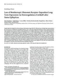
FIGURE LEGENDS: Figure 1. Splicing variants of AtGLR3.5. A) Alignment of N-terminal regions of the two AtGLR3.5 isoforms. Aminoacid sequences are shown.
Predicted mitochondrial targeting sequence (TargetP) is in red box. According to
ChloroP1.1, the chloroplast targeting sequence is 82 aminoacid long. B) Subcellular localization of the AtGLR3.5 isoforms with predicted mitochondrial (isoform 1) and chloroplast (isoform 2) targeting. Expression of pGREAT-2x35S-GLR3.5v1::DsRed2 (upper part) and pGREAT-2x35S-GLR3.5v2::EGFP (lower part) in 4 week-old Arabidopsis leaves.
The
isoform1-DsRed2 fusion protein is located in highly motile structures resembling mitochondria (see video 1 and 2). Bar: 20 µm. Results shown are from 2 independent experiments and are representative of 3 independent experiments giving the same results.
Figure 2. Mitochondrial localization of splicing variant 1.
Downloaded from www.plantphysiol.org on December 11, 2014 - Published by www.plant.org Copyright © 2014 American Society of Plant Biologists. All rights reserved.
22 A) Relative expression of the two transcript variants. Analysis of the relative amount of each variant in different tissues/organ of 4-week old Arabidopsis Col-0 plants. Actin 2 (At3g18780) was used as reference gene. B) Transcript analysis of Arabidopsis SALK mutant atglr3.5-1. PCR was performed with primers reported in Table I. Lower panel: positive control of transcript content (actin). This result was confirmed also on another set of plants. C) AtGLR3.5 isoform 1 was expressed in vitro using a Wheat Germ Lysate Kit. Empty reaction mix (WGL) and mix following expression of the protein (G35V1) were loaded on SDS-PAGE and assayed with anti-His tag and anti-GLR3.5 antibodies; D) Western blot of mitochondria purified from wild-type and knockout plants (atglr3.5-1) (100 µg each) assayed with anti-GLR3.5 antibody. The membrane shown in the lower part was colored with Ponceau Red and shows comparable loading. Results in C) and D) are representative of 3 experiments.
Figure 3. Topology of GLR3.5 and differences in mitochondrial dimensions in the absence of GLR3.5. A) Confocal images of protoplasts expressing GLR3.5-SypHer and mitochondrially targeted GFP (left and right panels, respectively). Lower left panel shows the effect of acidification on SypHer fluorescence ratio (via addition of nigericin to the cells) (n=4 ±SEM). Lower right panel: predicted topology (ATD: amino-terminal domain, S1 and S2: glutamate-binding domains). B) to E) show TMRM staining of intact 10-day-old seedling roots from WT, KO and silenced plants (#1 and #2), respectively. F) Statistical analysis of distribution of mitochondria size revealing significant differences (**p